
記住我

The present study was designed as a prospective clinical imaging study on patients with SLL tears diagnosed earlier on direct MR arthrography. Before the study, institutional review board approval (Ethical Committee, Medical Faculty, Heinrich-Heine-University, Düsseldorf, Germany, study number 2019-590) and individual written informed consent were obtained. The minimum patient number was determined as 10 based on the initial four patients, using dedicated online software (http://www.statstodo.com) and the following statistical parameters: power 0.8; probability of type-I-error 0.01; assumed effect size 1.4 [defined as the mean paired difference divided by the expected standard deviation], two-tailed procedure.
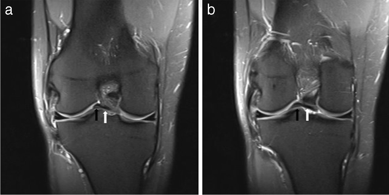
Consequently, ten patients (male: 8; female: 2; mean age: 41.2 ± 18.1 years, age range: 24 – 66 years) with partial or complete SLL tears were included (Table 1). Isolated tears of the dorsal or volar component of the SLL complex, defined as a discontinuous appearance of the ligament and consecutive entry of contrast agent, were defined as partial SLL tears. High signal intensity within individual ligament components was not considered diagnostic of a partial SLL tear. Partial SLL tears (n = 6 [3 right, 3 left]) involved the dorsal and volar components in 4 and 2 patients. In contrast, complete SLL tears (n = 4 [2 right, 2 left) were presumed when both components appeared discontinuous.
Table 1 Patient demographics and clinical MR imaging informationThe real-time MRI studies were performed 21 ± 10 months (range 7–35 months) after the initial injury and 10 ± 11 weeks (range 2–33 weeks) after the direct MR arthrography. At the time of the real-time MRI studies, all patients still suffered from mild or moderate chronic wrist pain. To ensure the absence of tear progression or re-injury to the symptomatic wrist, all patients underwent a thorough medical review of their clinical history, a physical examination to detect any potential indications of recurrent injury to the affected wrist, and a thorough imaging evaluation utilizing morphologic, static MR imaging.
Each patient’s contralateral wrist served as the reference. Consequently, acute or chronic wrist pain or a history of wrist trauma or surgery were defined as exclusion criteria. In the following, “injuredpartial” and “injuredcomplete” refer to the wrists of patients with partial and complete SLL tears, respectively, while “controlpartial” and “controlcomplete” refer to the respective (healthy) contralateral wrists.
MR imagingIn the same session, all patients underwent morphologic standard (i.e., static) and real-time (i.e., dynamic) MRI of both wrists, i.e., the injured and the healthy side. MR images were acquired on a clinical 1.5T scanner (MAGNETOM Avantofit, Siemens Healthineers, Erlangen, Germany) using two different imaging setups (Fig. 1).
Fig. 1
Details of the MR imaging setups. A) During static imaging, morphologic sequences were acquired with a standard 4-channel flex coil centered around the wrist. B-D) Setup for real-time MRI measurements: The MRI-compatible motion device is shown without (B) and with (C) a patient’s right forearm and hand. The hand was positioned on the mobile sliding plate (#) and, thus, guided along a pre-defined semi-circular range on the immobile base plate (x). Tourniquets were used to immobilize the forearm (*). The loaded and operational device (D) was covered with an 18-channel body coil (§) placed on top of the transparent spacers and centered around the wrist. A 32-channel spine matrix coil was positioned underneath the device – left out for clarity
First, static imaging was performed using a 4-channel flex coil (Flex Coil small, Siemens Healthineers) wrapped around the wrist for an optimized signal-to-noise ratio (Fig. 1A). Patients were placed in a prone, head-first position with the examined arm stretched out (“superman position”). Coronal, axial, and sagittal proton density-weighted (PDw) fat-saturated (fs) sequences and axial T2-weighted sequences were obtained for morphologic evaluation (Table 2).
Table 2 Acquisition parameters of MRI sequencesSecond, for dynamic imaging by real-time MRI, wrists were positioned on a custom-made MRI-compatible motion device that guides the wrist along a pre-defined semi-circular range and standardizes the motion plane during active radioulnar motion (Fig. 1B) [16]. The hand was placed on a mobile sliding plate with an under-surface covered with synthetic polytetrafluoroethylene (“Teflon”, DuPont, Wilmington, DE, US) to reduce friction. The wrist was centered on the pivot point, allowing for a semi-circular motion of 60° on each side. Two tourniquets were used to attach the forearm to the device, thus decreasing adaptive forearm motion (Fig. 1C). The loaded motion device was positioned centrally in the scanner’s bore with an 18-channel body coil (Body 18, Siemens Healthineers) centered on the radiocarpal joint and placed on top of the device (Fig. 1D). A 32-channel spine matrix coil (Direct connect spine 32, Siemens Healthineers) was positioned underneath the device.
Other than the SLL tears described above, no additional ligamentous and osseous injuries of the carpus were found in patients. D.B.A. (board-certified clinical radiologist with six years of experience in musculoskeletal imaging) assessed all static MRI examinations performed in this study while remaining blinded to the diagnosis obtained from the previously acquired direct MR arthrography, the surgical findings, and also to the information which wrist served as the healthy control and which wrist was the injured wrist during the interpretation process, thereby ensuring an unbiased assessment of the static MR images.
Real-time MRIFor real-time MR imaging during active radioulnar abduction, 300 images and 15 pre-scans were acquired of each patient’s wrists in 30 s. We used a temporally optimized radial FLASH real-time MRI sequence validated before [16] that is characterized by pronounced radial undersampling, resulting in a temporal resolution of 95 ms per image (Table 2). Patients were instructed to avoid out-of-plane motion, including pro- or supination. All patients were instructed to continuously perform active radioulnar motion at a standard frequency of 15 s per complete cycle, i.e., from maximum radial abduction to maximum ulnar abduction and vice versa. Two complete cycles were acquired per patient. The coronal image plane was defined in the neutral position and aligned with the center of the scapholunate joint as defined on sagittal and axial scout views. On average, magnet time was 30 minutes per patient, i.e., 15 min per wrist.
Quantitative analysis of the scapholunate and lunotriquetral jointsFor the evaluation of the dynamic MR images, we employed a previously developed and validated framework to quantify scapholunate and lunotriquetral joint widths [16]. This framework employs an automated process for the semantic segmentation of the wrist and forearm, utilizing a convolutional neural network. Based on the segmentation outlines, advanced post-processing techniques are applied to determine the joint widths as a function of wrist angle. Notably, owing to the entirely automated nature of this process, no radiologist was engaged in the quantitative analysis of the dynamic MR images, thus ensuring an unbiased assessment of the dynamic MR images.
First, using a Gaussian anti-aliasing filter, the images were bi-quadratically interpolated from 168×168 pixels to 336×336 pixels. Then, the outlines of the distal radius, distal ulna, scaphoid, lunate, triquetrum, hamate, capitate, trapezium, and trapezoid were automatically segmented using a U-net based convolutional neural network. The segmented outlines of the scaphoid, lunate, and triquetrum were smoothed and connected using a centreline to determine scapholunate and lunotriquetral joint widths automatically. The scapholunate joint width was determined as the distance between the intersections of the centreline with the scaphoid’s ulnar cortex and the lunate’s radial cortex. Correspondingly, the lunotriquetral joint width was determined as the distance between the intersections of the centreline with the lunate’s ulnar cortex and the triquetrum’s radial cortex.
Additionally, the wrist angle was determined using minimal bounding boxes around the distal carpal bones, i.e., hamate, capitate, trapezium, and trapezoid, and around the bone contours of the distal radius and the distal ulna. Subsequently, the angle between the centers of the two bounding boxes was quantified as the wrist angle [16]. The wrist angles were grouped into 5° intervals for visualization and comprehensibility.
Statistical analysisStatistical analyses were performed by L.M.W. using GraphPad Prism (v9.5.0, San Diego, CA, US). Data are presented as mean ± standard deviations. Differences (Δ) in joint widths were determined between the injured wrist and the same patient’s contralateral healthy wrist as a function of wrist position (intraindividual). Absolute joint widths were comparatively evaluated between patients (interindividual). Assuming normal distributions of the scapholunate and lunotriquetral joint widths, the respective widths were compared group-wise across the entire range of motion (ROM) using parametric tests. Mixed effects analysis-of-variance (ANOVA) was used for the group-wise analysis of joint widths across the entire ROM. In contrast, two-way ANOVA was used for the subsequent group-wise comparison of injured and healthy wrists (intraindividual). Mixed-effects ANOVA was also used for comparing joint widths between groups at distinct ROM intervals (interindividual), followed by select Tukey’s multiple comparison posthoc tests. To reduce the number of statistically significant yet (most likely) clinically irrelevant findings, we set the significance level to p≤0.01. Multiplicity-adjusted p-values are reported to account for the multiple comparisons involved.
留言 (0)