Overview of Coccydynia
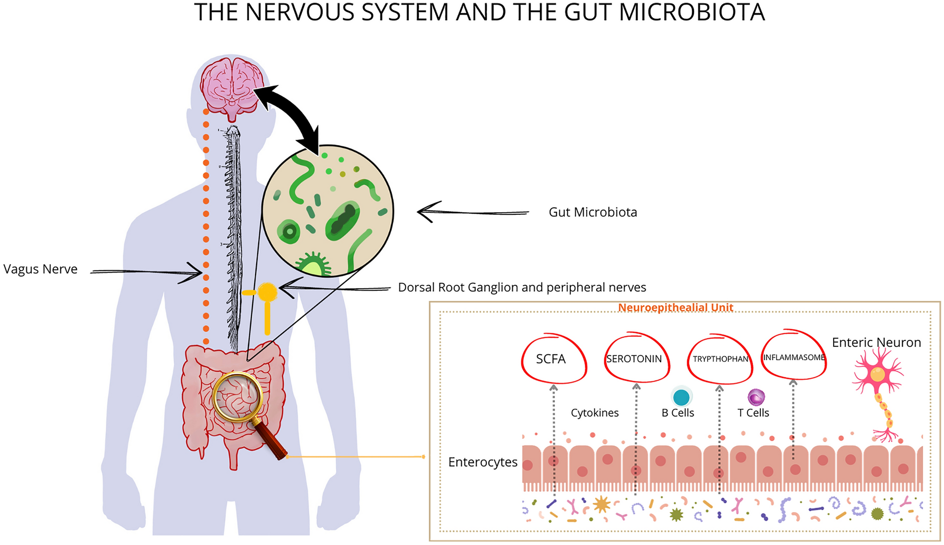
Coccydynia or coccygodynia is a debilitating condition that affects many individuals and can cause significant discomfort and decreased quality of life. The coccyx is located at the bottom of the spine and is susceptible to injury or damage, resulting in pain. It can be caused by a variety of traumatic or non-traumatic factors [1]. When there is an injury, inflammation, or irritation in the coccyx region, nociceptors, specialized pain receptors, are activated. These nociceptors send signals to the nearby somatic nerves. The somatic nerves then transmit these nociceptive signals to the central nervous system, where they are processed, leading to the perception of pain. The ganglion impar or Walther ganglion, which is in the retroperitoneal space behind the rectum around the sacrococcygeal joint or directly in front of the coccyx, may also be involved in the transmission of pain signals [2]. The coccygeal plexus plays a crucial role in providing sensory input from the coccyx and comprises the 4th and 5th sacral nerves and the coccygeal nerve. Neuropathic pain is caused by damage or irritation of these nerves. Additionally, pain in the coccygeal area may also stem from pelvic organs, as pain signals travel through the superior hypogastric plexus consisting of sympathetic fibers. Moreover, the pelvic splanchnic nerves, which carry parasympathetic nerves from the second, third, and occasionally fourth sacral segments, can also convey this type of pain [3, 4].
Refractory coccydynia is characterized by persistent pain in the coccyx that does not respond to conservative or non-surgical treatment approaches and can cause significant discomfort and decreased quality of life. Several studies have shown that chronic pain is linked to synaptic plasticity and alterations in the central nervous system (CNS), impacting neural areas that regulate pain. This condition involves structural and functional changes in the corticolimbic brain region [5,6,7]. In some cases, there is a discrepancy between the underlying cause and the severity of pain. Central sensitization, characterized by amplified neural signaling in the CNS, is considered a potential explanation, leading to hypersensitivity [8, 9].
Management of coccyx pain involves a comprehensive approach of pharmacological and non-pharmacological interventions. The choice of treatment depends on the severity of the condition and the patient’s response to previous treatment [10, 11].
Pharmacological treatments often include the use of nonsteroidal anti-inflammatory drugs (NSAIDs) along with topical analgesics. Opioid medications may be prescribed, particularly in cases of severe pain. In patients with pelvic hypertonia or muscle spasms, muscle relaxants and benzodiazepines are also prescribed. Antidepressants and anticonvulsants are effective for neuropathic and CNS hypersensitivity pain. A systematic review/meta-analysis highlights palmitoylethanolamide (PEA) as a well-tolerated and efficacious treatment for chronic neuropathic and inflammatory pain [12,13,14]. Non-pharmacological treatments emphasize conservative approaches including ergonomic adaptations, physical therapy, manipulation, various injections, and alternative therapy.
Physical therapists may employ modalities and techniques to reduce pain, improve mobility of the coccyx and surrounding tissue structures, and educate patients on lifestyle adjustments such as postural education, work ergonomics, and defecation mechanics. In addition, soft tissue mobilization and manual therapy may help to reduce muscle spasms and enhance the flexibility of surrounding tissues [15, 16].
Local injections around the coccyx, trigger point injections, caudal epidural injections, ganglion impar blocks, and radiofrequency ablation (RFA) can also be administered to alleviate coccyx pain. These injections often contain local anesthetics and corticosteroids [17,18,19].
Some other therapies like extracorporeal shock wave therapy (ESWT), acupuncture, prolotherapy, and botulinum toxin injections have shown promise in treating coccyx pain [20,21,22,23].
In refractory cases, partial or complete coccygectomy may be considered as a last resort [24].
Overview of Neuromodulation Techniques
Neuromodulation (NM) is an umbrella term encompassing a spectrum of relatively new and minimally invasive techniques that have shown promising results in managing various chronic pain conditions. However, there is limited data regarding its efficacy in the treatment of coccydynia.
Several NM methods are available to manage chronic coccydynia, these include spinal cord stimulation (SCS), dorsal root ganglion stimulation (DRG-S), peripheral nerve stimulation (PNS), peripheral nerve field stimulation (PNFS), and sacral neuromodulation (SNM).
The traditional approach to SCS involves using 30–60 Hz of electrical stimulation to activate the dorsal columns, resulting in a tingling sensation or paresthesia in the painful areas of the body experienced by a patient and has been found to be effective in managing persistent pain conditions. SCS, rooted in gate control theory, stimulates non-nociceptive fibers in dorsal columns, closing a spinal gate to relieve pain. It triggers inhibitory interneurons, releasing GABA in the spinal dorsal horn [25,26,27]. Recent studies explore cortical and subcortical involvement. Neuroimaging shows cortical changes, suggesting direct effects from dorsal column stimulation or inhibition of nociceptive signals. Functional magnetic resonance imaging (fMRI) studies reveal modulation of brain regions linked to the spinothalamic tract responsible for pain transmission [28,29,30,31].
High-frequency SCS, also known as HF10, is an innovative form of neurostimulation treatment that employs the use of high-frequency stimulation at low amplitudes, ensuring that the stimulation remains below the sensory activation threshold and is paresthesia-free. Empirical observations have determined that the first electrode of one lead is usually placed at the top of the T8 vertebra and the last electrode of the second lead is placed at the bottom of the T11 vertebra with some overlap of the leads at the T9/T10 disc for managing back and leg pain [32]. The mode of operation involves reducing the activity of wide dynamic range (WDR) neurons, and by delivering high-frequency stimulation, it is possible to decrease WDR responsiveness and thereby alter the perception of chronic spinal pain. This method offers safe and long-lasting relief from pain in groups of individuals suffering from chronic pain [33,34,35].
Burst SCS is another technique that utilizes a burst frequency of 40 Hz and a pulse frequency of 500 Hz, by decreasing the amplitude to alleviate pain while minimizing or eliminating paresthesia [36].
DRG-S is a more focused type of NM due to the unique anatomy and physiology of the DRG. The DRG is located inside the neural foramen and the neurons in the DRG provide very specific dermatomal sensory information. Unique to the DRG is the thin surrounding layer of cerebrospinal fluid which prevents the propagation of current to the spinal cord or ventral root, which may explain the mechanism through which DRG-S relieves neuropathic pain [37,38,39].
PNS refers to the application of electrical stimulation to a particular nerve trunk through implanted electrodes placed under the skin to activate the region of affected nerves, cutaneous afferents, or the dermatomal distribution of the nerves instead of targeting the epidural space or a nerve bundle directly and the stimulation is directed towards nerves that converge back to the spinal cord [40].
PNFS also known as subcutaneous field stimulation is an emerging technology that utilizes electrical impulses to activate the region of affected nerves, cutaneous afferents, or the dermatomal distribution of the nerves instead of targeting the epidural space or a nerve bundle directly and the stimulation is directed towards nerves that converge back to the spinal cord. Some studies have shown the effectiveness of this modality in managing chronic pain [41, 42].
One specific form of NM is SNM, a type of nerve stimulation that aims to induce changes in the functioning of sacral nerves and the signals they transmit to the brain. SNM utilizes electrical stimulation to activate the afferent sacral nerve roots through the implanted electrode percutaneously and can be utilized through different percutaneous approaches such as retrograde lumbar, anterograde sacral hiatus, and transforaminal approaches. Each approach has its pros and cons. The retrograde approach is technically complex, posing risks like dural puncture and cerebrospinal fluid leakage. In contrast, the anterograde approach is simpler but can be challenging because of limited subcutaneous tissue, increasing the chance of skin erosion. The transforaminal approach minimizes dura-related risks compared to retrograde access, but it has a drawback of increased potential for lead migration compared to the sacral hiatus route [43,44,45,46,47,48,49,50].
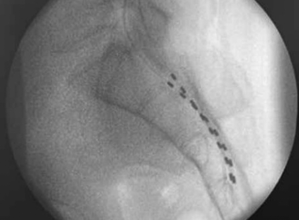
The process of NM consists of two main steps performed in an outpatient setting. Before undergoing a procedure, it is essential to conduct a psychological assessment. This assessment helps to understand how underlying psychosocial risk factors may impact clinical outcomes and aids in managing patient expectations. In the first step, patients undergo a trial stimulation, which allows them to assess the potential alleviation of pain. Trial therapy is more beneficial for predicting long-term success in patient selection compared to relying solely on clinical screening. This preference stems from the lower confidence associated with clinical screening alone, which can be influenced by various factors. The trial involves a minor surgical procedure where leads are inserted using an epidural Tuohy needle and are linked to a temporary external stimulator. If the therapy proves its effectiveness by typically achieving a more than 50% pain reduction, the next step involves the placement of a permanent system. This requires a small incision to implant the leads and to create a subcutaneous pocket in the buttock area. The leads are connected to an implantable pulse generator (IPG) to provide ongoing stimulation and are positioned within the pocket under fluoroscopy. Patients can also manage the therapy using a wireless programmer.
The procedures share similarities across different NM techniques. However, there are a variety of factors such as trial length and approaches, stimulation settings, the number of leads implanted, the placement of leads, and whether the device is rechargeable that distinguish the modalities from one another [42, 51,52,53,54].
The implanted NM system does not restrict the patient’s activities. However, it is crucial to consider that high-frequency diathermy and unipolar electrocoagulation are not recommended. Additionally, during extracorporeal shockwave lithotripsy, the focal point should not be near the neuromodulator or its lead. Ultrasound and radiotherapy in the area where the implanted components are located should also be avoided. NM should not be used during pregnancy [55].
Ensuring MRI safety for implantable devices is complex but crucial. Manufacturers’ guidelines are necessary for safe scans, and adherence to specific conditions is vital to mitigate risks [56, 57].
Like any medical procedure, NM is not without risks. Complications associated with NM include infection, device malfunction, lead migration, and nerve injury. However, the incidence of these complications is relatively low, and most patients tolerate the procedure well [58, 59].
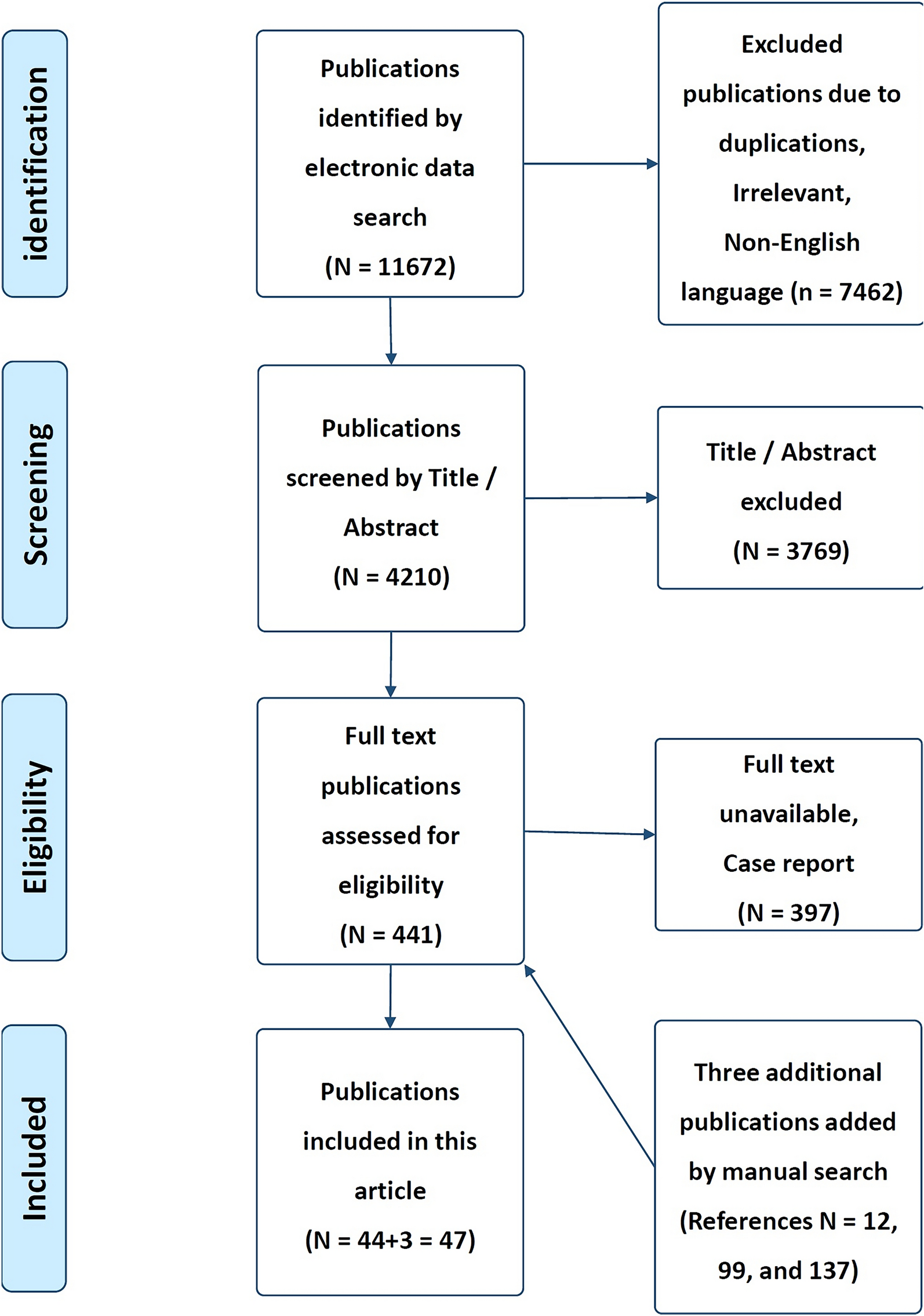
The aim of this review is to summarize the utility of current NM techniques in treating chronic refractory coccydynia and their respective effectiveness based on the available body of knowledge in the scientific literature at this time.

留言 (0)