
記住我
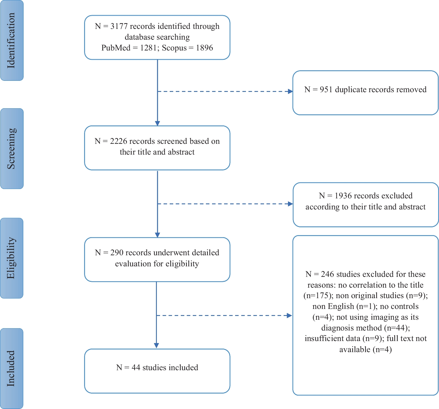
A total of 42 participants with 210 cervical intervertebral discs were included in the study. Out of these, 197 discs were finally analyzed while 13 were excluded due to sclerosis artifacts (n = 5) and severe narrowing of the disc space (n = 8). The participants’ demographics and clinical information are shown in Table 1.
Table 1 Characteristics of the participants (n = 42)Quantitative Image AnalysisDLIR-M and DLIR-H significantly improved the SNR and CNR of IVDs in 70 keV images compared with ASiR-V (all p < 0.001) (Table 2). The SNR values for ASiR-V, DLIR-M, and DLIR-H were 5.73 ± 2.5, 6.93 ± 2.8, and 8.43 ± 3.3, respectively, while the CNR values for ASiR-V, DLIR-M, and DLIR-H were 3.86 ± 1.2, 4.75 ± 1.8, and 5.78 ± 1.9, respectively.
Table 2 Quantitative image analysis among three reconstruction image setsIn water (iodine) image set, there was no significant difference among the ASiR-V, DLIR-M, and DLIR-H algorithm (all p > 0.05). However, the DLIR algorithm showed significantly lower SD and CV for WC compared with ASiR-V (all p < 0.001) (Table 2). Specifically, the SD values for ASiR-V, DLIR-M, and DLIR-H were 8.39 ± 2.5, 7.62 ± 2.5, and 6.9 ± 2.4, respectively, while the CV values for ASiR-V, DLIR-M, and DLIR-H were 0.00843 ± 0.0070, 0.00707 ± 0.0023, and 0.00635 ± 0.0020, respectively.
In water (calcium) or VNCa image set, the DLIR-H algorithm exhibited the lowest SD and CV. However, there was no statistically significant difference in SD and CV among DLIR-H, DLIR-M, and ASiR-V algorithms (all p > 0.05).
Qualitative Image AnalysisThe subjective score for diagnostic acceptability and conspicuity of IVDs of ASiR-V, DLIR-M, and DLIR-H image sets rated by two readers was shown in Table 3. Regardless of 70 keV images, water(iodine) images, or VNCa images, good to excellent interobserver agreement were noted (Table 3). Moreover, the interobserver agreement was highest in the reconstructed image set based on DLIR-H (0.74 for 70 keV; 0.76 for water (iodine); 0.83 for VNCa images).
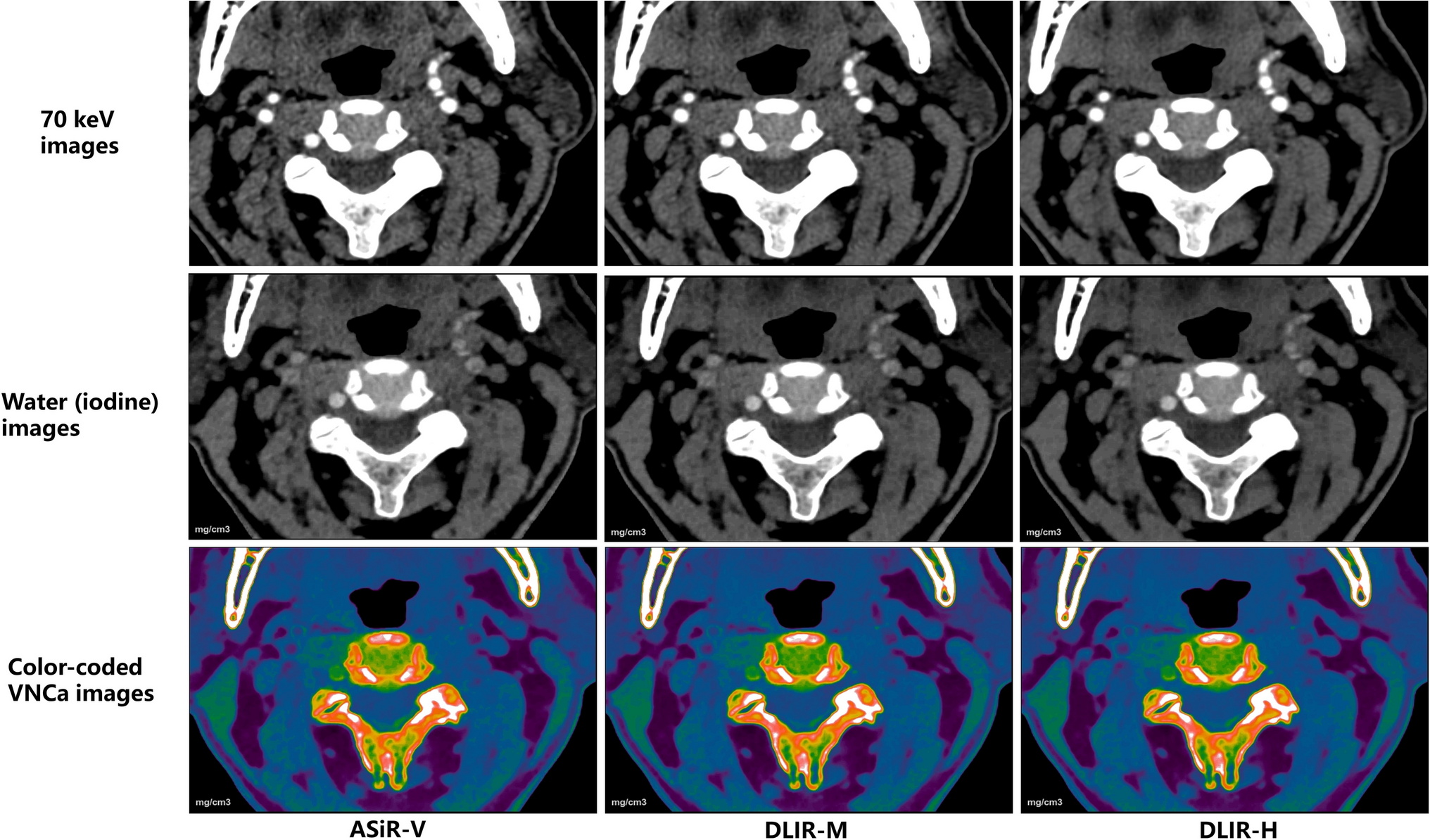
Table 3 Subjective ratings for qualitative image analysis by two reviewersThe comparison of qualitative analysis of three reconstruction image sets (ASiR-V, DLIR-M, and DLIR-H) by both readers was shown in Fig. 2. The DLIR algorithm improved the display and diagnostic acceptability of each IVD in both 70 keV (Fig. 1a, g) and water (iodine) image sets (Fig. 1b, h). This improvement was more pronounced as the strength of DLIR algorithm increases. In contrast, there were no significant differences observed for IVD display in color-coded VNCa image set using the three reconstruction algorithms (Fig. 1c, i).
Fig. 2
Box and dot plots compared image quality (diagnostic acceptability and conspicuity) of 70 keV (a, g), water (iodine) (b, h) and color-coded virtual noncalcium (VNCa) (c, i) image sets among different reconstruction algorithm, and iterative reconstruction-Veo (ASiR-V) (d, j), deep learning image reconstruction algorithm at medium level (DLIR-M) (e, k), at high level (DLIR-H) (f, i) among different image sets by both readers (a–f for reader 1; g–i for reader 2). **** indicates p < 0.001, *** indicates p < 0.01, and ns indicates means result was not statistical significance
Among three different image sets, the 70 keV image set showed the lowest subjective score. It was followed by the water (iodine) image set, while the color-coded VNCa image set was found to be most favorable for disc evaluation in both ASiR-V and DLIR-M algorithms (Fig. 1d–f, j–i). Using the DLIR-H algorithm, the grayscale water (iodine) image set showed comparable results to the color-coded VNCa image set in evaluating IVDs (Fig. 1f, i).
Diagnostic Accuracy for IVD Abnormalities per DiskAccording to the consensus of two senior radiologists, out of the 197 cervical intervertebral discs (IVDs) evaluated, 77 (39%) were classified as abnormal, while 120 (61%) were classified as normal. The abnormalities observed included bulging in 25 cervical IVDs (12.6%) and herniation in 52 cervical IVDs (26.4%). The diagnostic performance of 70 keV, water (iodine) and VNCa image sets using three reconstruction algorithms for detecting IVD abnormalities is summarized in Table 4. The VNCa image set showed superior diagnostic accuracy, sensitivity, specificity, positive predictive value, and negative predictive value compared to the 70 keV and water (iodine) image sets. Among three different algorithms used for the VNCa image set, DLIR-M demonstrated higher diagnostic accuracy than ASiR-V and DLIR-H. However, these differences were not statistically significant (compared to DLIR-H, p = 0.446; compared to ASiR-V, p = 0.675).
Table 4 Diagnostic efficiency of 70 keV images, water (iodine) images, and VNCa images of three reconstruction image sets for IVD herniation per diskThe inter-rater agreement for detecting IVD abnormalities using ASiR-V, DLIR-M, and DLIR-H was good (κ = 0.71, 0.72, and 0.69, respectively) for the 70 keV image set. It was excellent for the water (iodine) image set (κ = 0.91, 0.85, and 0.86, respectively) and the VNCa image set (κ = 0.86, 0.89, and 0.85, respectively).
留言 (0)