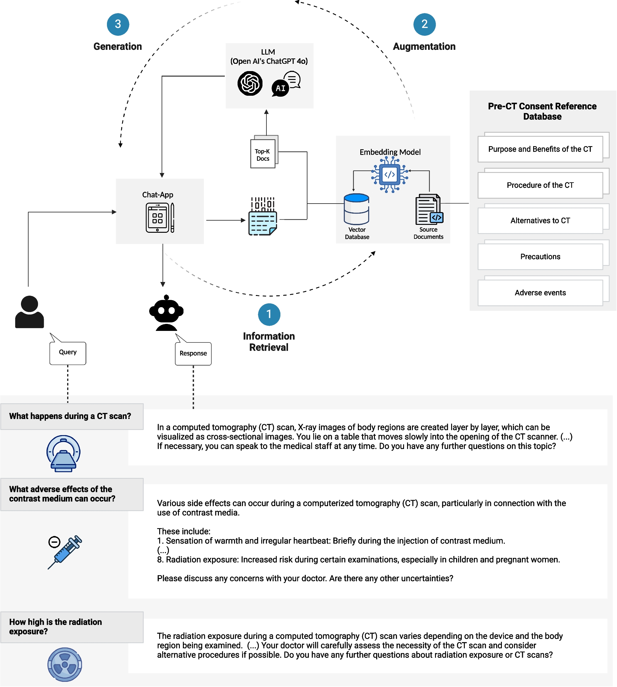
In this study, we evaluated the performance of a novel deep learning-based MCA by comparing IQ of 64-MDCT-acquired CCTA images. As in previous studies, the RCA and its segments 1 and 2 were found to be most prone to motion artifacts, as these are the most motile vessel segments [7]. MCA reconstruction had the greatest effect in these segments in improving IQ and reducing the total number of motion artifacts. Baseline IQ of LAD and LCx per-artery and per-segment was initially much better; MCA-improvement of LAD and LCx was negligible. On the per-patient level, we observed an overall improvement of IQ. By evaluating potential disturbers, we found a significant negative correlation between mean HR and IQ for RCA, LAD, and LCx in CA- and MCA-reconstruction. However, the influence of mean HR was strong in the CA-reconstruction and intermediate in the MCA-reconstruction of the RCA. Correlation between mean HR and IQ of LAD and LCx was weak in both CA and MCA. BMI, age, sex, and ΔHR had no significant impact on IQ.
Recently, various MCA-based approaches have been published to mitigate motion artifacts. Two vendor-specific MCA are currently available (2023): SnapShot Freeze (SSF) 1 and its successor SSF2 (GE Healthcare, Waukasha, WI, USA) [13, 20]. In the clinical setting, SSF1 improved IQ and interpretability in ≥ 64-MDCT independent of HR and BMI [21, 22]. In addition, good IQ was maintained even at high HR, allowing wider application of PGI leading to a lower total effective dose [21, 23]. Therefore, SSF1 is considered a useful tool to assist CCTA in CCS diagnosis [12]. Positive effects of SSF2 on IQ are even more profound compared to its predecessor [13]. Unfortunately, both MCA are vendor-specific and only applicable on vendor-specific CT scanners [17]. Besides SSF1 and 2, there have been several attempts to develop even more effective and widely applicable MCA [11]. However, most of these suffer from limitations due to high effective dose, poor performance at high or irregular HR, or long computation time [11, 16, 24, 25]. The recently introduced deep learning-based MCA might be a solution. Deep learning-based MCA can be applied post-acquisitionally without the need for raw data [26]. By this, they have a very short computation time and can be used vendor-independently [11, 15]. However, larger studies on the performance of deep learning-based MCA are still scarce. Therefore, their clinical applicability cannot yet be assessed although phantom studies are promising [11, 15, 25].
In this study, we have found that the applied deep learning-based MCA Deep PAMoCo improves the IQ of 64-MDCT-acquired images [13, 15]. By this, the rate of non-diagnostic images and false-positive results could be remarkably reduced, especially at higher HR [22, 27, 28]. As CCTA is already considered to have a high-negative predictive value, this could further increase its validity for the diagnosis of CCS [1]. Especially regarding the limited temporal resolution of 64-MDCT, the presented MCA seems to be attractive to enhance 64-MDCT-acquired images. However, the applied MCA can also be expected to be useful in combination with high-end imaging technology, as high or irregular HR can also disturb ≥ 128-MDCT and DSCT imaging [29]. Besides IQ improvement, the tested MCA could also reduce the effective dose during CCTA, as PGI could be applied at higher HR, and by this more widely [21, 23]. However, as IQ still correlated with HR at an intermediate level, the presented MCA should be considered as a support and not as a substitute for HR control [30]. Finally, the tested MCA seems to be especially attractive in regard to its broad applicability due to its short computation time of 15s per entire CCTA image and its vendor-independent use [11, 15, 17]. Thus, the presented MCA resembles a low-effort software upgrade for CCTA imaging performed with a 64-MDCT.
This study has limitations. Firstly, since we wanted to test the ability of the MCA to compensate for motion artifacts and to improve IQ, patient data were not given in this trial. Secondly, in this study, we had to exclude eleven images completely and two partially because of stack transition, vessel calcifications, and medical devices (stents and pacemakers) producing massive artifacts. In addition, due to a lack of documentation, we were unable to determine BMI in 20 patients and mean HR, ΔHR, age, and sex in 9 patients. Thirdly, the evaluation of IQ was conducted by a sole professional. Consequently, we cannot provide an inter-observer agreement. Fourthly, the IQ assessment was conducted by employing a 5-point Likert score, consistent with previous research [19]. However, it is essential to note that there is no officially recommended approach for evaluating IQ, and therefore, the assessment lacks standardization. Consequently, the comparability with studies utilizing different assessment scores is restricted. Fifthly, the primary objective of this study was to evaluate the performance of the applied MCA in enhancing the IQ of real patient CCTA images. It is crucial to emphasize that the findings should not be generalized to other deep learning methods, given our limited study population and the focus on a sole MCA. Sixthly, this was a single-center study. We recommend further studies at other radiology centers to increase the power and validity of our findings. Moreover, as this study aimed to evaluate the impact of a deep learning-based MCA on IQ, we cannot draw conclusions regarding its clinical utility. Further research is needed to evaluate the impact of MCA on diagnostic accuracy e.g. using invasive coronary angiography as a reference. Thus, it would also be possible to evaluate the impact of vessel calcification on IQ and MCA-related effective dose reduction. Finally, we did not compare the tested MCA with vendor-specific or other MCA. Thus, we cannot determine the superiority of the presented MCA.


Comments (0)