Microstructural Changes in the Brainstem Auditory Pathway in Children With Hearing Loss
Objective
To assess the utility of diffusion tensor imaging of the auditory pathway in children with sensorineural hearing loss (SNHL).
Study Design
Retrospective cohort study.
Setting
A single academic tertiary children's hospital.
Patients
Sixteen pediatric patients with bilateral SNHL of at least moderate severity in the poorer ear (eight male; mean age, 5.3 ± 4.9 yrs). Controls consisted of age- and sex-matched children with normal hearing who were imaged for nonotologic, non-neurologic medical concerns and found to have normal magnetic resonance imaging (MRI).
Interventions
Three Tesla MRI scanners were used for diffusion tensor imaging.
Main Outcome Measures
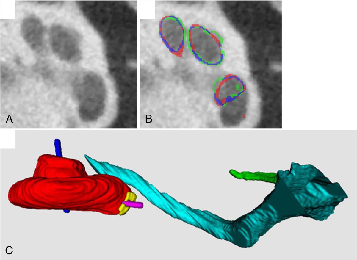
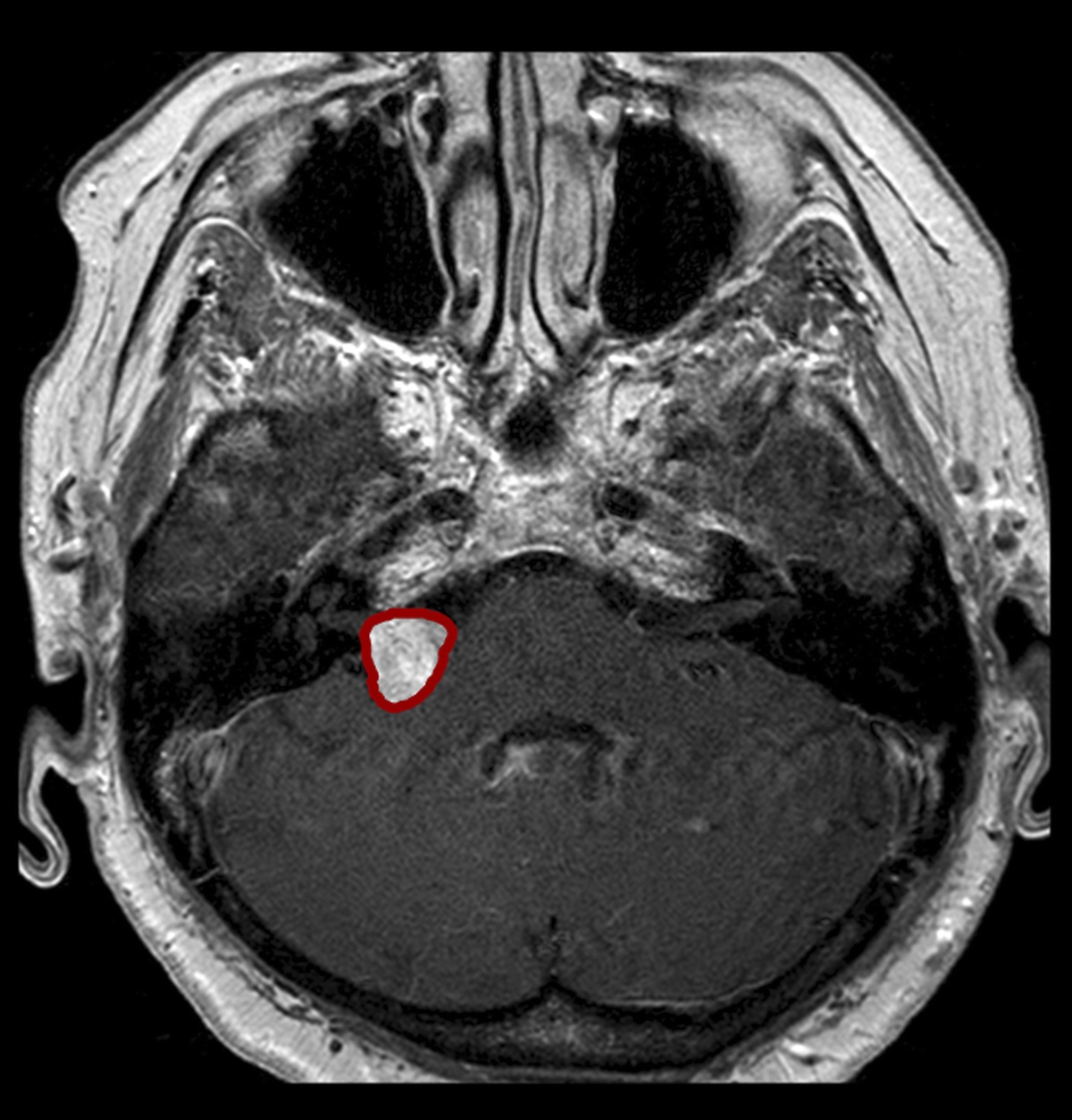
Quantitative diffusion tensor metrics were extracted from the superior olivary nucleus (SON), inferior colliculus (IC), and ipsilateral fiber tracts between the SON and IC delineated by tractography.
Results
We identified differences in fractional anisotropy of the SON between the SNHL cohort and controls (0.377 ± 0.056 vs. 0.422 ± 0.052; p = 0.009), but not in the IC. There were no differences in the mean diffusivity (MD) values in the IC and SON. Among younger children (≤5 yrs), MD was decreased in the SNHL cohort compared with controls in the IC (0.918 ± 0.051 vs. 1.120 ± 0.142; p < 0.001). However, among older children (>5 yrs), there were no differences in MD (1.124 ± 0.198 vs. 0.997 ± 0.103; p = 0.119). There were no differences in MD or fractional anisotropy in the white matter fibers of the IC–SON tract.
Conclusions
Our results suggest abnormal neural tracts along the central auditory pathway among children with SNHL. Longitudinal studies should assess the prognostic value of these MRI-based findings for assessing long-term outcomes and determining intervention efficacy.


Comments (0)