Social isolation, experienced during early development or in adulthood, has detrimental consequences for physical and mental health. Previous studies have shown that low social integration increases all-cause mortality by up to a 50%, which exceeds the risk associated with obesity, moderate alcohol intake, smoking, and a sedentary lifestyle (Holt-Lunstad et al., 2010). Clinical and pre-clinical studies suggest that social isolation is a risk factor for the development of Alzheimer's disease, Parkinson's disease, epilepsy, and depression (Holt-Lunstad et al., 2010; Leigh-Hunt et al., 2017; Valtorta et al., 2016). In the population-based Heinz-Nixdorf-Recall study, we found that social isolation predisposes to incident ischemic cardiovascular and cerebrovascular events and elevates mortality (Gronewold et al., 2021; Gronewold et al., 2020). Given the COVID-19 pandemic, which has resulted in repeated periods of quarantine and a loss of social contacts (Casale and Flett, 2020; Lin, 2020; Twenge and Joiner, 2020), there is a need to investigate the effect of social isolation on the ischemic brain tissue (Gronewold and Hermann, 2021).
Stroke is the leading cause of long-term disability and second leading cause of death worldwide, with a growing incidence in developing countries due to rapid growth and aging of the population (Feigin et al., 2021a; Tsao et al., 2022). Approximately one in six people dying from cardiovascular diseases are due to stroke and the large majority of all stroke cases are ischemic strokes (Tsao et al., 2022). Considering the dramatic impact of stroke on patient mobility and independence, along with its significant public health burden, it is critical to identify the mechanisms that contribute to stroke impairments and predict poor outcome (Feigin et al., 2021b; Leys et al., 2005). Previous studies in rodent models of focal and global cerebral ischemia indicated that social isolation increases brain injury, impairs sensorimotor recovery, and compromises synaptic plasticity (Craft et al., 2005; Karelina et al., 2009; O'Keefe et al., 2014; Venna et al., 2012; Venna et al., 2014a; Verma et al., 2016; Weil et al., 2008). Furthermore, social isolation was shown to alter the immune response to stroke by down-regulating brain IL-6 and upregulating peripheral blood IL-6 levels (Karelina et al., 2009) and to decrease neurogenesis by reduced BDNF levels (O'Keefe et al., 2014; Verma et al., 2016). While several studies have demonstrated the influence of social isolation on stroke outcome, there is currently a lack of systematic research examining the molecular mechanisms via which social isolation affects the brain.
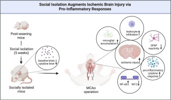
In this study, we characterized molecular signals and pathways influenced by social isolation using a broad set of immunofluorescence and immune array studies. Social isolation was initiated post-weaning (at the age of 3 weeks), and middle cerebral artery occlusion (MCAo) was induced at the age of 8 weeks. Our study shows that socially isolated mice had a worse outcome than paired housing mice with increased infarct size, brain edema, neuronal injury and immune cell infiltration. Proinflammatory cytokine/chemokine responses were increased in the brains of socially isolated compared with paired housing mice, presumably via mechanisms involving the deregulation of the transcription factors nuclear factor-ĸB (NF-ĸB) and nuclear factor erythroid related factor-2 (Nrf-2). Our study represents the first comprehensive investigation of the brain's inflammatory responses to social isolation in ischemic stroke.


留言 (0)