
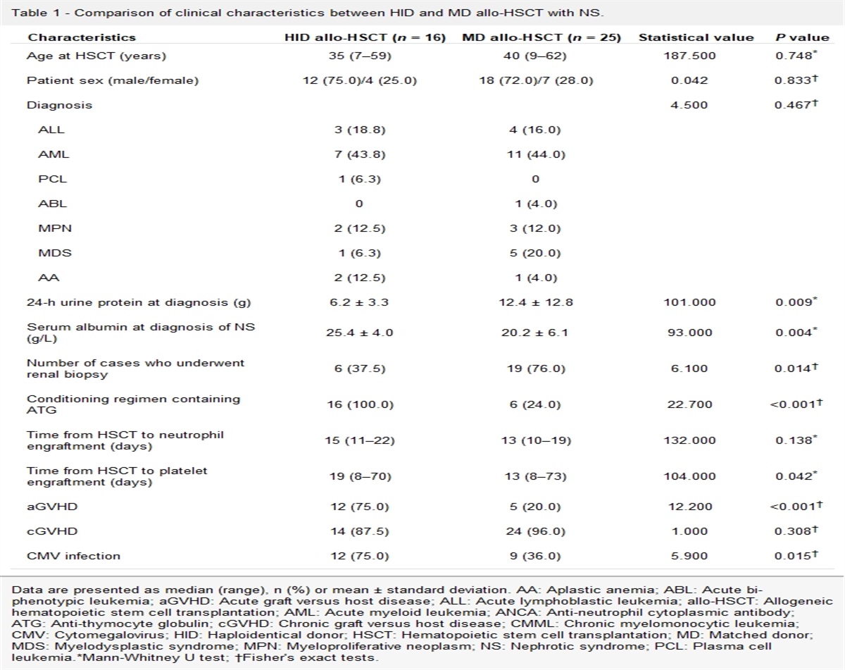
記住我

To the Editor: Normal-tension glaucoma (NTG) is a progressive optic neuropathy characterized by irreversible blindness with statistically normal intraocular pressure (IOP) (≤21 mmHg),[1,2] which makes early-stage diagnosis in the clinical practice difficult. Pathophysiological pathways underlying NTG include vascular dysregulation, toxic metabolite accumulation, and elevated translaminar cribrosa pressure difference (TLCPD).[3] Although various small animal models have provided evidence of different pathophysiological mechanisms, the complicated clinical phenotype cannot be fully imitated by secondary disease models, and the gap between rodents and humans weakens their translational potential. Thus, the pathological changes associated with NTG remain unclear.
Here we identified a rhesus macaque with a natural NTG phenotype and previous craniocerebral injury, and investigated this unique non-human primate (NHP) model of NTG by comparing the in vivo ocular parameters and histological changes with those seen in a normal macaque. We propose the theory that elevated TLCPD, defined as the difference between IOP and intracranial pressure (ICP), contributes to the pathological process of NTG. This study was approved by the Institutional Animal Care and Use Committee of the Zhongshan Ophthalmic Center (ethics approval number: 2020-168).
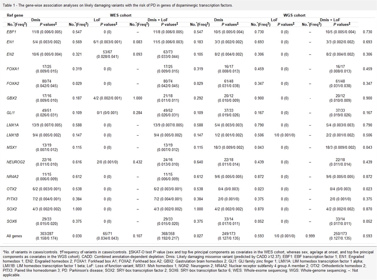
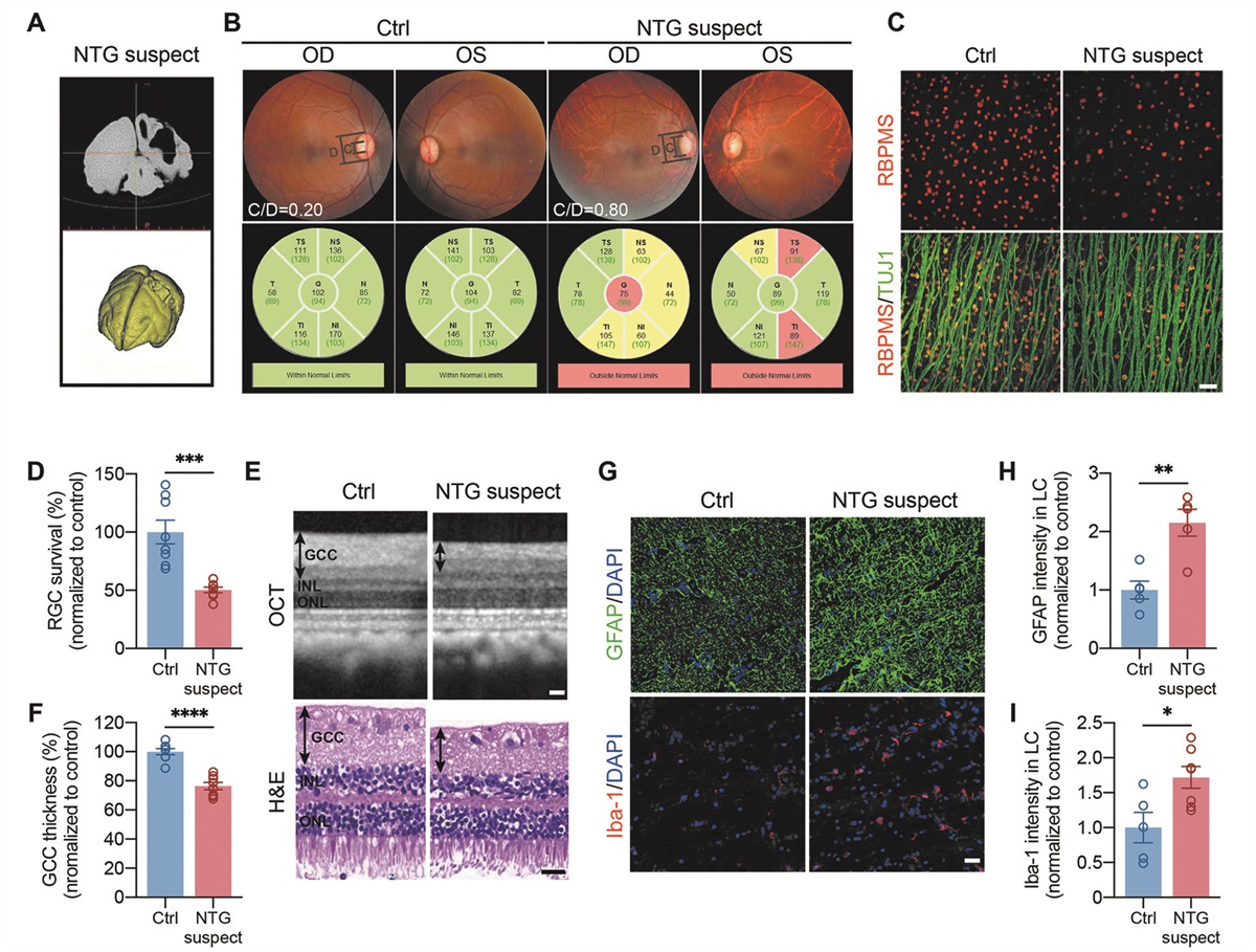
We accidentally discovered this NTG-suspect rhesus macaque with an unexpected old penetrating wound on its skull (observed during dissection) during a screening program for NTG [Supplementary Figure 1, https://links.lww.com/CM9/B676]. We reconstructed the three-dimensional structure of the lesioned brain, confirmed cerebral defects in the animal [Figure 1A], and matched it with a normal animal as control. Both macaques were female and approaching the age of 20 years; IOP ranged from 20 mmHg to 22 mmHg in both eyes [Supplementary Table 1, https://links.lww.com/CM9/B676]. Fundus images showed a cup-to-disc ratio (CDR) of approximately 0.20 in the normal macaque and a significantly deeper disc cup in the NTG-suspect animal (CDR = 0.80), with an enhanced light reflex and tessellated retina [Figure 1B]. Optical coherence tomography (OCT) images of NTG-suspect fundus also displayed a nasal shift of the central retinal vascular trunk [Supplementary Figure 2, https://links.lww.com/CM9/B676]. Figure 1B shows the peripapillary OCT outputs from both macaques. Nearly all (except the temporal and temporal superior) sectors on peripapillary OCT displayed an obvious reduction in RNFL thickness in the NTG-suspect macaque [Supplementary Table 2, https://links.lww.com/CM9/B676]. During the slit-lamp examination, both macaques presented an open anterior chamber angle within the normal depth [Supplementary Figure 3, https://links.lww.com/CM9/B676].
 Figure 1:
 Ocular parameters and histological changes in NTG-suspect and normal control macaques. (A) CT scanning images and 3D reconstruction of the craniocerebral injury in NTG-suspect macaque. (B) Fundus images and peripapillary OCT results displaying RNFL thickness. (C) Images of retinal wholemount stained for RGC markers TUJ1 (green) and RBPMS (red). Scale bar: 50 μm. (D) Images of OCT and H&E demonstrating GCC thickness. Scale bar: 50 μm. (E) Images of LC sections co-labeled with DAPI (blue) and GFAP (green) or Iba-1 (red). Scale bar: 50 μm. (F) Quantification of RGC survival rate normalized to control. n = 8 fields per retina. (G) Quantification of relative GCC thickness normalized to control. n = 8 sections per retina. Scale bar: 50 μm. (H,I) Quantification of relative fluorescent intensity of GFAP and Iba-1 staining normalized to control. n = 5–7 sections per eye. All statistical analyses were performed by unpaired t-test. *P <0.05, **P <0.01, ***P <0.001, and ****P <0.0001. All error bars represent mean ± SEM. CT: Computed tomography; Ctrl: Control;DAPI: 4',6-diamidino-2-phenylindole; GCC: Ganglion cell complex; GCL: Ganglion cell layer; GFAP: Glial fibrillary acidic protein; H&E: Hematoxylin-eosin staining; Iba-1: Ionized calcium-binding adapter molecule 1; INL: Inner nuclear layer; LC: Lamina cribrosa; NTG: Normal-tension glaucoma; OCT: Optical coherence tomography; OD: Oculus dexter; ONL: Outer nuclear layer; OS: Oculus sinister; RBPMS: RNA-binding protein with multiple splicing; RGC: Retinal ganglion cell; RNFL: Retinal nerve fiber layer; SEM: Standard error of mean; TEM: Transmission electron microscope; TUJ1: Tubulin β3; TUNEL: Terminal deoxynucleotidyl transferase mediated dUTP nick end labeling.
We then concentrated on changes in the lamina cribrosa (LC) area, a crucial anatomical site between the two pressure zones, to assess a potential elevated pressure gradient. Representative OCT images showed more serious optic nerve head and optic disc defects in the NTG-suspect macaque [Supplementary Figure 4, https://links.lww.com/CM9/B676], as well as significantly deeper cup depth (CD) and lamina cribrosa depth (LCD) and thinner prelaminar tissue thickness. The parameters of both axes (horizontal and vertical) demonstrated increases in CD, LCD, and posterior LC surface depth in the macaque with suspected NTG, consistent with the image observations. Furthermore, the adjusted LC curvature index was dramatically increased in the NTG-suspect macaque, along with a decrease in the Bruch's membrane opening minimum rim width [Supplementary Table 3, https://links.lww.com/CM9/B676].
To further investigate retinal ganglion cell (RGC) loss in the NTG-suspect animal, we first examined retinal cell apoptosis using a TUNEL assay. Compared to the control, the number of TUNEL-positive cells was significantly increased in the ganglion cell layer and inner nuclear layer of the NTG-suspect retina [Supplementary Figure 5A, B, https://links.lww.com/CM9/B676]. Next, we double-stained the retinal whole mounts with pan-RGC markers, tubulin β3 (TUJ1) and RNA-binding protein with multiple splicing (RBPMS). A marked decrease was observed in the number of TUJ1/RBPMS-positive cells in the NTG-suspect retina, and the survival rate of RGCs was reduced to approximately 50% compared to the control [Figure 1C,D]. These results indicate that the NTG macaque indeed suffered RGC loss by apoptosis. Consistent with this loss, ganglion cell complex thickness in the NTG-suspect was reduced to 75%, as detected by OCT examination and hematoxylin-eosin (H&E) staining [Figure 1E,F].
As increasing evidence demonstrates that neuroinflammation serves a vital role in glaucomatous retinopathy, we further tested glial activation by immunostaining of glial markers GFAP (glial fibrillary acidic protein; astrocytes) and Iba-1 (ionized calcium-binding adapter molecule 1; microglia). In retinal tissues, we detected no statistically significant changes in GFAP or Iba-1 staining [Supplementary Figure 5C–F, https://links.lww.com/CM9/B676]. However, in LC sections, both GFAP and Iba-1 staining intensities were significantly increased in the NTG-suspect [Figure 1G–I], suggesting that neuroinflammatory activation was more significant in the LC than in the retina.
Next, we monitored the ultrastructural changes using a transmission electron microscope to further investigate RGC death and LC structure modification. In the NTG-suspect macaque, we discovered double-membrane autophagy vesicles and shrinking mitochondria with shattered outer membranes. This indicated a higher level of autophagy formation in the RGC soma and the degradation of axonal mitochondria and obstruction of energy transportation in RGC axons, which eventually contributed to glaucomatous damage. In addition, we noticed a large number of onion bulb structures, the pathological hallmarks of demyelinating neuropathies in suspected NTG [Supplementary Figure 6, https://links.lww.com/CM9/B676].
In this study, we identified a special NTG-suspect macaque and investigated the possible connection between its glaucomatous damage and previous craniocerebral injuries. The results of in vivo and histological examinations confirmed glaucomatous changes in the NTG-suspect animal and significant deterioration of the LC area. Considering the animal's previous craniocerebral injury and more severe damage in the LC area, we propose the theory that the ICP caused by the old craniocerebral injury elevated the TLCPD and subsequently caused glaucomatous injury. Our preliminary studies have discovered a statistically lower ICP in patients with NTG compared to those with high-pressure glaucoma and normal controls.[2] However, it is impractical to perform a lumbar puncture to detect ICP for NTG screening in the clinical practice, and the reliability and accuracy of non-invasive examinations for ICP evaluation remain controversial. While relatively simple and reproducible animal models that produce low ICP in rats by constantly draining the cerebrospinal fluid merely mimic an acute condition for several hours,[4] distinct differences in the anatomical structure of the eyes between rodents and humans still exist. Therefore, natural low-ICP NHP models are the key to revealing the role of TLCPD in the generation of NTG.
However, direct ICP data were unavailable for our NTG-suspect macaque, because of the accidental discovery of its craniocerebral injury during dissection, which occurred during a screening for natural glaucoma NHP models. Considering the rarity of natural NTG NHP models, we decided to sacrifice the macaque instantly with the hope to gain insights into the potential mechanisms underlying NTG. Owing to the normal appearance and behavior of the NTG-suspect macaque, we therefore did not perform ICP examination before euthanasia. Nevertheless, we had reason to assume that the ICP was reduced after a previous craniocerebral injury due to the brain tissue defect and the penetrating wound on the animal's skull. Several studies have also supported the notion that decreased brain parenchymal volume is detected after traumatic brain injury.[5] The NHP results in this study are also similar to those found in other low-ICP animal models.[4] Our investigation is therefore of great significance, presenting another practical and clinical perspective on the TLCPD theory.
To summarize, we confirmed the glaucomatous damage in NTG macaques and proposed the theory that the reduction of ICP due to previous craniocerebral injury elevates TLCPD and subsequently causes glaucomatous injury. The proximity between NHPs and humans makes these observations valuable for understanding NTG. Studying our natural NTG-suspect macaque with craniocerebral injury provided us with new insights into TLCPD in the pathogenesis of NTG and, more importantly, our findings offer guidance for the clinical diagnosis and treatment of NTG patients.
Availability of data and materialsAll data generated during this study are available from the corresponding authors upon reasonable request.
AcknowledgmentsWe thank Dr. Wong from the Huazhen Laboratory Animal Breeding Centre for helping in the collection of monkey tissues, experimental advice, and providing reagents.
FundingThis work was funded by the National Key R&D Project of China (No. 2020YFA0112701), National Natural Science Foundation of China (No. 82171057), Science and Technology Program of Guangzhou, China (No. 202102010216), National Natural Science Foundation of China (No. GZR-2012–009), and Beijing Traditional Chinese Medicine Technology Development Fund Project (No. JJ-2018-50).
Conflicts of interestNone.
References 1. Shields MB. Normal-tension glaucoma: Is it different from primary open-angle glaucoma? Curr Opin Ophthalmol 2008;19: 85–88. doi: 10.1097/ICU.0b013e3282f3919b. 2. Wang NL, Friedman DS, Zhou Q, Guo L, Zhu D, Peng Y, et al. A population-based assessment of 24-hour intraocular pressure among subjects with primary open-angle glaucoma: The Handan eye study. Invest Ophthalmol Vis Sci 2011;52: 7817–7821. doi: 10.1167/iovs.11-7528. 3. Mao Y, Yang D, Li J, Liu J, Hou R, Zhang Z, et al. Finite element analysis of trans-lamina cribrosa pressure difference on optic nerve head biomechanics: The Beijing Intracranial and Intraocular Pressure Study. Sci China Life Sci 2020;63: 1887–1894. doi: 10.1007/s11427-018-1585-8. 4. Li XX, Zhang Z, Zeng HY, Wu S, Liu L, Zhang JX, et al. Selective early glial reactivity in the visual pathway precedes axonal loss, following short-term cerebrospinal fluid pressure reduction. Invest Ophthalmol Vis Sci 2018;59: 3394–3404. doi: 10.1167/iovs.17-22232. 5. Williamson MR, Colbourne F. Evidence for decreased brain parenchymal volume after large intracerebral hemorrhages: A potential mechanism limiting intracranial pressure rises. Transl Stroke Res 2017;8: 386–396. doi: 10.1007/s12975-017-0530-x.
留言 (0)