
Remember me


Central nervous system (CNS) tumors represented 1.6% of all new cancer cases worldwide according to Global Cancer Statistics 2020,[1] with glioma being the most common primary CNS tumor, accounting for approximately 75%.[2,3] The annual incidence of glioma in the United States between 2007 and 2016 was 7.3 per 100,000 individuals, 7.47 per 100,000 individuals in Korea between 2007 and 2016, and 8 per 100,000 individuals in China between 2016 and 2022.[4–7] At present, magnetic resonance imaging, histopathology, and molecular pathology are commonly used to diagnose and classify gliomas.[8] Despite a combination of surgery, radiotherapy, chemotherapy, targeted therapy and supportive care, the prognosis of glioma patients remains poor.[9,10] The worldwide median survival time for patients with low-grade glioma (LGG) is 5.6–13.3 years and for patients with high-grade glioma (HGG), it is only 12.2–15.4 months.[11–13]
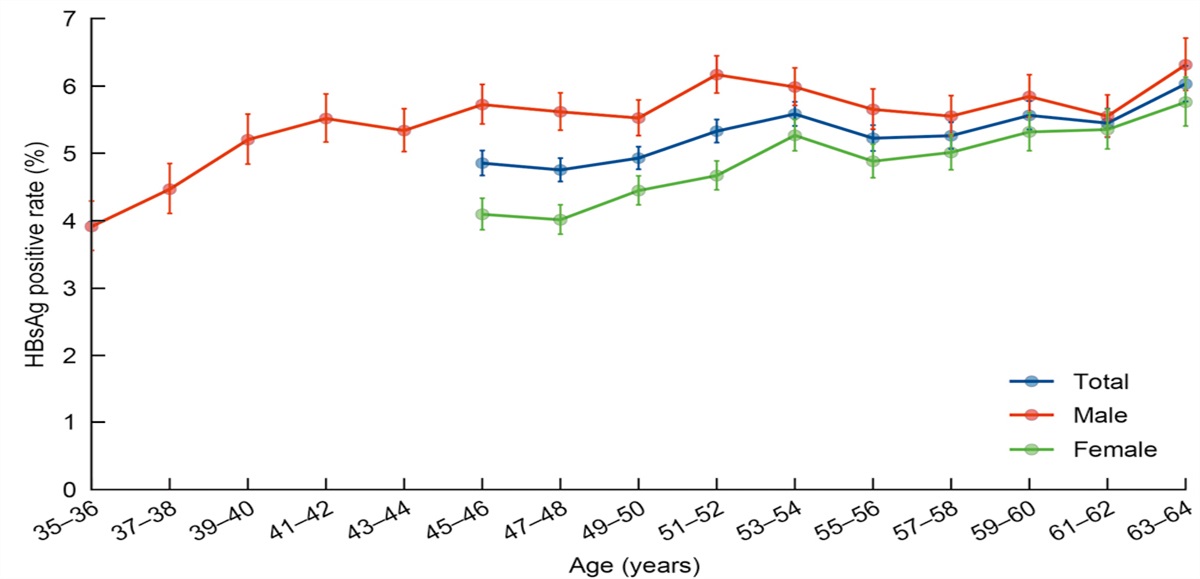
Stress is a regular part of daily life. Stress responses can have various effects on the body, and when the duration of the stressor is limited, it gives people an experience of excitement and accomplishment. It can also be defined as “acute stress”.[14,15] In addition, if the stressful situation is not solved for a long time, the body will enter an exhaustion stage, where cells cannot maintain normal functions.[14] This is known as chronic stress (CS). CS also increases the risk of mood disorders in glioma patients, such as depression and anxiety.[16,17] Several prospective cohort studies have shown that mood disorders are frequent complications of gliomas. Patients diagnosed with LGG often develop depression or anxiety, and patients with HGG are more likely to experience mood disorders after diagnosis.[18,19] Furthermore, a patient’s mental health problems worsen further during surgery and treatment.[20,21] Other cohort and clinical studies have also shown that depression is an important prognostic factor for the survival of glioma patients. Compared to HGG group without depression, HGG patients with depression have shorter overall survival.[11,22–24] In contrast, Arja et al[25] found that patients with preoperative depression had significantly shorter survival time than non-depressed patients in a subgroup of patients with LGG. However, they did not find this phenomenon in patients with HGG. It is likely that the different results of these observational studies were caused by the inconsistent types of questionnaires used [Table 1].
Table 1 - Progress in observational studies of the effects of chronic stress on glioma. Year Chronic stressors Patient status Questionnaire type to assess mood disorders WHO grade Effect References 2004 Depression Postoperative SF-36 WHO grade 4 ↓Survival rate [38] 2005 Depression Preoperative BDI WHO grade 1–2 ↑Death rate; [25] 2008 Depression Preoperative NA WHO grade 3–4 ↓Survival rate [39] 2017 Depression/anxiety Preoperative HADS-A/HADS-D NA –Survival rate [24] 2019 Depressive symptoms Postoperative BDI-II/TMTB WHO grade 4 ↓OS [40] 2020 Depression/anxiety Postdiagnosis PHQ-9/GAD-7 WHO grade 4 ↓OS [23] 2020 Stressful life Preoperative NA NA ↑Risk of brain tumors [41] 2020 Depression/anxiety Postoperative PHQ-9/GAD-7 WHO grade 4 ↓OS; ↑mortality [22] 2022 Psychooncological distress Postdiagnosis HADS-D/DT/Po-Bado WHO grade 2 ↓HRQoL [18] 2023 Depression/anxiety Postoperative HADS NA ↓OS [11]BDI: Beck depression inventory; BDI-Ⅱ: Beck depression inventory-second edition; DT: Distress thermometer; GAD-7: Generalized anxiety disorder 7-item; HADS: Hospital anxiety and depression scale; HADS-A: Hospital anxiety and depression scale-anxiety; HADS-D: Hospital anxiety and depression scale-depression; HRQoL: Health-related quality of life; NA: Not applicable; OS: Overall survival; PHQ-9: Patient health questionnaire 9-item; Po-Bado: Psychooncological base documentation; SF-36: 36-item short form survey; TMTB: Trail making test part B; WHO: World Health Organization; ↑: Increase; ↓: Decrease; –: No change.
Therefore, mood disorders such as anxiety and depression caused by CS are direct factors leading to glioma development and poor prognosis. The relationship between mood disorders and CS is bidirectional.[26,27] Numerous studies have shown that chronic stressors can induce mood disorders in laboratory animals and increase the incidence of mood disorders in humans.[28–31] However, it is unlear whether CS is a cause or consequence of glioma development and progression. Previous studies suggested that a patient’s emotional disturbance was caused by the negative impact of a cancer diagnosis or surgery.[32,33] However, the sudden onset of depression and anxiety may also be early symptoms of glioma.[34,35] Neuropsychiatric symptoms are the first clinical indication of a brain tumor in 18% of cases reported.[36] Although studies have speculated that mood disorders are risk factors for the development and progression of glioma, few studies have reported on the symptoms of mood disorders before diagnosis and related molecular mechanisms in glioma patients.
In this review, we unravel the intricate interplay between CS and gliomas. From the perspective of the hallmarks of cancer, we discuss the potential mechanisms of how CS affects the development and progression of gliomas.[37] We also describe CS as a promising target for the development of glioma therapy.
Chronic Stress, Stress System and GliomaThe influence of CS on humans and rodents is mediated by stress systems, which can be divided into upstream receptors and downstream effectors. The limbic system is the main information processing center for upstream receptors receiving and transmitting information through networks composed of different neurons.[42] Downstream effectors include the neuroendocrine system, which is one of the most prominent mechanisms of CS in oncology research. It is mediated mainly by two pathways, the sympathetic nervous system (SNS) and hypothalamic-pituitary-adrenal (HPA) axis.[43,44] Although there may be species-specific differences in the response to CS, studies in both humans and rodents have shown similar patterns of activation in this system.[45]
When an organism is exposed to CS, its sensory organs, including the eyes, nose, and skin, undergo physiological changes in response to stress signals. These signals are transmitted to the corresponding primary sensory cortex, which processes sensory information from the affected part of the body. The primary sensory cortical centers located in the parietal lobe are responsible for integrating stress signals and sensory information from other cortical regions of the brain. Subsequently, stress information is transmitted to the thalamus for integration via the cortico-thalamic circuits. The thalamus acts as a crucial relay station in the brain, which not only integrates sensory information, but also processes and transmits stress information to various brain regions involved in stress processing, including the prefrontal cortex, hippocampus, and amygdala. The prefrontal cortex plays a key role in regulating high-level cognitive processes such as decision-making and executive function in response to chronic stressors. Similarly, the hippocampus is responsible for the formation and storage of CS-related memory. The amygdala, an essential part of the limbic system, is involved in the processing and regulation of emotional responses to CS. The basolateral nucleus of the amygdala plays a key role in integrating emotional information from the thalamus. Finally, the central nucleus of the amygdala activates downstream neuroendocrine pathways to trigger physiological responses to CS [Figure 1A].[42,46–50]
 Figure 1: Processing of the stress system and its associated media. (A) The upstream signals of the stress system are mainly received and processed by different cortical areas and limbic systems through neural circuits for various types of stressors. The cortex communicates bidirectionally with the thalamus through a cortico-thalamo-cortical loop to make real-time judgments about information. The components of the limbic system, such as the thalamus, cingulate gyrus and hippocampus, exchange information through the Papez circuit, which plays an important role in the cortical control of emotion and memory storage. The hippocampus stores information transmitted multiple times by the Papez circuit and forms long-term memories. The medial prefrontal cortex–hippocampal circuit transmits information between the medial prefrontal cortex and the hippocampus. A neural circuit consists mainly of a population of neurons connected by synapses which perform specific function when activated. The CNS regulates the activity of the HPA axis by controlling the secretion of CRH and AVP in the PVN. As a result, the pituitary gland releases ACTH, which causes the adrenal cortex to release glucocorticoids. Simultaneously, the CNS activates the SNS, resulting in the secretion of E from the adrenal medulla. The secretion of acetylcholine by the vagal nerve binds to muscarinic receptors on chromaffin cells in the adrenal medulla, promoting the secretion of CAs. Sympathetic nerve endings also secrete small amounts of NPY. The vagal pathway acts as a bridge between the brain and the gut. (B) Several stress system molecules have been implicated in promoting glioma development and progression, including glucocorticoids, NE, E, Ach and other molecules secreted by the CNS. ACTH: Adrenocorticotropic hormone; Ach: Acetylcholine; AVP: Arginine vasopressin; CAs: Catecholamines; CNS: Central nervous system; CRH: Corticotropin-releasing hormone; CS: Chronic stress; E: Epinephrine; HPA axis: Hypothalamic-pituitary-adrenal axis; NE: Norepinephrine; NPY: Neuropeptide Y; PVN: Paraventricular nucleus; SNS: Sympathetic nervous system. The Figure was partly generated using Servier medical art repository (https://smart.servier.com), provided by Servier and licensed under a Creative Commons Attribution 3.0 unported license, and partly adapted from BioRender.com with permission.
Notably, stress information processing is not a simple concatenation of single lines from different brain sites, but a multidirectional collaboration through different neural circuits consisting of populations of neurons connected by synapses in multiple parts of the brain. CS can cause an imbalance between excitatory and inhibitory neurotransmission, leading to changes in the synaptic strength and stability. Studies in rats have shown that increased firing rates of excitatory neurons in the lateral and basolateral amygdala occurred under CS.[51,52] In mice, enhanced excitatory inputs to neurons projecting to the lateral habenula were observed in the ventral tegmental area, and tonic currents mediated by gamma-aminobutyric acid A receptor were continuously lost in projection neurons in the lateral amygdala. CS can also disrupt neuronal synaptic plasticity in the hippocampus and weaken synaptic connections in the prefrontal cortex.[53–56] Although most of these studies have been conducted on animals, changes in corresponding brain structures can also be found in patients with mood disorders, such as significant thinning of the medial frontal cortex, reversible damage to hippocampal morphology, and increased amygdala size. However, it is important to note that the effect of CS on brain structure is not absolute, as the perception of CS is subjective, and differences in brain structure and detection methods may affect the study results.[57,58]
Neural signaling activated by CS converges on the paraventricular nucleus (PVN), the control core of the downstream neuroendocrine pathways.[59] Activation of the HPA axis is triggered by release of corticotropin-releasing hormone (CRH) and arginine vasopressin (AVP) from neurons in the medial parvocellular PVN. CRH binds to the corticotropin-releasing hormone receptor 1 (CRHR1) on anterior pituitary corticotrope cells, leading to the synthesis and secretion of adrenocorticotropic hormone (ACTH). ACTH then acts on the adrenal cortex to stimulate the synthesis and release of glucocorticoids (GCs),[60,61] which are characterized by cortisol in humans and corticosterone in rodents.[62] These CS-activated neural signals also trigger activation of the SNS. Activation of the SNS results in the release of epinephrine (E) and a small amount of norepinephrine (NE) from the adrenal medulla. However, most NE is synthesized by the locus coeruleus in the brainstem and coreleased into the circulation with neuropeptide Y (NPY) during sympathetic activation.[63] The vagal nerve, a crucial component of the parasympathetic nervous system (PNS), plays a significant role in regulating both the HPA axis and SNS. Moreover, the vagal nerve is an essential center of the microbiota-gut-brain axis [Figure 1A].[64,65]
CS exerts a psychological impact on the body by acting through the CNS, while simultaneously exerting physiological effects through the neuroendocrine system and the coordinated activity of the SNS and PNS. The dysregulated secretion of stress signaling molecules, such as GCs, NE, and E, is a consequence of the persistent activation of the HPA axis and SNS during CS. High levels of NE, E and GCs can adversely affect immune function and lead to inflammation. In addition, high levels of GCs can cause neuronal dysfunction in the brain.[66,67] Dysregulated vagal inhibition is also observed in CS, whereby the cholinergic anti-inflammatory pathways acting via the vagal nerve are inhibited, resulting in reduced release of anti-inflammatory cytokines and an increase in inflammation in the body. This decrease in vagal tone can lead to increased intestinal permeability and may promote systemic inflammation.[65]
For many years, extensive research spanning several years has been dedicated to exploring the potential role of CS in promoting glioma development and progression through stress-related hormones that can weaken immune responses, allow tumor cells to evade immune system surveillance, and affect glioma proliferation and survival [Figure 1B].[68,69] However, little attention has been paid to the upstream mechanisms that contribute to glioma development and progression. Recent research has revealed that two-way communication between neurons and glioma cells influences glioma development and progression and that CS exacerbates this process by overstimulating neurons. Moreover, the brain-gut-microbiota axis, mediated by the vagal nerve, can modify microbiota and affect glioma growth.[68,70–75] Next, we discuss the specific molecular mechanisms underlying the impact of the CS stress system on glioma development and progression.
Mechanisms Linking Chronic Stress to the Hallmarks of Glioma Genomic instabilityInactivation of the tumor protein 53 (p53) is a critical event in the malignant transformation of most cells of origin.[76]In vivo studies of mice have confirmed that CS induced NE to stimulate G protein-protein kinase A (Gs-PKA) signaling through β2 adrenoceptors (β2-AR) to generate reactive oxygen species (ROS). This resulted in the accumulation of cellular deoxyribonucleic acid (DNA) damage. Furthermore, cytosolic β-arrestin1 (ARRB1) mediated NE-induced protein kinase B (AKT) and murine double minute 2 (MDM2) activation, whereas nuclear ARRB1 acted as a catalyst for MDM2-dependent p53 ubiquitination to promote p53 degradation and DNA damage in the frontal cortex of the brain.[77] Meanwhile, high levels of GCs during CS mediate the effect of CS on p53 through the induction of serum and glucocorticoid-regulated kinase 1 (SGK1), which in turn increases MDM2 activity and decreases p53 function [Figure 2A].[78] Accumulated clinical evidence also suggests that the loss of p53 function is a key initial event in glioma development.[79] However, it should be noted that p53 damage alone is not sufficient to cause glioma development, because mutations need to be maintained and accumulated throughout repeated cell divisions, leading to the acquisition of features including resistance to apoptosis and increased proliferation.[80] Although the frontal and temporal lobes of the human brain are high-risk areas for glioma growth, there is insufficient evidence that DNA damage in the frontal lobe has an impact on gliomas.[2]
 Figure 2:
 Mechanisms by which CS promotes malignant progression in glioma cell. (A) CS stimulates olfactory neurons to secrete IGF1, which binds to IGF1R on OPCs and activates classical RTK pathways. CS triggers the neuroendocrine system to secrete NE and GCs, which activate the PI3K-AKT and SGK1-MDM2 pathways to promote the p53 degradation. Meanwhile, NE also activates PKA to promote mitochondrial generation of ROS, which in turn promotes DNA damage. (B) NSCs give rise to NPCs, astrocyte progenitor cells and OPCs, which in turn each differentiate into neurons, astrocytes and oligodendrocytes. Two solid red lines (transdifferentiation) indicate cell types that have been recognized as cells of malignant glioma origin. (C) CS promotes abnormal signaling pathways in glioma cells, mainly through NE binding to β-ARs on the surface of glioma cells. GCs combined with intracellular GRs and a decrease in GABAergic interneuron activity could act synergistically. AC: Adenylyl cyclase; AKT: Protein kinase B; APC: Astrocyte precursor cell; ARRB1: β-arrestin 1; ATP: Adenosine triphosphate; cAMP: Cyclic adenosine monophosphate; β-AR: β adrenoceptor; Bax: B-cell lymphoma 2-associated X protein; BCL-2: B-cell lymphoma 2; BCL-XL: B-cell lymphoma-extra large; CD147: Cluster of differentiation 147; CDK4/6: Cyclin-dependent kinase 4/6; CS: Chronic stress; DNA: Deoxyribonucleic acid; EMT: Epithelial to mesenchymal transition; ERK: Extracellular signal-regulated kinase; GABAergic: Gamma aminobutyric acidergic; GC: Glucocorticoid; GR: Glucocorticoid receptor; Gs: G protein; HIF-1α: Hypoxia-inducible factor 1α; IGF1: Insulin-like growth factor 1; IGF1R: Insulin-like growth factor 1 receptor; LDHA: Lactate dehydrogenase A; MCT1/4: Monocarboxylate transporters 1/4; MDM2: Murine double minute 2; MEK: Mitogen-activated protein kinase kinase; MMP2/9: Matrix metallopeptidase 2/9; NE: Norepinephrine; NK cell: Natural killer cell; NPC: Neural progenitor cells; NSC: Neural stem cell; OPC: Oligodendrocyte precursor cell; P: Phosphorylation; PC/PE: Phosphatidylcholine to phosphatidylethanolamine ratio; PI3K: Phosphoinositide 3-kinase; PKA: Protein kinase A; p53: Tumor protein 53; Raf: Rapidly accelerated fibrosarcoma; Ras: Rat sarcoma virus; ROS: Reactive oxygen species; RTK: Receptor tyrosine kinase; SGK1: Serum and glucocorticoid-regulated protein kinase 1; Ubi: Ubiquitin; Twist1: Twist family basic helix-loop-helix transcription factor 1; +: Increase; -: Decrease. The Figure was partly adapted from BioRender.com with permission.
Unlocking phenotypic plasticityBecause there are histopathological similarities between gliomas and normal glial cells in the brain, gliomas are thought to originate from transformed neural stem cells (NSCs) or progenitor cells.[81,82] NSCs and oligodendrocyte precursor cells (OPCs) in the subventricular zone of mammals are considered to be major candidates for the cells of origin for glioma.[83] CS is one of the factors that promote the transdifferentiation of these cells [Figure 2B]. Transdifferentiation is a part of unlocking phenotypic plasticity. Unlocking phenotypic plasticity is one of the newest hallmarks of cancer.
In vitro studies showed that mouse NSCs were transformed into HGG upon the loss of p53, neurofibromatosis type 1 (Nf1) and/or phosphatase and tensin homolog (PTEN).[76] CS also promotes the secretion of growth factors by neurons to promote the transformation and malignant proliferation of oligodendrocyte precursor cell (OPC). Chen et al[84] reported that CS directly induces gliomagenesis. They found that mitral and tufted cells in the olfactory bulbs of mice secreted insulin-like growth factor 1 (IGF1) in response to olfactory stimulation. Through the IGF1–IGF1 receptor (IGF1R) axis, IGF1 may activate the classical receptor tyrosine kinase (RTK) pathway in OPCs conditionally knocked down for the tumor suppressor transformation-related p53 and Nf1 in the olfactory bulb. Ultimately, this molecular cascade leads to malignant transformation of mutated OPCs in the olfactory bulb.[85] Recent studies have also demonstrated that olfactory stimulation can lead to CS.[86,87] However, the specific mechanisms in humans need to be elucidated further.[86,88]
It should be noted that cells with germline mutations may not necessarily transform into cells of origin for gliomas. Instead, their descendant cells might be transformed and become cells of origin for gliomas.[84,89] However, these changes alone are not sufficient to promote glioma formation directly. Instead, they transform these cells into potential cells of origin that require further stimulation by other factors to develop into gliomas. This suggests that CS may act as a new risk factor for glioma development. There is limited experimental and clinical evidence for the relationship between sensory input from external stimuli and glioma development, but further studies are still needed to determine its existence. This area of research is of great interest because it may provide insight into the mechanism by which CS triggers glioma formation in the CNS [Figure 2A].
Sustaining proliferative signalingSustained proliferation is a fundamental characteristic of glioma cells. It has been shown that the serum levels of the stress hormones GC and NE are significantly increased by CS. NE and GC promoted the uncontrolled proliferation of gliomas in vivo,[70,73,74] and in vitro experiments using human glioma cell lines showed GC and NE promoted glioma cell growth by activating phosphoinositide 3-kinase (PI3K)/protein kinase B (AKT) signaling via binding to glucocorticoid receptor (GR) and β adrenoreceptors (β-ARs).[73,74] Simultaneously, β-ARs induced extracellular signal-regulated kinase (ERK) phosphorylation to affect the proliferation of glioma cells.[72] The PI3K/AKT pathway is an indispensable intracellular signal transduction pathway involved in cell growth, proliferation and survival/apoptosis. Furthermore, the ERK phosphorylation cascade is a central regulator of intracellular signaling. NE also directly promoted proliferation by regulating cyclin D1/cyclin-dependent kinase 4/6 (CDK4/6) expression in glioma cells [Figure 2C].[68,90]
Synapses, particularly glutamatergic synapses, were reported to form between neurons and glioma cells in human and mouse brains. Furthermore, α-amino-3-hydroxy-5-methyl-4-isoxazolepropionic acid (AMPA) receptor-mediated neuronal activity has been shown to induce glioma invasion and growth. During this process, neurons also released neuroligin-3 to activate the PI3K-mammalian target of rapamycin pathway in glioma cells and promote the proliferation of glioma cells.[91] Although there are no relevant studies demonstrating that CS promotes excitatory synapse formation, Tantillo et al[92] found that gamma aminobutyric acidergic (GABAergic) interneuron activity was reduced in mice after chronic visual deprivation, a specific form of CS, resulting in increased glioma proliferation in the primary visual cortex. Unlike excitatory information transmitted by glutamatergic synapses, GABAergic neurons are the main inhibitory neurons in the CNS.[93] Therefore, CS disrupts the balance between excitatory and inhibitory signals in the brain. Hyperexcitable environments may increase the sensitivity of cells to excitatory signaling, which may contribute to the establishment of excitatory synapses between neurons and gliomas.[94] Analogous to olfactory stimulation, the proliferation of gliomas induced by visual deprivation is region-specific. Recently, CS has been considered a cause of vision loss in glioma patients.[95,96] Despite observations of a relationship between visual deprivation, a specific form of CS, and glioma in mouse models, there is currently no evidence to suggest a similar relationship in human glioma cell lines [Figure 2C]. Additionally, stress hormones also synergize this process and stimulate the proliferation of glioma cells.
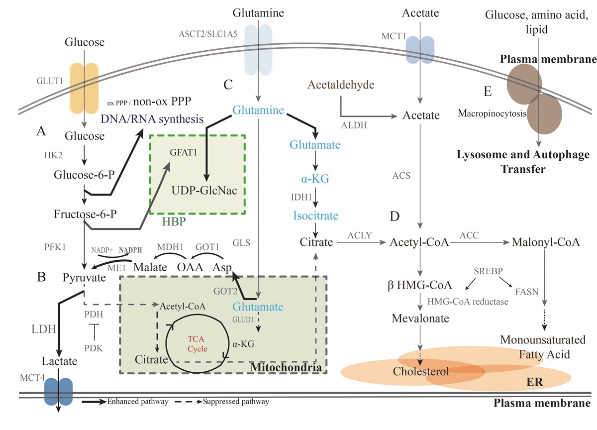
Reprogramming cellular metabolismCS reprograms glioma cell metabolism and increases lactate production. Dong et al[70] demonstrated that NE promoted lactate dehydrogenase A (LDHA) expression through the β-ARs-extracellular signal-regulated kinase 1/2 (ERK1/2)-hypoxia inducible factor 1-alpha (HIF-1α) pathway, resulting in a lower intracellular potential of hydrogen (pH) in human glioma cell lines. At the same time, NE also activated the specificity protein 1 (SP1) region of the cluster of differentiation 147 (CD147) promoter through the β-ARs-ARRB1-ERK1/2 pathway, promoting the expression of CD147, which assists monocarboxylate transporters 1/4 (MCT1/4) in transporting lactate out of glioma cells. The lactate, transported out of glioma cells by MCT1/4, further decreased the pH in the extracellular environment and indirectly promoted the proliferation of gliomas. Notably, a decrease in the pH of the extracellular environment may also disrupt homeostasis in the TME, and changes in the TME were closely related to each step of tumorigenesis.[97,98] However, further experiments are required to demonstrate whether this process occurs in vivo [Figure 2C].
Resisting cell deathDong et al[68] also found that NE inhibited apoptosis by increasing B-cell lymphoma 2 (BCL-2)/B-cell lymphoma-extra large (BCL-XL) expression in glioma cells. In addition, Vlad et al[99,100] found that caspase 3 levels were significantly decreased in the hippocampus of mice with CS compared with control mice [Figure 2C]. Glioma cells acquire resistance to apoptosis to support their proliferation. However, there is currently limited experimental evidence of the beneficial effects of CS on cell resistance to death and further animal studies and clinical studies are required to support this hypothesis.
Activating invasion and metastasisβ-ARs on glioma cell are activated by catecholamines (CAs). It was reported that activation of β-ARs increased the expression of matrix metallopeptidase 2 (MMP-2) and matrix metallopeptidase 9 (MMP-9), members of the matrix metalloproteinase (MMP) family through ERK1/2 activation. Increased expression of MMP-2 and MMP-9 promoted human glioma cell line invasion.[100] MMPs can trigger the remodelling of basement membrane components and extracellular matrix molecules, which greatly facilitates glioma invasion.[101,102] GCs increased the fluidity of membranes in rat glioma cell lines by reducing the ratio of phosphatidylcholine (PC) to phosphatidylethanolamine (PE) in their cellular phospholipid composition.[74] Membrane fluidity is a key physical property that determines the metastatic potential of cancer cells. However, several groups have shown that PC levels were significantly higher in patients with gliomas than in normal subjects.[103] The observed variations can be attributed to differences in cell lines. NE also promoted human glioma cell line migration by upregulating the expression of the twist family basic helix-loop-helix transcription factor 1 (Twist1), which activated epithelial-mesenchymal transition. However, the specific mechanism involved is unknown [Figure 2C].[75] Experimental evidence is required to confirm the mechanism by which CS promotes glioma migration, because clinical data do not show a correlation between CS and increased glioma invasion.
Inflammation and tumor-promoting inflammationInflammation, especially a variety of inflammatory factors and inflammatory cells, which are indispensable components of the tumor microenvironment (TME), is considered a significant factor that contributes to glioma development.[104] Several studies have linked CS to inflammation.
Microglia are innate immune cells of the CNS that regulate brain development, neuronal network maintenance and injury repair. They secrete cytokines, chemokines, prostaglandins and ROS, and help direct the immune response.[105] Limatola et al[69,106] found CS activated microglia in mice in the TME of gliomas. Microglia do not exhibit highly ramified morphology and show a decrease in the length of cell branches. CS also regulated the activity and gene expression of other immune cells in glioma microenvironments such as glioma-associated myeloid cells and CD11b+ dendritic cells. Furthermore, glioma cells under CS secreted chemokines and cytokines involved in glioma progression, such as C-C motif chemokine ligand 2 (CCL2), C-X-C motif chemokine ligand 10 (CXCL10) and interleukin 6 (IL-6), to recruit microglia with tumor-promoting phenotypes and promote glioma progression. Moreover, the tumor-promoting microglia phenotypes decreased the accumulation of naturalkiller (NK) cells in the glioma microenvironment [Figure 3A].[69,106]
 Figure 3: Effects of chronic stress on immune cells and gut microbiota. (A) CS activates resting microglia to become either anti-inflammatory or pro-inflammatory microglia, predominantly pro-inflammatory. Pro-inflammatory microglia recruit IL-1β+ immune cells such as monocytes and neutrophils into the brain. Recruited monocytes and neutrophils secrete pro-inflammatory factors that promote glioma development, such as TNF-α, EGF, HGF, IL-1β and IL-8. At the same time, glioma cells secrete VCAM-1 to promote monocyte adhesion to glioma cells. Neutrophils bind to G-CSF secreted by glioma cells to promote their further proliferation. Anti-inflammatory microglia block communication between pro-inflammatory microglia and natural killer cells, inhibiting natural killer cells from eliminating glioma cells. Dashed black lines represent processes suppressed by CS and solid black lines represent processes facilitated by CS. (B) The gut-brain-microbiota axis and CS. CS has been shown to alter the species and abundance of the gut microbiota. Specifically, Lactobacillus, Bacteroidia and Actinobacteria have been found to decrease in number, while pathogenic bacteria increase in the intestines. The brain receives information from the gut through the ENS via the SNS and PNS. Reductions in Lactobacillus have been linked to increased PI3K-AKT signaling in gliom
Comments (0)