Kinetics of RNA-LNP delivery and protein expression
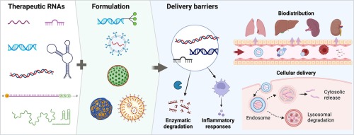
Lipid nanoparticles (LNPs) provide a facilitating platform for mRNA-based delivery [1], [2], [3]. The tremendous success of mRNA based vaccination during the SARS-CoV2 pandemic also boosted the perspectives of mRNA-LNP-based therapies in a wide range of applications, including cancer immunotherapies [4], [5], CAR-T cell-based immunotherapies [6] and CRISPR-based gene editing [7]. As personalized gene therapies advance, the demand for an efficient, broadly applicable and reliable mRNA delivery platform grows. LNP formulations stand out by unique properties such as defined size, colloidal stability, low immunogenicity and the possibility of cell-specific delivery via surface functionalization. These properties are in parts the result of rational design approaches [8] and high-throughput screening of libraries of lipid-like compounds [9], [10]. Current LNP formulations are composed of four lipid components: (i) ionizable lipid, (ii) helper lipids, e.g., DSPC (1,2-distearoyl-sn-glycero-3-phosphocholine), (iii) PEG (polyethylene glycol)-lipid, and (iv) cholesterol. It is understood that the favourable properties come about by optimal choice of these lipid components that ensure self-assembly into a well-defined core–shell architecture. Typically, PEG-lipid and helper lipids, such as DSPC and cholesterol, form a surface monolayer that stabilizes LNP size, while the ionizable lipid, cholesterol and nucleic acids reside in the core [2], [11], [12]. LNP manufacturing employs efficient condensation and encapsulation of negatively charged nucleic acid cargo with ionizable lipid at low pH via rapid microfluidic mixing, which promotes homogeneous self-assembly of lipid nanoparticles by fast solvent exchange [13]. Particle size and stability is adjusted via modification of core-lipids to shell-lipids ratio [12]. LNP formulations govern their ability to efficiently mediate cellular uptake via plasma proteins and subsequent release of nucleic acid to the cytosol. The LNPs were developed and first optimized for siRNA delivery [8], [14], [15]. Interestingly the same LNP formulations with only small adjustments proved also highly efficient for mRNA delivery [9], [16]. In this review we will focus on mRNA-LNPs, but refer to siRNA-LNPs for comparison or in cases where corresponding data are not available for mRNA-LNPs. Over the past decade substantial progress has been made in optimizing LNP formulations turning both siRNA and mRNA LNP-based delivery into a manageable platform technology. However, quantitative pharmacokinetic modelling of LNP delivery and profound understanding of the delivery mechanism at the molecular level are still in their infancy.
The aim of the present review is to present a quantitative reaction kinetic framework of gene delivery and to collect kinetic rates for the various sub-steps leading to gene expression. We begin with an abstraction of nucleic acid delivery as a chain of transfer processes. Next, we introduce common reporter readouts and single-cell time lapse imaging. We then show that time courses of reporter gene expression reflect the reaction kinetics of a simple translation model. Single-cell expression exhibits distinct onset-times that indicate the delivery time as the period from LNP administration to mRNA release. We highlight factors that influence the uptake-rates and the existence of a window of opportunity in endosomal release. Experiments using multiple fluorescence markers or multiple mRNAs reveal distinct event-time correlations. We discuss codelivery of different mRNA reporter constructs and provide an outlook how timing can be modulated to achieve controlled gene expression.


留言 (0)