Reagents
THP-1 cells were obtained from ATCC. The reagents used included phosphate-buffered saline (PBS, Gibco, Shanghai, China, Cat# C10010500CP), Opti-MEM (Gibco, Shanghai, China, Cat# 31985070) and hygromycin B (Sigma‒Aldrich, Shanghai, China, Cat# V900372-1G). The national standard for bacterial endotoxins is LPS, which was obtained from Escherichia coli O55:B5 [9,000 endotoxin units (EU)/vial, batch 150800-201601, identical to the 2nd international WHO standard for endotoxin 94/580 from E. coli O113:H10] and was provided by the National Institutes for Food and Drug Control (NIFDC). Additional reagents included LPS (Sigma‒Aldrich, Shanghai, China, Cat# L2654-1MG, Cat# L6529-1MG, Cat# L4391-1MG, Cat# L4516-1MG, Cat# L2755-10MG, Cat# L5886-10MG, Cat# L7170-1MG, Cat# L4641-1MG, Cat# L6143-1MG, Cat# L7895-1MG, Cat# L4268-10MG), LTA (Sigma‒Aldrich, Shanghai, China, Cat# L3265-5MG), zymosan (Sigma‒Aldrich, Shanghai, China, Cat# Z4250-1G), peptidoglycan (Sigma‒Aldrich, Shanghai, China, Cat# 69554-10MG-F), lectin (Sigma‒Aldrich, Shanghai, China, Cat# L8754-5MG), β-1,3-glucan (Sigma‒Aldrich, Shanghai, China, Cat#89862-1G-F), RPMI 1640 (Gibco, Shanghai, China, Cat# C11875500BT), foetal bovine serum (FBS, Excell, Suzhou, China, Cat# FSP500), HEPES (Sigma‒Aldrich, Shanghai, China, Cat# H0887-100 ml), penicillin‒streptomycin (BasalMedia, Shanghai, China, Cat# S110JV), L-glutamine (Gibco, Shanghai, China, Cat# 25030081), bovine calf serum (BCS, Gibco, Shanghai, China, Cat# A3520502), mouse IgG (Jackson ImmunoResearch, West Grove, PA, USA, Cat# 015-000-003), human FcR blocking solution (Shanghai Maokang Biotechnology Co., Ltd., Shanghai, China, Cat# MX1505-50T), F(ab’)2 fragment goat anti-mouse IgG (Jackson ImmunoResearch, West Grove, PA, USA, Cat# 115-606-071), propidium iodide (Sigma‒Aldrich, Shanghai, China, Cat# P4170-10MG), paraformaldehyde (Biyuntian, Shanghai, China, Cat# P0099), Fixable Viability Stain reagent (BD Cat# 564997), Triton X-100 (Solarbio, Beijing, China, Cat# T8200), PE-mouse IgG1 (BioLegend, San Diego, CA, USA, Cat# 400114), PE-mouse anti-human CD14 (BioLegend, San Diego, CA, USA, Cat# 367104), Bright-Glo luciferase assay reagent (Promega, Madison, WI, USA, Cat# E2650), polymyxin B (Sigma‒Aldrich, Shanghai, China, Cat# P1004-10MU), pyrogen-free water for the BET (Zhanjiang A&C Biological Ltd., Zhanjiang, Guangxi, China), group A & C meningococcal polysaccharide vaccine (Yuxi Walvax Biotechnology Co. Ltd., Yuxi, China, batch No. D201805064), basiliximab (Novartis Pharma Stein AG, Beijing, China, batch No. SFT91), rabies vaccine (Vero cells) for human use, freeze-dried (Liaoning Chengda Biotechnology Co. Ltd., Shenyang, China, batch No. 201804084), Japanese encephalitis vaccine (Vero cells), inactivated (Liaoning Chengda Biotechnology Co. Ltd., Shenyang, China, batch No. 201709B16), insulin aspart injection, (Beijing Double-Crane Pharmaceutical Co. Ltd., batch No. 20161203), human albumin (Nanyue Biopharming Co. Ltd., China, batch No. 201805016) and recombinant human erythropoietin injection (CHO cell) (Harbin Pharmaceutical Group Biological Engineering Co. Ltd., Harbin, China, batch No. 20170404).
Consumables
The consumables used included 96-well plates [Corning, NY, USA, Cat# 3917 (flat bottom, white polystyrene, tissue culture treated), Cat# 9018 (flat bottom clear, polystyrene, high binding surface)], 96-well plates (JET Biofil, Guangzhou, China, Cat# TCP002096, U-bottom clear), 0.20 µm syringe filters (Millipore, Shanghai, China, Cat# SLLGX13NL) and human IL-1β, IL-6 and TNF-α ELISA kits (R&D, Shanghai, China, Cat# DY201-5, Cat# DY206, Cat# DY210-05). Other reagents/materials were purchased as sterile and free of pyrogens, and the glassware was baked at 250 °C for 1 h.
Construction and transfection of the plasmid pCM1.1_luciferase (luc)_NFκB_hygromycin (hygro)
Overlap PCR was used to synthesise the NF-κB response element (5_-TCCTCGGAAAGTCCCCTCTGAGATCCTCGGAAAGTCCCCTCTGAGATCTCAGAGGGGACTTTCCGAGGA-3_), which was inserted into the multiple cloning site ahead of the minimal promoter region in the plasmid pCM1.1_luc_hygro and then DNA sequencing was used to identify the positive clone (named pCM1.1_luc_NFκB_hygro).
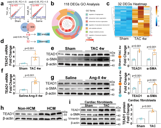
THP-1 cells (ATCC) were transfected with pCM1.1_NF-κB_luc_hygro by electroporation and then cultured in complete medium (RPMI 1640 supplemented with 10% FBS, 25 mM HEPES, 1% penicillin‒streptomycin and 2 mM L-glutamine) at 37 °C in an atmosphere of 5% CO2 aerobically for 48 h. Then, the cells were cultured in complete medium supplemented with 200 μg/ml hygromycin B for 2–3 months and the hygromycin-resistant cells were sorted by limited dilution to obtain monoclonal cells, which were screened for luciferase activity by treatment with LPS. The resulting positive clones (named THP-1_NF-κB_luc_cells) were routinely maintained in complete medium supplemented with 200 μg/ml hygromycin B.
Procedure for the novel pyrogen test
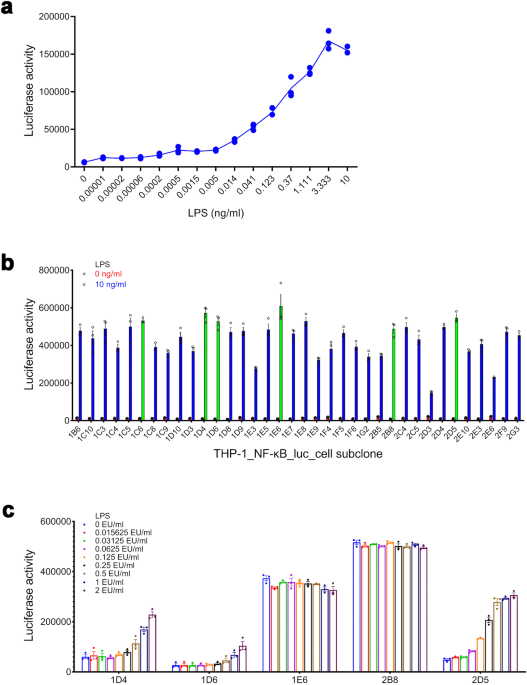
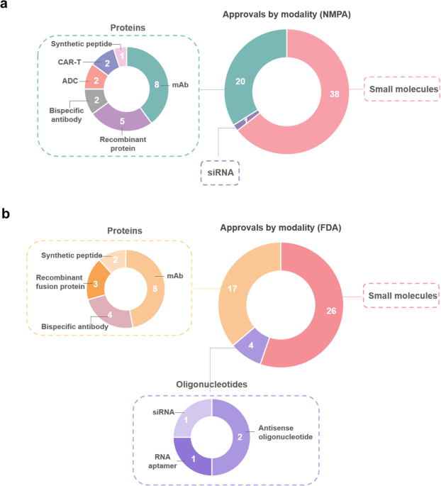
THP-1_NF-κB_luc_cells in the logarithmic growth stage were collected by centrifugation (200 × g for 5 min), resuspended in the assay medium (RPMI 1640 supplemented with 1% FBS) at the needed concentration and seeded into 96-well plates (50 μl/well). Sample solutions prepared with the assay medium were added to 96-well plates (50 μl/well, n = 3 or 4). The plates were aerobically incubated at 37 °C in an atmosphere of 5% CO2 for the indicated period. After incubation, Bright-Glo luciferase assay reagent was added to 96-well plates (100 μl/well), which were subsequently shaken for 2 min. Finally, the fluorescence intensity was determined using a microplate reader (SpectraMax i3X) and expressed as relative light units (RLUs).
FACS
For cell-surface staining, THP-1 cells were harvested by centrifugation (200 × g for 5 min), washed twice with ice-cold PBS, resuspended in PBS containing 4% BCS, 200 μg/ml mouse IgG and human FcR blocking solution at a concentration of 2 × 10⁶ cell/ml and aliquoted into 96-well plates (50 μl/well), which were placed on ice for 15 min. Then, the cells were stained with mouse anti-TLR1 (Invitrogen, Shanghai, China, Cat# 16-9911-82), TLR6 (BioLegend, San Diego, CA, USA, Cat# 334708), TLR10 (BioLegend, San Diego, CA, USA, Cat# 354604) and PE-mouse anti-human CD14 (BioLegend, San Diego, CA, USA, Cat# 367104), with PE-mouse IgG1 as an isotype control. Meanwhile, the cells were stained with mouse anti-TLR2 (R&D, Shanghai, China, Cat# FAB2616R), TLR4 (R&D, Shanghai, China, Cat# FAB6248A) and TLR5 (R&D, Shanghai, China, Cat# FAB6704R), and AF647-mouse IgG1 was used as an isotype control. The cell-antibody mixture was incubated on ice for 45 min in the dark, washed three times with PBS containing 4% BCS and resuspended in 200 μl of 5 μg/ml propidium iodide in PBS to stain dead cells. Data were collected using a BD FACS Celesta system and analysed with FlowJo software. For intracellular staining of TLRs [e.g., TLR3 (Invitrogen, Shanghai, China, Cat# 12-9039-82), TLR7 (R&D, Shanghai, China, Cat# IC5875P), TLR8 (R&D, Shanghai, China, Cat# IC8999R) and TLR9 (R&D, Shanghai, China, Cat# IC36582R)], the cells were stained with fixable viability stain reagent (BD, Cat# 564997), which is a kind of live/dead dye. Then, the washed cells were fixed in 4% paraformaldehyde for 15 min, washed three times with PBS containing 0.2% Triton and blocked; detection was performed as described above.
Statistical analysis
All experiments were repeated three times. The data are expressed as the mean and standard deviation (SD) of the mean. The data were compared between groups using Student’s t test.

留言 (0)