Materials and Reagents
The FAAH inhibitor (URB597) (No. HY-10,864), the ER stress inhibitor and inducer, [benzenebutyric acid (4-PBA) (No. HY-A0281) and thapsigargin (TG) (No. HY-13,433), respectively], and a selective CB2 antagonist (6-lodopravadoline, AM630) (No. HY-15,421) were all purchased from the Med Chem Express (MCE, Shanghai, China). The drugs were dissolved using 10% dimethyl sulfoxide, 40% polyethylene glycol-300, 5% Tween-80, and 45% saline according to the manufacturer’s instructions for in vivo experiments. The doses of agents were selected as previously reported (Wang et al. 2017a, 2022; Reddy et al. 2019; Pawar et al. 2022). Briefly, URB597 at a concentration of 2 µM for cell experiments, was administered via intraperitoneal (i.p.) injection at 0.3 mg/kg/day for animal experiments, 4-PBA at a concentration of 4 µM for cell experiments, was administered i.p. to rats at 40 mg/kg/day, and TG was used at a concentration of 0.02 µM for cell experiments. Rabbit monoclonal/polyclonal antibodies against NeuN (24307T), glial fibrillary acidic protein (GFAP, 80788T), caspase-9 (9508T), β-tubulin (2146 S), and phospho-PERK (Thr980, 3179 S) were from Cell Signaling Technology (Danvers, MA, USA). Antibodies against 78-kDa glucose-regulated protein (GRP78) (ab212054), protein kinase R-like ER kinase (PERK) (ab229912), CB2 (ab35601), and GAPDH (ab8245) were from Abcam (Cambridge, MA, USA). Antibodies to Iba-1(DF6642) and C/EBP-homologous protein (CHOP, DF6025) were from Affinity (Cincinnati, OH, USA). Antibody to translocase of the outer mitochondrial membrane 20 (TOMM20) (WH0009804M1) was from Sigma-Aldrich (St. Louis, MO, USA). Antibodies to β-Arrestin-1 (sc-53,780) and cytochrome-c (Cyt-c, sc-13,156) were from Santa Cruz Biotechnology (Shanghai, China). The Alexa 488/594-conjugated goat anti-rabbit/mouse antibody was from Invitrogen (Carlsbad, CA, USA). The antibody to ATF6 (AF6243), the enhanced BCA protein assay kit (P0010), the Annexin V-FITC apoptosis detection kit (C1062), the ATP assay kit (S0026), the total superoxide dismutase (SOD) assay kit (S0109), the catalase assay kit (CAT, S0051), the lipid peroxidation malondialdehyde (MDA) assay kit (MDA, S0131S), and the dihydroethidium (DHE, S0063) and Nissl staining solution (C0117) were from Beyotime Biotechnology (Shanghai, China).
The CCH Model and Treatment Groups
Sprague-Dawley male rats (1-month-old, 150 − 180 g) were from the experimental animal center of Shanghai Sippr-BK Laboratory Animals (Shanghai, China). They were housed in a SPF animal center with a room temperature of 24 °C and 60% humidity, with free access to food and water during a 12 h light/dark cycle. CCH was induced by bilateral common carotid artery occlusion (BCCAO) as described in our previous studies (Wang et al. 2017a, b). After 2 weeks of acclimatization, the rats (age, 6-weeks-old; body weight, approximately 200 g) were initially anesthetized with 5% isoflurane in 70% nitrogen and 30% oxygen, then maintained using 2% isoflurane in 0.5 L/min oxygen. A midline cervical incision was performed to expose the bilateral common carotid arteries, which were carefully double-ligated with 5 − 0 silk sutures. Sham-operated animals were not subjected to carotid artery ligation.
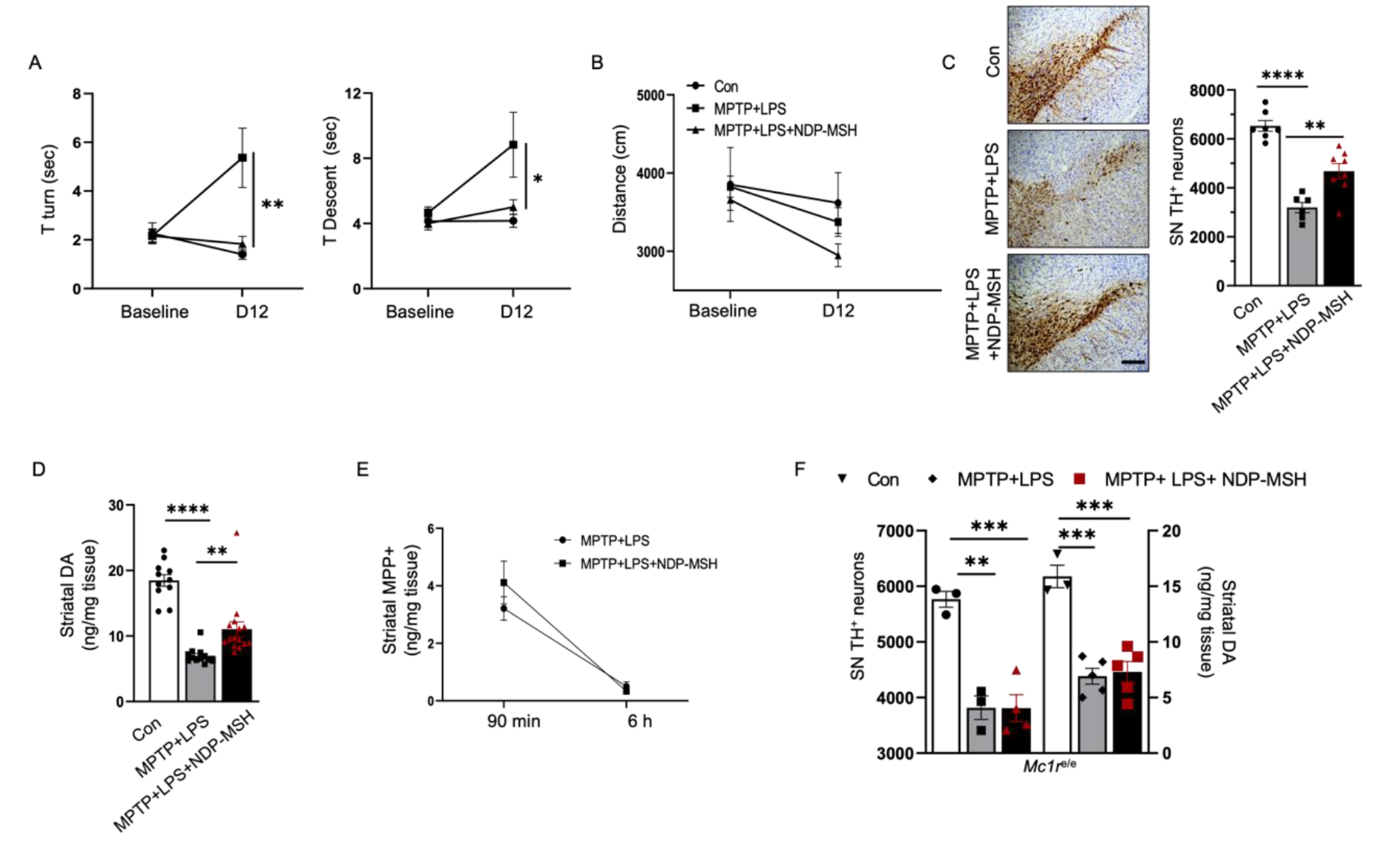
CCH rats were then randomly divided into five treatment groups: (1) the BCCAO group (M), (2) the BCCAO + URB597 group (MU), (3) the BCCAO + 4-PBA group (MP), (4) the BCCAO + URB597 + 4-PBA group (MUP), and (5) the BCCAO + URB597 + 4-PBA + AM630 group (MUPA) (n = 8 in each group). Four rats were dead after BCCAO surgery. Rats received daily injections of URB597 (0.3 mg/kg/day, i.p.), 4-PBA (40 mg/kg/day, i.p.), and AM630 (1 mg/kg/day, i.p.) for 4 weeks in the MU, MP, MUP, MUPA groups, respectively. Rats in the Sham and M groups (n = 8 in each group) received daily injections of an equal amount of vehicle. Rats were decapitated 2 h after the last injection and the brains were immediately removed for experiments, or stored at -80 °C.
The Oxygen-Glucose Deprivation (OGD) Model and Treatment Groups
The mouse hippocampal neuronal HT22 cell line was purchased from a public cell bank (ATCC, Manassas, VA, USA). The cells were cultured in Dulbecco’s Modified Eagle Medium (DMEM) (Beyotime Biotechnology) supplemented with 10% fetal bovine serum (HyClone, Ogden, UT, USA) and 1% penicillin/streptomycin (HyClone) in an incubator (Heraeus, Hanau, Germany) at 37 °C in 5% CO2. For OGD, HT22 cells were seeded in 96-well plates at a density of 1 × 10⁵ cells/mL and were cultured in glucose-free DMEM at 37 °C in 0.5% O2, 94.5% N2, and 5% CO2 for 4 h. The cells were then incubated in a maintenance medium for 24 h under normal conditions before subsequent experiments.
HT22 cells in the control group were treated identically except that they were not exposed to OGD. Experimental treatment groups were as follows: (1) the control group (Con), (2) the OGD group (OGD), (3) the OGD + 4-PBA (4 µM) treatment group (4-PBA), (4) the OGD + URB597 (2 µM) treatment group (URB), and (6) the OGD + TG (0.02 µM) treatment group (TG).
Cell Viability Assay
Cell viability was assessed using the 3-(4,5-dimethyl-2-thiazolyl)-2,5-diphenyl-2-H-tetrazolium bromide (MTT) assay as previously reported (Chang et al. 2021; Wang et al. 2022). A solution with 20 µL MTT [5 mg/mL MTT in phosphate-buffered saline (PBS), pH 7.4] was added to each group. Then, neurons were incubated for 4 h at 37 °C in 5% CO2. The absorbance (OD) was measured spectrophotometrically at 490 nm on a microplate reader (Epoch; Bio-Tek, Winooski, VT USA).
Measurement of Oxidative Stress and ATP
Oxidative stress and ATP levels were evaluated by determination of SOD, CAT, MDA, and ATP using commercial kits. All assays were performed using a microplate reader according to the manufacturer’s instructions (manufacturer and address) (Wang et al. 2020a).
DHE Staining
The intracellular ROS were detected using DHE staining. Cells were plated in 24-well plates, then fixed with formaldehyde for 30 min, stained with 30 µM DHE staining at room temperature for 5 min, then checked with an immunofluorescence assay using ImageJ software (Version 1.46r; National Institutes of Health, Bethesda, MD, USA) (Gao et al. 2019).
Annexin-V-FITC Flow Cytometry Analysis
Cell apoptosis was quantified using an Annexin V Apoptosis Detection kit according to the manufacturer’s instructions (manufacturer and address). Briefly, 10 µL propidium iodide and 5 µL Annexin-V-FITC solution were added to HT22 cells, followed by incubation for 15 min in the dark at room temperature. Finally, the cells were collected into flow cytometry tubes, and cell apoptosis was measured using a flow cytometer at 488 nm.
Nissl Staining
Coronal slices (10 μm thick) were used to estimate hippocampal neural tissue damage using Nissl staining as earlier reported (Xu et al. 2018). Briefly, the coronal cryosections of the brain were stained with 0.75% cresyl violet, dehydrated using graded alcohol percentages (70%, 95%, and 100%), and placed in xylene. The slices (n = 3) were visualized using a microscope (BX53; Olympus, Tokyo, Japan) at ×200 by an investigator blinded to the identities of the treatments.
Morris Water Maze (MWM)
The MWM was used to measure hippocampus-dependent learning and memory, as previously described (Wang et al. 2017a, 2021c). Rats were tested in a circular tank, 1.5 m in diameter with a platform of 14 cm in diameter below the water surface (1.5 cm). Animals were trained for 4 days with four trials per day by looking at the platform in the tank with water (25 ± 1 °C) (n = 8 rats per group). On day 5, the platform was removed and rats were allowed to swim freely for 60 s in the probe trial. A video camera and Human Visual System Image Software (HVS Image, Hampton, UK) were used to observe and record the times spent in the target quadrant, platform position crossings, and the swimming pattern of each rat.
Transmission Electron Microscopy (TEM)
Brains were prepared for TEM analysis to determine ultrastructural changes, using previously reported procedures (Wang et al. 2017a, b). Briefly, tissues were dehydrated by alcohol and embedded with a mixture of acetone and ethoxylate resin (Ladd Research Industries, Burlington, VT). Brain sections, cut to a thickness of 600 nm using the LKB Huxley ultramicrotome, were placed on copper grids, then stained with uranyl acetate and lead citrate (Wang et al. 2017b). Finally, the ultrastructure changes of organelles in the hippocampus CA1 area were observed using a transmission electron microscope (CM-120; Philips, Amsterdam, The Netherlands). The degree of mitochondrial damage and the ultrastructure changes of mitochondria-associated ER membranes (MAMs) were semi-quantitatively analyzed by one investigator, blinded to the identities of the treatment groups, according to published guidelines as shown in Table 1 (Flameng’s score) (Flameng et al. 1980; Paillusson et al. 2017; Ouyang et al. 2022).
Table 1 The criteria for scoring the neuronal mitochondria injury using TEM (Flameng et al. 1980)Immunofluorescence Staining
Brain sections or HT22 cells were fixed in precooled 4% paraformaldehyde for 20 min, permeabilized with 0.1% Triton X-100 (Sigma-Aldrich) for 30 min, then blocked with 5% bovine serum albumin (BSA) in PBS for 30 min at room temperature. The fixed tissues and cells were incubated with primary antibodies against GRP78 (1:300), TOMM20 (1:300), NeuN (1:300), Iba-1 (1:300), or GFAP (1:300) in 5% BSA overnight at 4 °C(Jin et al. 2018; Wang et al. 2022). Subsequently, samples were counterstained with 4´,6-diamidino-2-phenylindole after incubating with deconjugated secondary antibodies for 2 h in the dark. A fluorescence microscope (Zeiss, Jena, Germany) was used to photograph the immunofluorescent images. Finally, three fields of view in each group were used to estimate the mean fluorescence intensity, by an investigator blinded to the identities of the groups.
Western Blotting and Immunoprecipitation
Proteins from neuronal tissues in the hippocampus CA1 area were extracted and quantified using a total protein extraction kit (BC3711; Solarbio, Beijing, China) and a BCA protein assay kit (P0012S; Beyotime) according to the manufacturer’s protocols. Equal amounts of protein were separated by SDS-PAGE on a 6 − 12% polyacrylamide gel, then transferred onto polyvinylidene difluoride (PVDF) membranes. The PVDF membranes were subsequently probed with primary antibodies against the following proteins overnight at 4℃: GRP78 (1:1,000), ATF6 (1:500), PERK (1:1,000), p-PERK (1:1,000), CB2 (1:500), β-Arrestin-1(1:500), CHOP (1:500), Cyt-c (1:500), caspase-9 (1:1,000), GAPDH (1:5,000), and β-tubulin (1:5,000) used as an internal loading control. Following washing in PBS, the membranes were incubated with secondary antibodies conjugated with horseradish peroxidase, at room temperature for 1 h. The western blot protein bands were visualized using the enhanced chemiluminescence system (Millipore, Watford, UK) and quantified by ImageJ software.
For immunoprecipitation, 300 µg of protein extract was incubated with the antibodies against CB2, β-Arrestin-1, or unspecific IgG at 4 °C overnight, and protein-A/G agarose was added for another 2–3 h at 4 °C (Wang et al. 2021a). The immune precipitates were centrifuged, washed, suspended, and subjected to western blotting analysis.
Statistical Analysis
Each experiment was conducted at least in triplicate. The data are presented as the mean ± standard deviation (SD) and were analyzed by SPSS statistical software for Windows, version 20.0 (SPSS, Chicago, IL, USA). The repeated-measures mixed analyses of variance (ANOVA) with Tukey post-hoc test was used to assess group and training day differences. One-way analysis of variance, followed by the Tukey post-hoc test was conducted to evaluate statistical differences among the experimental groups. Significance was defined as P < 0.05.


留言 (0)