In this REPAIR Echo sub-study, correlation and agreement analyses both demonstrated a good relationship between selected echo and cMRI variables, and showed that echo is a valuable complementary method for the assessment of macitentan treatment effect on RV structure and function in patients with PAH. Macitentan treatment led to improvement in RV structure and function as assessed by echo and cMRI, and improved hemodynamics and functional variables in patients with PAH. Results from this post hoc analysis were consistent with those observed in the REPAIR primary analysis that demonstrated improvements in RV functional and hemodynamic parameters following macitentan treatment assessment by cMRI and RHC, respectively [15].
Most echo variables of cardiac structure and function in this sub-study improved after treatment with macitentan, including those with known prognostic value in PAH, such as RVSV, LVSV, LVEDV, RVFAC, TAPSE, and 2D GLRVS [16,17,18,19,20,21,22]. Of note, the change in RVSV measured by echo surpassed the threshold of a minimally important difference (10 mL) [23], with an increase of 11.6 mL at week 26 for the Echo subgroup after treatment with macitentan. This was consistent with the change in RVSV as assessed by cMRI previously reported in the REPAIR study [15]. Improvements were also observed for LVSV and LVEDV, with increases of 9.6 mL and 15.1 mL respectively, at week 26 compared to baseline. In this analysis, macitentan also improved the key prognostic parameters RVFAC and TAPSE [19,20,21], with RVFAC increasing by 7.5 and 7.1% at weeks 26 and 52, respectively, compared to baseline. The mean increase in TAPSE above 17 mm observed at weeks 26 and 52 is associated with better systolic function and survival [2, 3].
Measures of 2D GLRVS are particularly useful for the evaluation of the function of the right heart and a marker of subclinical worsening, as it shows changes before other more conventional parameters deteriorate [24]. The results from our analysis suggest macitentan improved 2D GLRVS, as a worse 2D GLRVS (≥ − 15.5%) has been shown to independently predict adverse clinical events and death in patients with PAH [22].
Mitral flow (E/A ratio) improved at week 26, suggesting that improvements in RV function are associated with normalization of LV filling patterns, likely as a result of reduced leftward septal bowing [25]. These data indicate that echo can be sensitive enough to show clinically relevant treatment effects in these parameters.
Given the unique RV anatomy, the estimation of 2D echo parameters relying on volume modelling by simple geometrical assumptions can be challenging [26, 27]. These challenges highlight the importance of central assessments of echo parameters if used as endpoints in clinical trials, as this has been shown to reduce the variability of the measurements obtained [28]. In this study, assessment of imaging data for each individual patient was performed batch-wise at the same time by the same reviewer.
Assessment of the structure and function of the heart plays an essential role in the follow-up of patients with PAH. Although cMRI is considered the gold standard technique for assessing RV structure and function, it is not always feasible and available. In its absence, echo can help clinicians to assess response to therapy at clinical follow-up. In a prospective study of 27 patients with PAH, 3D reconstruction of 2D transthoracic echo images to measure RVSV, RV end-diastolic volume (RVEDV), RV end-systolic volume (RVESV) and RVEF correlated well with cMRI assessed using Pearson’s correlation coefficient [12]. Similar observations were seen in a prospective study of 30 patients with PAH, which showed a strong correlation between 2D GLRVS by echo and RVEF by cMRI (r = 0.69) [29].
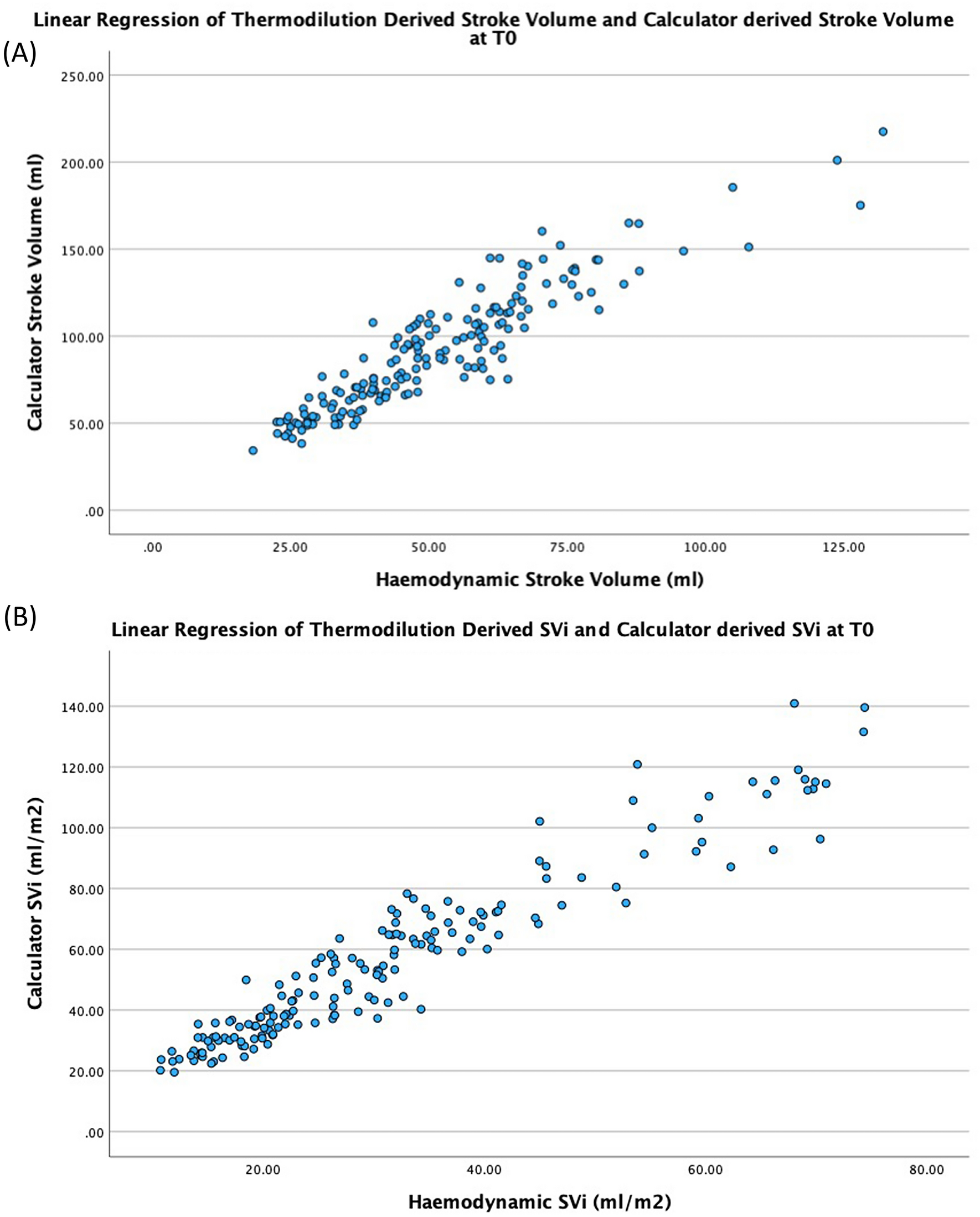
Our analysis found a strong correlation in three of the four variables measured by echo and cMRI, including LVSV by echo and cMRI, LVEDV by echo and cMRI, and 2D GLRVS by echo and corresponding RVEF by cMRI in patients with PAH. However, only a moderate correlation was observed for RVSV assessed by echo and cMRI for the patients in our study. The difference in the level of correlation for RVSV by echo and cMRI between this analysis and the aforementioned study by Bhave et al. [12], could be due to their use of 3D reconstruction of 2D transthoracic images, in comparison to 2D echo used in this study.
In our study, the Bland–Altman analysis showed a good agreement for LVSV measured by echo and cMRI. RVSV reflected an overestimation compared to cMRI, as shown by the mean RVSV at baseline of 68.4 mL measured by echo and 48.9 mL by cMRI. The overestimation of RVSV by echo could be explained by the underlying pathophysiology of this patient population and the method of choice to assess the RVSV. In this study, RVSV by echo was assessed using pulmonic valve Doppler and pulmonary artery annulus. This method offers an indirect way of measuring the RVSV as it relies on the assumption that the cross-sectional area of the pulmonary artery annulus remains constant during the cardiac cycle, which may not always be the case in patients with PAH. In contrast, cMRI provides a direct measurement of the RVSV using 3D images without the need for geometric assumptions [30]. However, cMRI can underestimate the RVSV by flow, for example, by the occurrence of off plane orientation of the slice as well as turbulent flow leading to further underestimation [31]. Yet, it is difficult to deduce with any degree of certainty what the true reasons are for this overestimation.
The most frequent AEs reported for the Echo subgroup; peripheral edema, headache, and dizziness, were comparable to those previously reported in the REPAIR study [15]. Altogether, these results add to the evidence supporting the beneficial effects of macitentan either initiated as monotherapy or as part of an initial or sequential combination therapy regimen, and also demonstrate the value of echo measures to capture such improvements.
Due to the post hoc nature of this study, some potential biases exist; no adjustments were made for multiplicity, patients may not have had complete data for all variables, and there was limited interpretation of an increase and/or decrease in specific echo variables due to a lack of published validated thresholds in PAH. Furthermore, very few patients in the REPAIR study experienced disease progression and therefore no assessment could be made on the use of echo and cMRI variables in measuring PAH worsening. A number of echo variables had a considerable level of missing data, mainly attributed to absent calibrations for M-mode and Doppler images, absent calibrations for time-based measurements, and poor resolution/quality which made some images unmeasurable. Lastly, the Echo subgroup sample size was small (however, observed cMRI, RHC, and functional results were consistent with the larger REPAIR safety set), and some echo-assessed variables, including tricuspid regurgitation peak jet velocity, were only reported for a small number of participants.


Comments (0)