Solid formulations, such as tablets and capsules, undergo disintegration after administration as a function of formulation and capsule. Disintegration begins in the oral cavity for oral disintegration tablets, the stomach for immediate release (IR) forms, stomach or intestine for sustained release (SR) forms, and the intestine for enteric-coated (EC) forms. Following disintegration, drugs are released from each disintegrated formulation and dissolved in the gastrointestinal (GI) fluid according to the formulation characteristics. An SR strategy is often applied to guarantee to maintain settled plasma drug concentration and induce longer pharmaceutical effects (Takagi et al., 1987, Suleiman et al., 1989, Neill et al., 2008, Preechagoon et al., 2010). EC formulations are generally used to prevent drug degradation from the strong acidity in the stomach (Tjandramaga et al., 1984, Pilbrant and Cederberg, 1985). Polymers with specific dissolution properties are used for SR and EC formulations so that the desired release properties are attained. Water-soluble polymers are generally contained in amorphous solid dispersion (ASD) formulations that improve the solubility of poorly water-soluble drugs, enhancing oral drug absorption (Yamashita et al., 2003, Six et al., 2005, Jang and Kang, 2014, Wiederhold, 2015).
For pharmaceutical companies, estimating drug absorption profiles in humans is a key factor in the success of oral formulation development. Compound absorption profiles are determined by the dissolved concentration in GI fluid, absorption clearance, and effective transit time through the absorption site. Therefore, the drug release time profile of formulations, defined in this study as in vivo performance, should be identifiable in the GI tract and reflect EC, SR, and ASD formulations. To evaluate the in vivo performance of orally administered forms, various in vitro systems are used that reflect in vivo dissolution of drugs from formulations (Dickinson et al., 2012, Tsume et al., 2020, Silva et al., 2020, Silchenko et al., 2020, Masada et al., 2021). As these each have different advantages, they help to compare in vivo performance among formulations. However, this performance must be demonstrated by in vivo studies evaluating the plasma concentration-time profile. In the case of a lack of correlation between formulation in vitro and in vivo testing, the cause must be identified, although this is often no simple task.
Several non-invasive imaging techniques are reported to assess the GI behavior of formulations and oral absorption processes. For example, gamma scintigraphy with 111In and 99mTc can reveal the GI transit of an orally ingested solution or formulation in addition to evaluating various effects (Christensen et al., 1985, Wilson et al., 1989, Basit et al., 2001). Positron emission tomography is also useful in quantitatively evaluating GI movement, intestinal absorption, and pharmacokinetics of orally administered drugs using 11C or 19F-labeled compounds (Yamashita et al., 2011, Kataoka et al., 2012, Takashima et al., 2013). Magnetic resonance imaging can also quantitatively evaluate formulation disintegration and behavior in the GI tract (Takeshita et al., 2017, Grimm et al., 2019, Sager et al., 2019, Sulaiman et al., 2022). Although these technologies have certain advantages, the requirement for markers such as radio-labeled compounds result in time-consuming and costly processes.
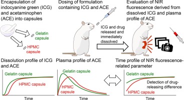
Near-infrared (NIR) imaging is a light-based imaging technique that detects the in vivo distribution of NIR probes after administration. This is possible because tissue and skin is transparent to the emitted NIR light. This characteristic allows NIR imaging to assess time-dependent changes in tissues (Luo et al., 2011, Cilliers et al., 2017, Borlan et al., 2021). NIR imaging is also useful in developing oral dosage forms to understand mechanisms of disintegrants and tablet disintegration process in vitro (Smetiško and Miljanić, 2017, Baranwal et al., 2019, Jiménez-Romero et al., 2020, Ojala et al., 2020). While several studies report the application of NIR focused on intestinal injury (Kwon and Sevick-Muraca, 2011, Tahara et al., 2018, Yamaguchi et al., 2021), there are no reports exploring NIR imaging for formulation performance in vivo.
In this study, we performed in vitro and in vivo experiments to demonstrate the feasibility of NIR imaging to evaluate the in vivo performance of orally administered formulations. First, the NIR probe characteristics were investigated in both solution and solid form. Secondly, the GI movement of the dissolved NIR probe and corresponding abdominal and intraperitoneal NIR fluorescence intensities were evaluated. Thirdly, two capsule formulations made from different materials containing the solid probe and drug were prepared. Finally, NIR fluorescence imaging and blood sampling were performed on rats administered with each formulation. Then the relationships between the time profiles of the NIR fluorescence-derived parameters and drug plasma concentration were assessed.


留言 (0)