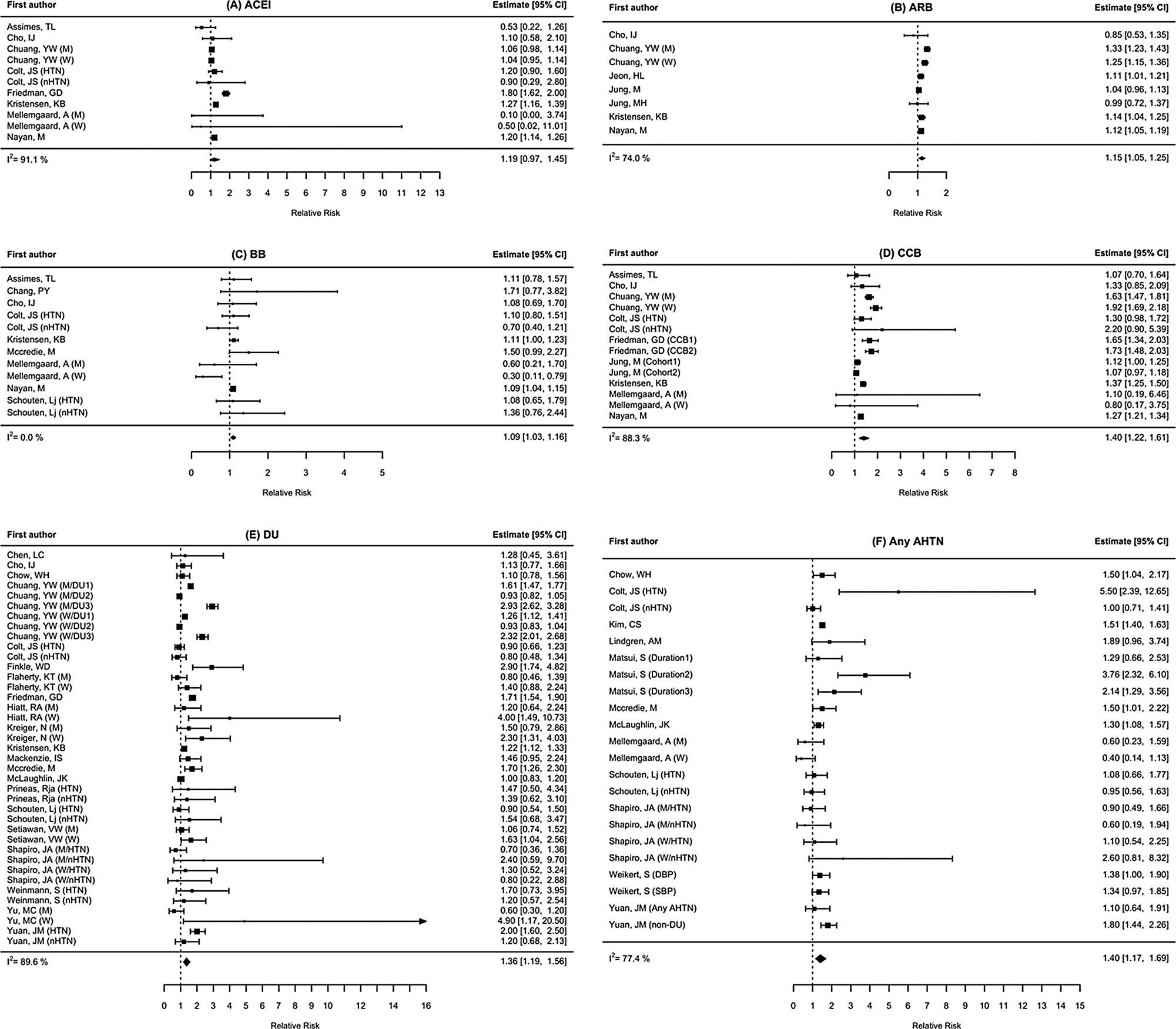
Head and neck squamous cell carcinoma (HNSCC) arising on the mucosal surfaces of the oral cavity, sinonasal cavity, pharynx, and larynx represents the eighth most common cancer worldwide, with an estimated annual incidence and mortality of around 880,000 and 445,000 cases, respectively [1]. Tobacco, excessive alcohol consumption, and human papillomavirus (HPV) infection are the main risk factors of HNSCC. The prognosis for HPV-associated oropharyngeal cancer is more favorable due to a better response to chemotherapy and radiotherapy, and fewer comorbidities [2, 3], whereas the prognosis for recurrent or metastatic (R/M) HNSCC is poor. In 2008, the addition of cetuximab to conventional platinum/5-fluorouracil (5-FU) chemotherapy (EXTREME regimen) improved overall survival as seen in long-term responders, although it was a marginal improvement (48 months progression-free survival (PFS) rate < 3%) [4, 5]. Immune checkpoint inhibitors of programmed cell death protein 1 (PD-1) are an important breakthrough with prolonged duration of response and survival observed in both first- and second-line therapies (5 year-overall survival (OS) rate 15.4% for pembrolizumab monotherapy for the combined positive score (CPS) ≥ 1 arm from the KEYNOTE 048 cohort, Makoto Tahara et al. ESMO 2022) [6, 7]; but, this duration of response is observed in < 20% of the patients. The only predictive marker currently available is the expression of PD-L1, according to the CPS, which has been shown to correlate with survival [7]. However, strong primary/secondary resistance is observed, and tumor response is unpredictable (with an objective response rates (ORR) of 23.3% for pembrolizumab monotherapy in the CPS ≥ 20 arm). Consequently, patients with severe symptoms awaiting an urgent therapeutic response require treatments including chemotherapy to reduce life-threatening progression at the cost of increased toxicity [7]. In addition, a growing number of studies are reporting a long-term survival benefit from cetuximab-based chemotherapy after failure of immunotherapy [8,9,10]. Therefore, immune checkpoint inhibitors appear to be the only current treatment providing effective clinical responses. These observations underline the main challenge in cancer immunotherapy: optimizing long-term survival requires personalized approaches based on composite biomarkers to define the optimal therapeutic strategy and discover new drugs that can generate immunity in patients who do not have a strong immune response [11]. The development of biomarkers and new therapeutic agents to overcome primary/secondary resistance requires an optimum knowledge of the complex interactions between cancer and the immune system involving molecular and cellular drivers of immune escape. This is considered to be a major challenge facing cancer immunotherapy [11]. However, this objective is complicated by the difficulty of studying not just immune cells but rather an “orchestra of immune and non-immune players” evolving during systemic cancer therapy (Girolami et al. 2023 Journal of Personalized Medicine). Currently, the classification of tumor immunity is almost exclusively based on the spatial distribution of CD8 + T cells in the tumor microenvironment (TME), which are targeted by immune checkpoint inhibitors of PD-1 thereby enhancing their anti-tumor functions [12]. A gradient of three immunophenotypes associated molecular pathways is observed: inflamed tumors including tumor infiltrating lymphocytes (TILs) and B cells in the TME in close proximity to tumor cells; immune-excluded tumors including immune cells distant from tumor cells in the tumor stroma; and immune desert tumors devoid of T cell infiltration. Using gene expression of HNSCC tumor data obtained from The Cancer Genome Atlas (TCGA) database, another immune-infiltrating signature-based classification was recently proposed with three immunophenotypes including other immune cells: cold, lymphocyte (enrichment for CD4 + T cells, CD8 + T cells, and B cells) similar to the previous classification, and myeloid dendritic (DC) signatures (enrichment of neutrophils, macrophages, monocytes, DCs, and T regulatory (Treg) cells) [13]. Thus, classifying TME based on immune cell infiltration alone excludes major actors in cancer immunity such as stromal cells including carcinoma associated fibroblasts (CAF) or tertiary lymphoid structures (TLS) representing promising targets for cancer treatment [14]. Notwithstanding these limitations, classification based on immune cells infiltration provides a reference framework for research describing specific molecular mechanisms for each immunophenotype such as interferon-gamma (IFN-γ) signaling associated with inflamed tumors and transforming growth factor-β (TGF-β) signaling with immune-excluded tumors. Immunotherapy resistance observed in cold tumor may be related to impaired antigen processing (Human leukocyte antigens (HLA) and/or β2 microglobulin loss or downregulation, and elimination of neoantigens through copy-number loss), loss of T-cell priming mainly because of the inhibition of T cells generation by DCs in desert tumor, or the absence of preexisting antitumor T cells infiltration (T-cell-excluded tumors) [15]. Therefore, to enhance antitumor immune responses, next generation immunotherapy should promote lymphocyte activation by tumor antigens and antigen-presenting cells (APC), differentiation and infiltration of lymphocytes in the TME, and tumor cell recognition. However, new therapeutic drugs still have limitations in achieving long-term responses and survival in the majority of patients. This is illustrated in HNSCC by the absence of new therapeutic agents since the arrival of PD-1/PDL-1 blockade drugs and the large number of failures in clinical trials testing combination treatment with immune checkpoint inhibitors in recent years: INDUCE-3 and INDUCE-4 trials investigating feladilimab (inducible T cell co-stimulatory agonist) in combination with pembrolizumab (press release GSK April 14, 2021), INTERLINK-1 study evaluating monalizumab (inhibitor targeting natural killer cells group 2A) in combination with cetuximab (press release Innate pharma Jan 8, 2022), CP-MGA271-06 study with enoblituzumab (anti-B7-H3 mediating antibody-dependent cellular cytotoxicity (press release MacroGenics Jul 8, 2022), Active8 trial with motolimod (TLR8 agonist), and ATHENA trial evaluating the combination of atezolizumab and bevacizumab [16, 17]. Although there are promising studies with drugs targeting immune checkpoint receptors (TACTI-002 Part C: A phase II study of eftilagimod alpha, a soluble lymphocyte-activation gene 3 (LAG-3) protein and pembrolizumab), or immunomodulators (BCA101, bifunctional epidermal growth factor receptor (EGFR)/TGFβ inhibitor and pembrolizumab), these are preliminary studies with a small sample size and a tumor response of less than one in two patients [18, 19].
The immune microenvironment is a dynamic structure in a complex interaction with an evolving cancer, but is affected by specific oncology treatments, particularly the immune checkpoint inhibitors. Therefore, a differential evolution of the TME between responders and non-responders induced by cytotoxic T-lymphocyte–associated antigen 4 (CTLA-4) and PD-1 blockade have been observed in a longitudinal study of metastatic melanoma patients [20]. Significantly, differences in TME composition such as number of CD4 + or CD8 + T cells are increased during on-treatment than before suggesting that predictive biomarkers based on immune signatures should be evaluated in on-treatment tumor samples. Few other recent studies have confirmed temporal changes of the TME during PD-1 inhibitor therapy between immune subpopulations but also for tumor mutational burden, T-cell receptor (TCR) repertoire, immune-related genes, proliferation-associated and chemokine genes, neoantigen immunogenicity landscapes, and tumor clonal populations [21,22,23,24,25]. However, how immune checkpoint inhibitors shape the TME and how they interact with cancer cells remain poorly understood. Therefore, the majority of studies only provide a snapshot of the immune landscape because dynamic profiling of the TME requires biological samples not often available in clinical practice. Profiling of the TME during anti-PD-1 immunotherapy may be essential to accelerate the understanding of the mechanisms that underlie primary and secondary resistance [26,27,28]. Besides existing immunotherapy, studies also suggest the importance of a deeper understanding of dynamic neoantigen profiles for vaccine development [26]. Targeting antigen selection to ensure high immunogenicity and tumor-specificity is a complex process considering mutation calling, clonality, HLA typing and binding affinity, antigen processing, and similarity to self [29]. Genomic and transcriptomic data may facilitate this selection step by prioritizing the antigens maintained despite tumor evolution and neoantigens silenced in immune-excluded TME [26]. This latter mechanism could explain tumor responses observed on salvage chemotherapy after progression with immune checkpoint inhibitors used in first-line treatment, but our proposal in this study is yet to be investigated [30]. Collecting consecutive clinical tumor biopsies remains a challenge when the treatment is ongoing, suggesting that basic research in cancer immunology and the development of cancer immunotherapy agents are largely based on treatment-naive tumors. However, most patients with incurable HNSCC eligible for immune checkpoint inhibitors had locoregional recurrence occurring in a previously irradiated area, which probably induce a change in TME. However, in the case of HNSCC, tumors are often visible and palpable, and biopsies can therefore be easily processed, allowing a longitudinal TME profiling.
A differential TME-related gene expression analysis between anti-PD-1/PD-L1 immunotherapy responders and patients with a progressive disease requires the use of breakthrough technologies. Indeed, deep analysis of the main cellular subsets including tumor cells, tumor-associated stromal cells, and tumor-infiltrating immune cells should consider their position, their organization in the TME, their state, function and cellular phenotype, their interaction with neighboring cells, and the changes over time caused by immunotherapy. Among these new technologies, single-cell RNA sequencing (scRNA-seq) offers the highest cellular resolution but requires tissue dissociation resulting in the loss of spatial information. Spatial transcriptomics (ST), named “Method of the Year 2020” by Nature Methods, is another powerful technique. It preserves the spatial context of biological data and allow the visualization of gene expression distributions. It is suitable for small samples but is limited by low cellular resolution [31, 32]. Thus, scRNA-seq and ST are two complementary technologies that will be performed in this study. Differential gene expression analysis between responders and non-responders to anti-PD1/PD-L1 immunotherapy will focus on immune-related genes such as immune checkpoint genes, but also on genes that play important roles in TME including hypoxia-related genes, ΔNp63 (potentially implicated in the recruitment of myeloid-derived suppressor cells and overexpressed in HNSCC), immunogenic cell death genes, and mitophagy-related genes, which may be targeted to improve the effectiveness of cancer immunotherapy [33,34,35]. A particular focus will be made on IFN-γ mediated signaling that may result in both protumoral (immunosuppression, angiogenesis, and tumor cell proliferation) and antitumoral (tumor cell killing, T cell activation, immune cell proliferation, and promotion of antigen presentation) activities indicating the emphasis on its therapeutic modulation [36]. These conflicting activities may be related to the duration and the importance of IFN-γ signaling which are affected by tumor burden, immune cell infiltration characteristics, and cancer treatment (immunotherapies and chemotherapies) [36]. Although producers of IFN-γ and the different effects of IFN-γ in the TME are well documented, lots of questions remain on its involvement in the primary/secondary resistance and on its complete and long-lasting response to immune checkpoint inhibitors. Understanding these mechanisms also requires dynamic analysis of IFN-γ production in the TME undergoing immunotherapy.
Thus, analysis of biopsy samples of patients undergoing immunotherapy might contribute to a better understanding of resistance process but with inherent limitations. These include the invasive nature of the analysis with uncertainties related to patients compliance issues and importantly, the intra-tumor heterogeneity with disparate levels of immune infiltration leading to underestimation of the tumor genomics landscape. Conversely, liquid biopsy may overcome tumor heterogeneity in addition to being noninvasive, allowing continuous monitoring of circulating tumor DNA (ctDNA), circulating tumor cells (CTCs), exosomes, miRNAs, circulating immune cell, and TCR repertoire [37]. Studies have already documented the prognostic ability of ctDNA to detect HNSCC recurrence and/or metastasis [38]. However, there are no prevalent hotspot mutations in HNSCC, only a wide spectrum of mutations including for TP53 tumor suppressor gene which is the most frequent of all somatic genomic alterations in HNSCC [39]. Thus, ctDNA monitoring requires personalized trackable mutations.
The same would apply to miRNAs, a class of endogenous non-coding RNAs involved in post-transcriptional regulation of gene expression and playing an important role in chemo-resistance of cancer cells [40]. Cancer-associated circulating miRNAs are secreted both by immune and tumor cells, suggesting potential predictive value and therapeutic interest [41, 42]. Lastly, understanding the role of circulating cytokines in cancer immunotherapy was a major development that led to the approval of recombinant IFN-α and interleukin (IL)-2 for the treatment of several malignant diseases [43]. Current challenges include suppression of immunosuppressive cytokines, limitation of systemic proinflammatory effects, and to a lesser extent, biomarker development [43]. Thus, tumoral immunity is characterized by a complex interaction between the TME and the peripheral immune system that requires a deeper understanding. Regarding liquid biopsy, the proposed study will focus on the exploration of circulating miRNA-mediated immunomodulation and circulating cytokines.
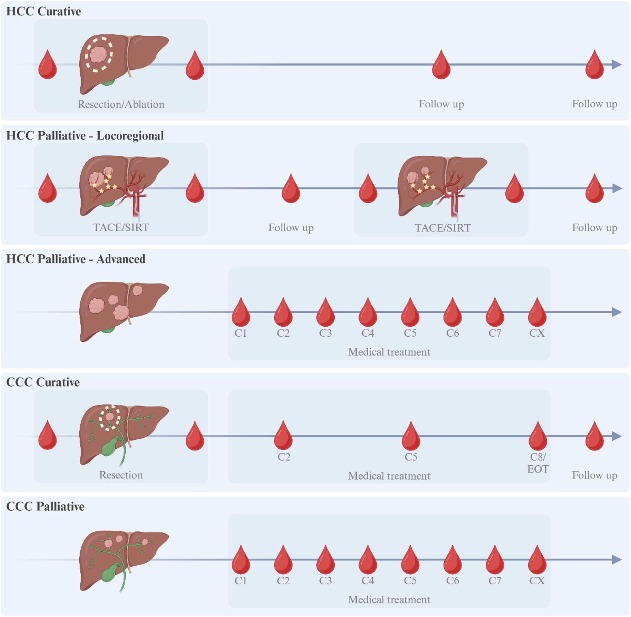
In this single-center prospective study, we propose to combine liquid biopsy, tissue biopsy, and clinical data at key time points during the management of patients undergoing anti-PD-1 immunotherapy for R/M HNSCC. We will compare the evolution of gene expression profile between responders and non-responders, using spatial transcriptomics analysis among other technologies.


Comments (0)