2.1 Samples acquisition
A total of 927 HCC samples were used in this study, including 342 from The Cancer Genome Atlas-LIHC cohort (TCGA, https://xenabrowser.net/), 260 from the LIRI-JP cohort (ICGC, https://dcc.icgc.org/), 10 from the GSE149614 cohort, 320 from the IMvigor210 cohort and 50 from the HCC clinical samples. In Supplemental Table S1, data on the pathology of HCC patients was shown. Samples with insufficient clinicopathological traits or 30-day survival rates were ruled ineligible. To make up for the dearth of normal sample instances in the TCGA dataset, 110 normal samples from the GTEx dataset were employed. The log2 (FPKM + 1) transformation was used to standardize the transcriptome data. The batch effects between the normalized data from the TCGA and GTEx were corrected using Combat from the R package "SVA." To accurately assess the quantity of TEX, the immune cell abundance identifier (ImmuCellAI, 2020.02) was used [13].
2.2 qRT-PCR and immunohistochemistry (IHC) assay
As was done before [14], the qRT-PCR method was used to ascertain the mRNA expression of the genes covered in this investigation among tumor tissues and corresponding paracancerous tissues from 50 HCC patients. Primer sequences are displayed in Table S2. IHC tests using anti-PAFAH1B3 (Proteintech, China) or anti-CD8 (Abcam, UK) antibodies were separately performed on paraffin-embedded tissues from HCC patients. The slides were incubated with secondary antibodies and imaged using a Leica DM 2500 microscope. The immunostaining intensity of the required proteins was evaluated and graded separately by two distinct observers. To differentiate between low/loss (≤ 4) and high (> 4) expression of PAFAH1B3 in HCC and corresponding paracancerous tissues, a final score derived from the sum of the extent of expression score (no positive cells = 0, < 10% = 1, 10–50% = 2, positive staining of > 50% = 3) and intensity score (negative = 0, weak = 1, moderate = 2, strong = 3) was employed.
2.3 Assay for double-label immunofluorescence
The TSA Fluorescence Kit was utilized to conduct double-labeled immunofluorescence staining on the aforementioned HCC tissue samples. In summary, tissue slices were pre-stained with CY3 and Alexa Fluor 488 after being treated with primary antibodies anti-PAFAH1B3 (Proteintech, China) and anti-PD1 (Abcam, UK). After that, CaseViewer software was used to scan and analyze the parts. Lastly, the degree of PAFAH1B3 + PD1 + cell infiltration in HCC tissue samples was noted.
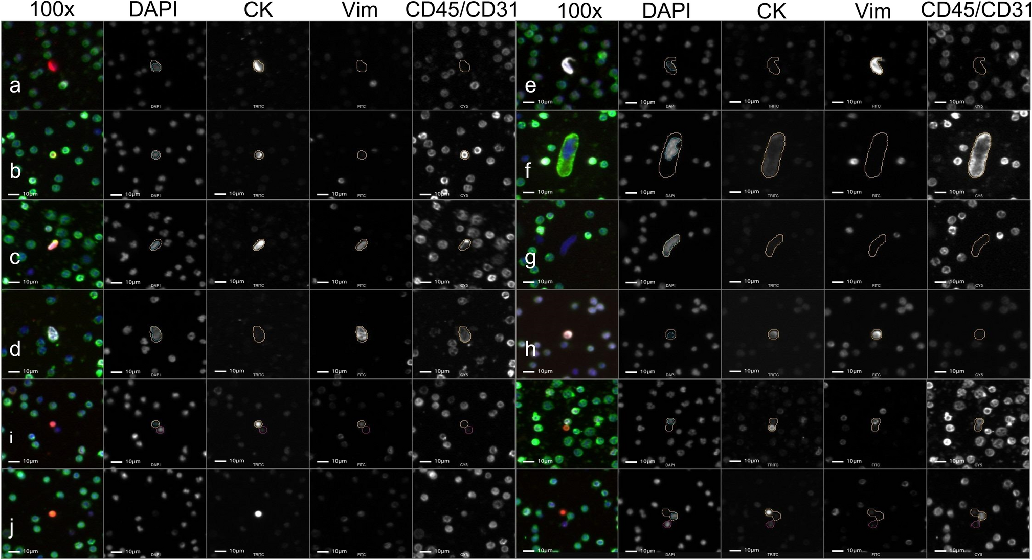
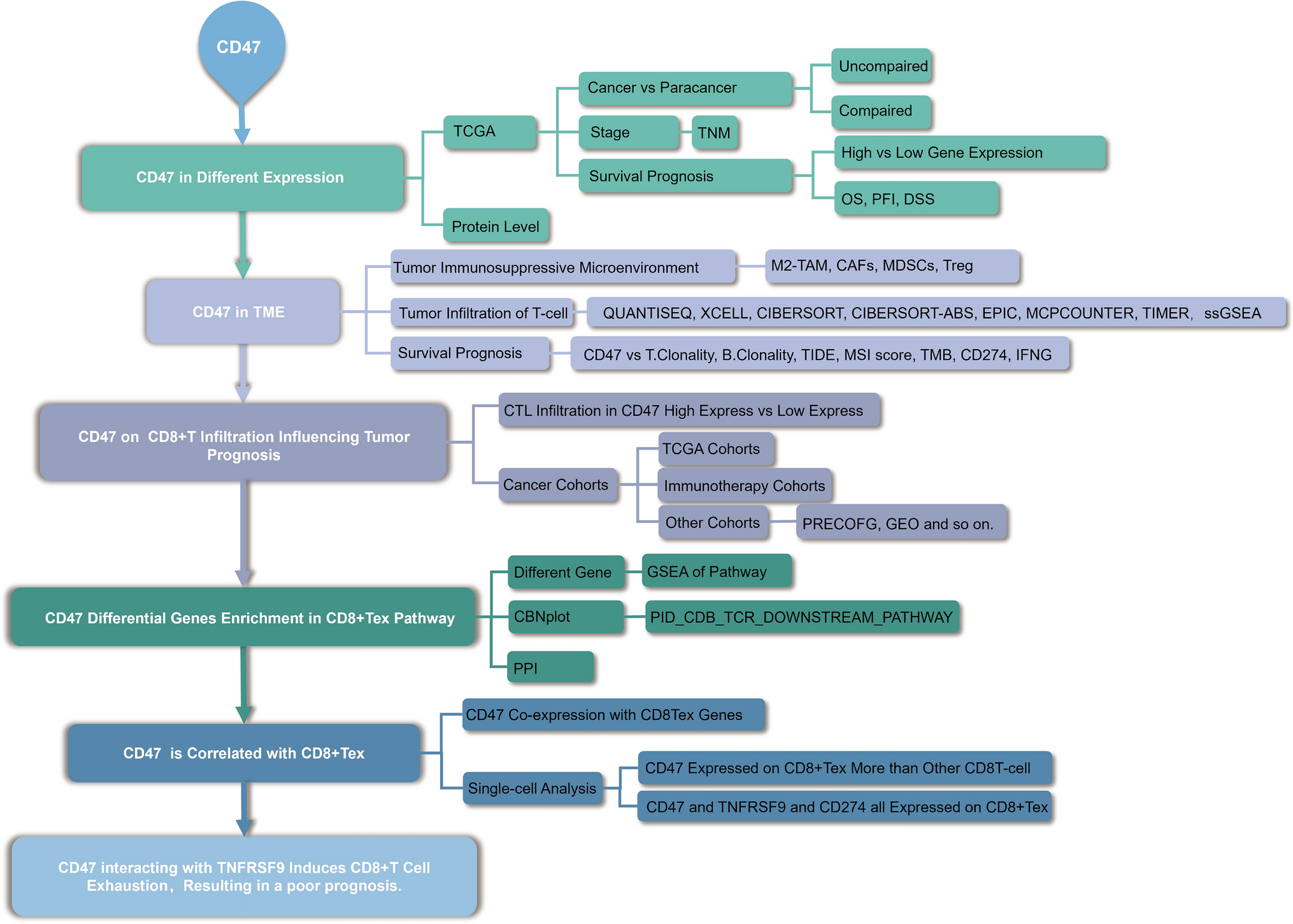
2.4 TEX identification in the single-cell sequencing data
Quality control and cell screening were carried out on the GSE149614 dataset using the Seurat package [15] as reported by the previous study [16], yielding a total of 31,396 cells and 17 distinct subgroups for additional analysis. These distinct groupings were identified based on several marker genes (Figure S1). We picked out each NK/T cell separately and binned them once again with dim = 50 and resolution = 0.1. Finally, TEX was identified according to the TEX marker (HAVCR2, TIGIT, CTLA4, LAG3, and PDCD1).
2.5 The pathway richness analysis
To assess the variation in biological processes between the PAFAH1B3-high and PAFAH1B3-low subgroups in the TGCA cohort, we used GSVA analysis with the R package 'GSVA'.
2.6 Comprehensive analysis of the effectiveness of immunotherapy
We looked at the differences in gene expression between the PAFAH1B3-high and PAFAH1B3-low subgroups for numerous immune checkpoint inhibitors (ICIs). The prognostic utility of PAFAH1B3 for immunotherapy was further demonstrated using an anti-PD1/PD-L1 inhibitor cohort (IMVigor 210) with relatively extensive transcriptome data and information on immunotherapy response.
2.7 Potential tumor-sensitive drug prediction
We especially looked at the connections between 216 medicines in the CellMiner database and PAFAH1B3 expression [17]. A drug is considered tumor-sensitive if it has a Pearson correlation coefficient of more than three-tenths and its adjusted P-value is less than one in a thousand. The half-maximal inhibitor dosage (IC50) of the targeted medication was then predicted using gene expression levels to demonstrate therapeutic sensitivity. Furthermore, to confirm the effect of the screened tumor-sensitive drugs on PAFAH1B3, we constructed PAFAH1B3 low-expressing cell lines according to the previous study [12], which were incubated with tumor-sensitive drugs at 100% humidity, 37 °C, 5% CO2 in the recommended fetal bovine serum containing 10% fetal bovine serum (FBS, Sangon Biotech, China) in DMEM medium (Sangon Biotech, China) for colony formation assay. The colonies were fixed and stained with crystal violet (Sangon Biotech, China) in 10% ethanol for 5 min. Finally, the cell colonies were imaged and counted.
2.8 Statistical analysis
R-4.2.1 performed all the statistical analyses. For quantitative data, the Wilcoxon rank sum test was used to analyze the statistical significance of variables with non-normal distributions, while the Student's t-test was used to check the statistical significance of variables with a regular distribution. All comparisons were two-sided with an alpha level of 0.05, and the false discovery rate (FDR) for multiple hypothesis testing was corrected using the Benjamini–Hochberg method.

留言 (0)