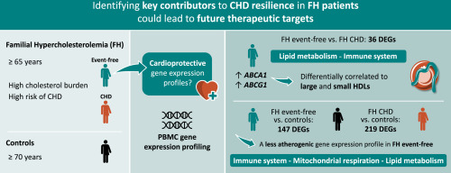
The association of lipoprotein (a) with coronary artery calcification: A systematic review and meta-analysis
Coronary artery disease (CAD) is a critical cardiovascular disease (CVD) and the leading cause of death worldwide [1]. Atherosclerosis, associated with the pathogenesis of CAD, is characterised by the accumulation of large amounts of lipids, fibre components, and calcification in arteries [1]. Coronary artery calcification (CAC) is a pathophysiological feature of atherosclerosis and closely associated with atherosclerotic plaque burden [2]. The Agatston score is a semi-automated tool utilised in computed tomography (CT) to quantitatively assess calcified coronary plaques, evaluating cardiovascular risk in asymptomatic individuals [3]. Each lesion with an area ≥1 mm2 and radiological attenuation >130 Hounsfield units is scored, combining the volume and density of CAC [4]. Higher scores correlated with increased CAC volume and density. The Agatston score, endorsed by international guidelines, is a valuable predictor of CAD and all atherosclerotic CVD (ASCVD) events and a personalised management strategy for the primary prevention of ASCVD [4]. Notably, a CAC score >300 is a strong predictor of CAD incidents [5]. Although asymptomatic patients with CAC or progression of CAC are at risk for stroke and other cardiovascular outcomes, the risk factors for CAC have not been fully elucidated. Therefore, identifying the risk factors associated with CAC is essential to explore further ways to prevent or inhibit calcification, thereby reducing ASCVD risk.
Elevated lipoprotein (a) [Lp(a)] level is an the independent risk factors for atherosclerosis [6]. The main constituents of Lp(a) are primarily composed of a low-density lipoprotein-like cholesteryl ester-rich core, apolipoprotein B-100, apolipoprotein (a), and oxidized phospholipids [7]. Lp(a) is small in diameter (<70 nm), can cross the endothelial barrier freely, and be retained in the arterial wall, inducing endothelial dysfunction and inflammation in the vessel [8]. Additionally, a possible atherogenic mechanism is oxidized Lp(a) entering macrophages through receptors and gradually forming foam cells, which are precursors of atherosclerosis [6].
Recent evidence suggests that increased Lp(a) levels are positively associated with aortic valve calcification (AVC) risk [9]. Further, CAC and AVC measurements and quantitative lesion definitions were similar, and many shared ASCVD risk factors appear in the preliminary stages of CAC and AVC pathogenesis [10]. Previous studies have identified the relationship between Lp(a) levels and CAC; nevertheless, the results have been inconsistent [[11], [12], [13], [14], [15]]. Thus, this systematic review and meta-analysis identified the relationship between Lp(a) and CAC by exploring the association between elevated Lp(a) and CAC prevalence, the relationship between Lp(a) level and CAC prevalence, and the correlation between elevated Lp(a) and CAC progression.


留言 (0)