The current study was a randomized prospective comparative study conducted on 40 patients with BAVFP who were subjected to endoscopic/assisted microscopic posterior cordotomy from 2019 to 2023 after approval of the institutional review board. Informed written consent was taken from every patient before participation in the study.
Participants in the trial had to have moderate to severe stridor at least 12 months after a diagnosis of bilateral abductor vocal fold paralysis with a glottic chink of 3 mm or smaller. The study excluded patients with insufficient cardiopulmonary reserve, unilateral vocal fold paralysis, laryngeal masses, age under 20, persistent chronic aspiration, or unfitness for surgery.
The patients of the study were alternatively allocated into two equal groups. Group A included 20 patients treated with radiofrequency. Seven of them were tracheostomized preoperatively. Group B included 20 patients treated with controlled coblation using electrosurgical ablation probe. Six patients were tracheostomized. Patients of both groups were subjected to the following protocol.
The three authors of the study worked at two different centers with one author at one center using radiofrequency and the other two authors at another center using coblation. Both teams used the same approach (Kashima’s procedure) for posterior cordotomy despite the difference in the used device.
Preoperative assessment
A thorough history taking, general examination, ENT examination, and flexible laryngoscopy were all part of the pre-operative assessment routine. Prior to surgery, patients were evaluated for their level of dyspnea, which was divided into four categories: none, mild dyspnea (when there was no restriction on daily activity), moderate dyspnea (when there was a mild restriction on daily activity), severe dyspnea (with stridor), and very severe dyspnea (when there was respiratory difficulty requiring tracheostomy). Patients were assessed by the modified Arabic voice handicap index 10 (VHI-10) questionnaire [5] for the assessment of voice and 8 point penetration aspiration scale [6] for assessment of aspiration.
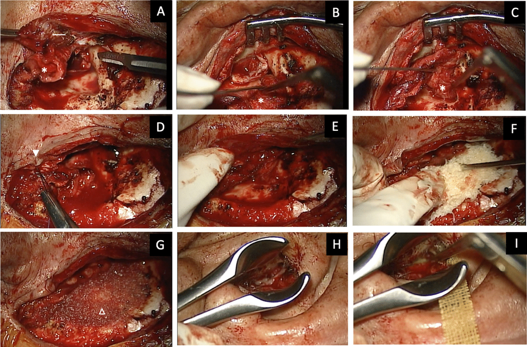
Surgical technique (Kashima’s procedure)
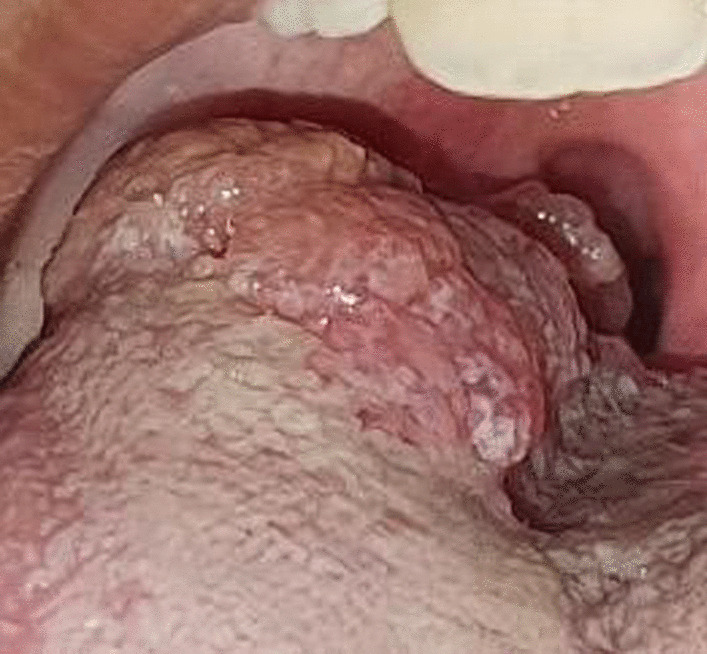
After the cricoarytenoid joints were palpated intraoperatively, the optimal side for cordotomy was decided upon based on the side with the least range of motion. Surgeon preference was considered if there was no difference between the two sides. Under general anesthesia, the procedure was conducted, and either a tracheostomy tube or a small endotracheal tube (5 or 5.5 microlaryngeal tube) was used to maintain the airway.
The glottic gap was opened with the use of a Kleinsasser laryngoscope. At the back of the membranous vocal fold, a transverse incision was created. With the frequency set to 4 MHz and the passive electrode applied to the shoulder region, the incision in group (A) was made using an Ellman Radiosurgical Instrument adjusted in the partially rectified mode (50 percent cut, 50 percent coagulation), with the frequency setting. A specially designed needle electrode was used to make the incision. An Arthro Care ENT Coblator was used to make the incision in group (B) to resect soft tissue. Both cauterization and coblation were employed as settings. The suggested settings for a laryngeal wand were Cauterization—3 (non-plasma setting) and Coblation—7 (plasma setting). PROcise LW was employed by the coblation wand. The shaft could be bent. It was equipped with a screen electrode that could quickly debulk the target tissue. Its flexible shaft conformed to the anatomy of the patient.
The vocal ligament and the muscle fibers of the thyroarytenoid muscle were completely divided, and the incision continued laterally until it reached the inner perichondrium of the thyroid cartilage. The incision started anterior to the vocal process of the arytenoid cartilage without exposing the cartilage. Since the electrode moved slowly, the cutting was accomplished by the electrode’s energy effect rather than the surgeon’s physical strength. If bleeding occurred, it was stopped by suction diathermy and bipolar laryngeal cautery or by exerting pressure with gauze soaked with adrenaline (1:1000).
Postoperative management
All patients received dexamethasone intravenously throughout the procedure to lessen laryngeal edema. To reduce fibrosis, this was repeated twice within 48 h of the surgery. This was then followed by a 5-day course of oral steroids (prednisolone), which was then decreased over the following 5-day period. Strong anti-reflux drugs were also administered for 15 days following surgery. After a weaning test, patients who had a tracheostomy were decannulated.
Postoperative assessment
During the follow-up periods, an endoscopic examination was performed to check for any complications (such as edema, granulations development). The glottic chink, grade of dyspnea, and presence of aspiration were evaluated as early as two weeks after the subsidence of postoperative oedema to evaluate the outcomes of the operation and the feasibility of decannulation in tracheostomized patients. Voice rehabilitation needed a prolonged period of follow up with assessment at 2 weeks and 3 months postoperative.
Statistical analysis
Statistical analysis was done by SPSS 25 (IBM Corp., Armonk, NY, USA). Quantitative variables turned to be non-normally distributed using Kolmogorov–Smirnov. Quantitative data were presented as mean, standard deviation (SD) and range. Preoperative and postoperative data were compared using Wilcoxon Sign Rank test. While the quantitative data of both groups were compared using Mann–Whitney U test. Qualitative variables were expressed as number and percentage and were compared using Chi square test. P value less than 0.05 was considered statistically significant. While p value less than 0.001 was considered highly significant.


留言 (0)