
Remember me


Data collection will take place in the University Hospital of the Technical University (TU) Munich, Klinikum rechts der Isar, Germany. Three departments are involved in data collection: Department of Nephrology (lead), Department of Neurology, and Department of Ophthalmology. Recruitment of patients will take place in the post-COVID outpatient clinic at the University Hospital of the Ludwigs-Maximilians-University (LMU) Munich, Campus Großhadern, in general practitioners and via social media.
Inclusion criteria:
i.Patients with PCS (positive PCR or positive rapid antibody test ≥3 months) with a currently existing, PCS-typical complaint complex, ongoing for at least 2 months that cannot be explained by an alternative diagnosis.
ii.Control group (CR): participants recovered from SARS-CoV-2 infection (positive PCR or positive rapid antibody test ≥ 3 months) without any residual symptoms.
iii.Healthy cohort (CN): no history of SARS-CoV-2 infection (exclusion via measurement of disease specific antibodies).
Exclusion criteria:
i.Missing or incomplete consent form.
ii.Age < 18 years.
iii.Pregnancy.
iv.Malignancy.
v.Diseases associated with a significant change in life expectancy.
vi.Autoimmune diseases of the rheumatological type.
vii.Cataract.
viii.Epilepsy.
ix.Glaucoma.
Measurements are carried out by experienced examiners in the departments of nephrology, neurology and ophthalmology. Informed consent is obtained by a clinical study investigator prior to inclusion. A structured medical history and the entry criteria are re-evaluated. A written consent form approved by the ethics committee of the Technical University of Munich, School of Medicine, is used to obtain informed consent.
To establish a PCS biobank, we ask patients to donate their biospecimens. For this purpose, we use an information sheet approved by the ethics committee of the Technical University of Munich, School of Medicine.
OutcomesPrimary outcomePCS patients show an impaired retinal vessel responsiveness and microcirculation when compared to a SARS-CoV-2-infection-recovered cohort. To investigate ED, both parameters (vMax and aMax) are important [32]. In a recent publication, we could show that vMax is a predictor for all-cause mortality in end-stage renal disease (ESRD) [33]. In addition, we will compare parameters of retinal microcirculation. To analyze microvascular function, central retinal artery equivalent (CRAE), central retinal vein equivalent (CRVE), and arteriolar–venular ratio (AVR) are important variables. Narrower CRAE and wider CRVE have been shown to be predictors for cardiovascular mortality [34]. Therefore, our primary endpoint will focus on differences in both dynamic retinal vessel analysis (DVA, vMax and aMax) and static retinal vessel analysis (SVA, CRAE, CRVE, and AVR) between our PCS cohort and fully SARS-CoV-2-infection-recovered patients.
Secondary outcomes:
(1)To determine whether PCS patients have an impaired retinal vessel responsiveness at baseline compared to the CN cohort. DVA parameters, including aMax and vMax, will be measured at baseline, and mean or median values will be calculated for each cohort, respectively.
(2)To assess whether SVA parameters of retinal vessel analysis (including CRAE, CRVE and AVR) are altered in the PCS cohort compared with CN cohorts. The baseline, mean or median values of these parameters will be calculated for each cohort, respectively.
(3)To investigate whether PCS patients show an improved retinal vessel responsiveness and static parameters after 6 months compared to baseline parameters. The static and dynamic parameters will be measured at baseline and follow-up and mean or median values will be calculated for each cohort, respectively.
(4)To assess whether symptom severity of PCS correlates with impaired retinal vessel responsiveness and static parameters of retinal vessel analysis. The symptom severity measured with patient-reported outcome measures (PROMs), along with the baseline static and dynamic parameters, will be assessed, and mean or median values will be calculated.
(5)To examine whether patients with PCS show a chronic immune activation and a changed amount of circulating angiogenic cells (CAC) and circulating endothelial progenitor cells (CEC) in fluorescence-activated cell sorting (FACS) analysis compared with the CR and CN cohort. At baseline, percentage of CAC and CEC will be measured and mean or median values will be calculated for each cohort, respectively.
(6)To assess whether epithelial and endothelial cells show a change in surface markers and inflammation markers when incubated with recombinant SARS-CoV-2 S1 subunit protein and/or patient serum from the PCS cohort, in cell culture experiments.
(7)To determine whether PCS patients show elevated markers of ED (sICAM, sVCAM, Thrombomodulin, P-Selektin, ADMA, SADMA, Endothelin-1, ACE-1, ACE-2, ANG-2, Pentraxin-3, GDF-15) compared to the CR and CN cohort. At baseline, mean or median values of these markers will be calculated for each cohort, respectively.
(8)To investigate whether PCS patients show elevated markers of chronic inflammation (INFß, TNFα, IFNy, Il-8, Il-6, Il-1ß, Mcp1, Il-10) compared to the CR and CN cohort. At baseline, mean or median values of these markers will be calculated for each cohort, respectively.
(9)To examine whether PCS patients show a reactivation of Epstein–Barr virus (EBV). At baseline, PCR of EBV DNA will be measured and mean or median values will be calculated for each cohort, respectively.
(10)To assess whether patients with PCS show changes of the retinal vasculature as measured by OCT angiography. At baseline, mean values of retinal vessel densities of the superficial and deep vascular complex as well as size of the foveal avascular zone in both eyes will be measured for each cohort, respectively.
(11)To investigate whether PCS patients with impaired retinal vessel responsiveness show an autonomic dysfunction characterized by low cortisol levels. At baseline, mean or median values of cortisol levels will be measured for each cohort, respectively.
(12)To explore whether PCS patients with fatigue fulfill diagnostic criteria of ME/CFS. At baseline, the Canadian Criteria of CFS score and the handgrip strength will be measured and mean or median will be calculated)
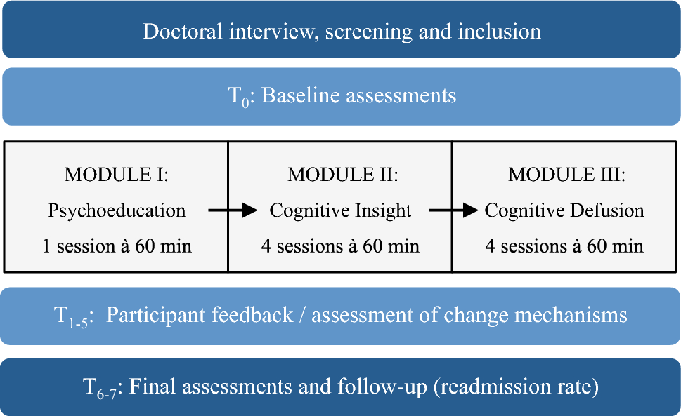
Participant timelineRecruitment for our study is scheduled to begin in October 2022, with the first patients expected to be measured by the end of 2022 or early 2023. In addition to measuring the PCS cohort, CR participants will be recruited and measured, with completion of baseline measurements (T0) planned by June 2023. In the PCS cohort, measurements will be repeated after 6 months (T1), with completion planned by the end of September 2023. One year after the initial measurements, we will conduct a telephone survey to assess any residual symptoms and treatment needs. Data analysis and especially data quality control will continuously happen after measurement of the first patient. Cell culture experiments are ongoing starting with incubation of retinal endothelial cells with SARS-CoV-2 spike antigen (S1) followed by incubation with patient serum (Fig. 1).
Fig. 1
Participant timeline and overview of conducted measurements. Figure shows baseline (T0) and follow-up (T1) measurements that are conducted in the “All Eyes on PCS” study. The figure provides a visual representation of the planned measurements and includes sample images of key assessment tools, including adaptive optics (AO), dynamic and static retinal vessel analysis (DVA, SVA) and OCT angiography (OCTA), and patient reported outcome measures (PROMs)
For a detailed list of the variables to be measured, the departments involved in the study, and the relevant hypotheses, please refer to Table 1.
Table 1 Detailed list of measured variablesSample sizeIn the PCS and CR cohort, we plan to recruit 100 participants. The SARS-CoV-2-infection-naïve cohort is defined as no history of SARS-CoV-2 infection (exclusion via measurement of specific antibodies) and consists of an already established, pre-pandemic healthy cohort and a cohort recruited during the pandemic.
Primarily, a comparison of the group means regarding the dilation of the retinal vessels will be performed using an analysis of variance (ANOVA) (significance level α = 5%).
To estimate the required sample size, we will assume a standard deviation within groups of 2%, based on the largest normative sample published to date, which is consistent with previous healthy control studies in the “diseased vs. healthy controls” group comparison. [39]. A biologically relevant mean difference of aMax between groups is assumed to be 1%—specifically, 2.5 vs. 3.5% dilation for PCS vs. CR. Similar differences of this magnitude have been observed in comparable groups with other disease entities, such as heart failure [40]. With these assumptions, we estimate that 78 evaluable patients per group will be needed to reject the null hypothesis (equality of all group means) with a power of 90%. To account possible dropouts or measurement errors, 100 patients per group should be included in the study. If the global null hypothesis is rejected based on ANOVA, pairwise group comparisons will be made at a significance level of α = 5% as a final test procedure. Power analysis was done by BH using nQuery (version 9.1.0.0).
RecruitmentTo achieve an adequate recruitment number, we cooperate with the post-COVID outpatient clinics of the LMU University Hospital (KA, HS). Via social media of the Klinikum rechts der Isar, flyers will be additionally posted online and distributed. To provide adequate information for participants and patients, we are creating a website with further information (https://www.mri.tum.de/alleyesonpcs). Patients recruited via social media who wish to participate in the study must complete a screening form to ensure that only those with PCS are enrolled. Potential study participants will be discussed using the screening form in a weekly meeting with the chief investigator, PI, and doctoral students and will be included or excluded based on a majority vote. In particular, the temporal relationship between acute SARS-CoV-2 infection and the onset of PCS symptoms but also alternative diagnoses are discussed. Enrollment of participants will be conducted by a physician from the Department of Nephrology.
Data collection and managementWe have written “standard operating procedures (SOPs)” to ensure standardization of processes. SOPs were translated and can be found in the supplements. Study instruments include:
Questionnaires: Only standardized and established questionnaires are used to assess patient-reported outcomes (“PROMs”).
Laboratory tests: Routine parameters are measured by our in-house clinical chemistry department.
DVA and SVA analysis: Data acquisition will be performed by following the SOPs (Online Resource 1). All examiners will be trained by a single experienced supervisor and must reach high image accuracy and quality in at least ten volunteers. Examiners do not perform data analyses and are only responsible for data acquisition. Data analysis will be performed by independent scientists and will be compared afterward. One rater will perform a quality evaluation and exclude insufficient measurements. Vessel response curves will be evaluated using the cumulative scoring method. The score ranges from 0 to 5. Retinal images with insufficient quality (< 2.5) will be discussed with a second rater and then excluded.
OCT and OCT angiography: Measurements and data analysis are performed by the Department of Neurology (Online Resource 2).
AO: Measurements and data analysis are undertaken by the Department of Ophthalmology (Online Resource 3).
Handgrip strength test: Data acquisition is done by following the SOP “Handgrip strength test” (Online Resource 4).
To promote participant retention, we contact the participants 2 days before their follow-up appointment to ensure that they remember the follow-up visit. Detailed information of PCS patients which were lost to follow-up will be provided.
Data management and confidentialityData entry only takes place in the University Hospital of the TU Munich on an assigned key-locked study computer. Excel sheets used for data collection are key-locked and standardized. All collected data will be verified by an independent second scientist (double data verification) and checked for plausibility (e.g., range checks for clinical data) (Online Resource 3). Data which are collected analog e.g., questionnaires and clarification sheets, are stored in a locked room in the University Hospital of the TU Munich. We have developed a checklist for each admitted participant to ensure all necessary information is collected, and in the event of missing data, we have established a protocol for conducting a standardized evaluation (Online Resource 3). Clinical data will be stored on a dedicated study server and in non-pseudonymized form on hard drives (accessible only to study physicians and persons directly involved in the study). Blood samples are labeled with the patient ID and will be stored in the routine nephrology laboratory and in the nephrology research laboratory in locked rooms.
PseudonymizationFor further analysis, all patient data are undergoing double pseudonymization. Patient-related data such as age and gender will be stored in a list, with a sequential study number assigned to each patient. The clinical data are exclusively assigned to the study numbers in a separate list. Both lists are password protected. The list containing the patient-related data and study numbers is kept separately from all other lists by the study director in a password-protected computer. Clinical data and informed consent forms are kept for 10 years, respectively.
Upon revocation of informed consent, all data will be deleted and printed records and collected samples will be destroyed.
Storage of biological specimens for genetic or molecular analysis in this trialAfter recruitment, 40 ml of blood is initially collected from patients and sent directly to the nephrology laboratory. There, a portion of the blood is sent for analysis by clinical chemistry, and another portion is further processed. Peripheral blood mononuclear cells (PBMCs) are isolated from the processed blood according to protocol and stored at – 80 °C. In addition, samples of serum and whole blood are also frozen at -80 °C for future analysis; however, are not stored longer than 10 years.
Statistical methodsAll statistical analysis will be carried out using R in the current version implemented in RStudio and will be supervised by BH. Used R Packages and their version numbers will be reported in the peer-reviewed manuscripts.
We plan to match our PCS cohort with the CR recovered and CN cohort at least 1:1 based on age and gender. The success of the matching is checked by the matchbalance function and results will be published in the supplements. To create a baseline description before and after matching, Table1 function is used. Depending on the distribution of the values aMax and vMax, a parametric (t test) or non-parametric test (Mann–Whitney test) is used to compare the means or medians, respectively. Boxplots will be plotted with the ggplot2 function. To evaluate a possible function as a biomarker in PCS, receiver operating characteristic (ROC) curves and the area under the curve (AUC) will be calculated using the plotROC function. Correlations will be visualized with the corrplot function. In general, Spearman or Pearson coefficients are calculated depending on the relationship between the two variables. To check for confounding, we will use linear regression models. Interim analyses are planed after completion of baseline recruitment. All participant scientists will have access to the evaluated data. Data will be published after baseline evaluation and after completed follow-up. To perform subgroup analyses, a test for interaction is performed using ANOVA and then visualized using the interaction.plot function. Information and baseline characteristics of patients lost to follow-up will be compared with those who completed the study using non-parametric or parametric tests, respectively. Upon reasonable request, access to the raw data and statistical codes used will be granted to the reviewers in the peer-review process when needed. Another sharing is not intended.
Oversight and monitoringChief investigator: Weekly meeting via Zoom with the study base team and PIs. Responsible for overseeing the trial in general.
Principal investigator (PI): Responsible for organizing and coordination of different involved departments and recruitment process in PCS ambulances. Responsible for data management and storage and final data analysis.
RVA-team: Monthly meeting of the three supervisors of RVA (RG, KK, TK). Quality control, data management,and updating on newest developments in the trial. Maintenance of the RVA machine.
Head of biobank: Processing and storage of bio samples. Maintenance of databank.
Study base team: Responsible to carry out technical measurements and blood sample drawing. Arrangement of study appointments. Not primarily involved in data analysis.
Data management is carried out by the PI who directly reports to the chief investigator. Data verification is done by the head of biobank which also carries out data collection and maintenance.
However, in case of RVA parameters, data analysis will be carried out by three independent principal investigators (RG, KK, TK). Data are checked frequently for validity and quality. If abnormalities occur, they are discussed between these three PIs and report to the chief investigator.
If we plan to amend the study protocol (e.g., new measurement of certain parameters), we will consult the ethical review board of TU Munich to discuss the feasibility of implementation. If the amendment is feasible, we will notify all participating researchers of the change. If the change affects study participants, we will inform them of the possible change and obtain new written informed consent. There are no publication restrictions, and we will publish the results in an appropriate peer-reviewed journal.
Comments (0)