
Remember me


Tranexamic acid (TXA) is a synthetic lysine derivative that exerts antifibrinolytic effects by inhibiting activation of plasminogen1,2. TXA has been extensively applied in a variety of orthopaedic surgical procedures, especially total joint arthroplasty and spinal surgery, to minimize perioperative blood loss and postoperative transfusion3,4. The safety and efficacy of TXA have led to recent interest in its administration during arthroscopy. Prior research has demonstrated that TXA can significantly reduce hemarthrosis following anterior cruciate ligament (ACL) reconstruction, which is a major complication that can cause pain, arthrofibrosis, and disruption of rehabilitation5–7.
Topical administration of TXA has demonstrated efficacy comparable to intravenous (IV) routes, with no safety concerns8. Some common medical conditions such as renal impairment and cardiovascular diseases may preclude the IV administration of TXA9. Additionally, the topical use of TXA is advantageous due to its ease of administration, ability to allow maximum concentration at the bleeding site, and minimal systemic absorption10,11. In a study by Chiang et al., intra-articular injection (100 mg/mL) of TXA in patients undergoing ACL reconstruction resulted in decreased pain and a lower grade of hemarthrosis in the early postoperative phase without side effects6. However, the safe dose of TXA that can be administered in the knee joint during ACL reconstruction remains unclear, with concerns over potential toxicity to articular cartilage and meniscus10,12–14. Tuttle et al. observed that 25 mg/mL of TXA had no impact on murine chondrocyte viability, whereas 100 mg/mL resulted in no viable bovine chondrocytes15. Parker et al.16 reported up to 97% cell death at a concentration less than half of the 100 mg/mL administered by Chiang et al.6. Ambra et al. detected no chondrotoxicity after exposing osteochondral plugs to TXA concentrations of 1, 2, and 4 mg/mL for 6 hours17. McLean et al. found 100 mg/mL of TXA to be cytotoxic to human periarticular tissues18. Notably, these studies were conducted using in vitro or ex vivo models that could not replicate the dynamic environment in a knee joint. Thus, further research is required to determine the in vivo toxicity of TXA.
In arthroscopic surgery, unlike total joint replacement, resident cartilage and meniscus are directly exposed to TXA during topical administration. Given the limited regenerative capacity of these tissues, intra-articular inflammation, and altered biomechanics after ACL reconstruction, even mild toxicity to these intra-articular tissues could be devastating to the knee joint19,20. Therefore, prior to the routine use of intra-articular TXA injection for the prevention of hemarthrosis after ACL reconstruction, the safety of TXA for cartilage and meniscus must be well established. It is important to acknowledge that ACL reconstruction might not always fully restore knee joint biomechanics and stability, especially in cases with concomitant meniscal injuries, leading to posttraumatic osteoarthritis20,21. Given this limitation, the objective of this study was to examine the safety of a single intra-articular injection of TXA in a destabilized knee joint. We hypothesized that TXA at a concentration exceeding 50 mg/mL would accelerate joint degeneration in rats with ACL rupture. To replicate the degeneration of articular cartilage and meniscus resulting from compromised knee joint stability, we utilized an ACL transection model instead of an ACL reconstruction model.
Materials and Methods Study DesignOne hundred and twenty Sprague-Dawley male rats at the age of 12 weeks (weighing ∼360 g) were used in this study (Fig. 1). All animal surgical procedures were in accordance with the guidelines of the Animal Experimentation Ethics Committee (AEEC) of our institution. After a 7-day acclimatization period, the animals were randomly allocated into 5 groups based on the concentration of TXA in saline solution that was to be administered: 0 mg/mL (saline solution only), 20 mg/mL (G20), 50 mg/mL (G50), 100 mg/mL (G100), and 150 mg/mL (G150). The concentrations of TXA in this study were determined based on previous in vitro and clinical studies6,11. The short-term effects of TXA on cartilage and meniscus were evaluated by cell viability assessment 24 hours after injection (n = 6 in each group). The animals were killed for histological analysis at 2, 4, and 8 weeks postoperatively (n = 6 in each group at each time period).
 Fig. 1:
 Flowchart of the study design. All groups received intra-articular injection of 30 μL of saline solution with varying concentrations of TXA, with a total dose of 0, 0.6, 1.5, 3.0, and 4.5 mg of TXA in the saline, G20, G50, G100, and G150 groups, respectively. ACL = anterior cruciate ligament.
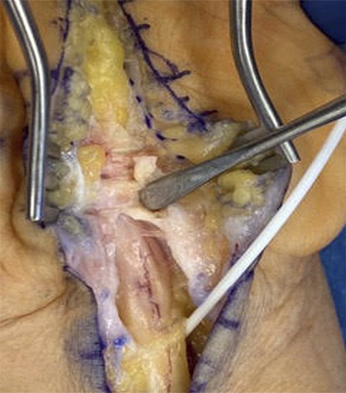
Surgical ProceduresThe rats were anesthetized with ketamine (75 mg/kg) and xylazine (10 mg/kg) via intraperitoneal injection. To access the left knee, we made a 3-cm medial parapatellar incision and exposed the intercondylar area by laterally subluxating the patella. Following a previous study, we transected the ACL using micro-scissors and confirmed complete transection with the anterior drawer test22. After closing the joint capsule, a Hamilton syringe with a 30-gauge needle was used to administer a single intra-articular injection of 30 μL of saline solution with varying concentrations of TXA prior to skin closure23. The knees were passively flexed and extended 5 times to distribute the solution within the synovial cavity after all injections. Postoperative pain control was performed with buprenorphine, and all rats were housed individually and allowed to move and feed freely.
Chondrocyte and Meniscal Cell Viability AssessmentAt 24 hours post-TXA injection, under a dissecting microscope, a 1-mm-thick section of cartilage tissue was harvested from the medial condyle of the distal femur, perpendicular to the long axis of the femur as illustrated in Figure 2-A. An intact medial meniscus was also collected. The harvested cartilage section and meniscus were subsequently subjected to cell viability assessment using the LIVE/DEAD Viability/Cytotoxicity Assay Kit (Invitrogen), as previously reported24. During the dissection, samples were frequently sprinkled with saline solution to prevent dehydration. The cartilage and meniscus samples were then viewed with fluorescence microscopy (Leica DM5500). To quantify live and dead cells, the centers of the cartilage of the medial femoral condyle and inner halves of the menisci were identified and imaged (Fig. 2-B). The obtained fluorescent images were divided into 300 × 300-µm rectangles. From these rectangles, 5 individual 300 × 300-μm regions were then randomly selected for further analysis. Using ImageJ software (National Institutes of Health), we followed standard particle analysis protocols to manually adjust the threshold and quantify the number of live and dead cells in each region.
 Fig. 2:
 Sample collection for the cell viability assay. Fig. 2-A The anatomical plane (white rectangle) for sectioning of the cartilage from the medial femoral condyle for examination. This plane is approximately perpendicular to the long axis of the femur. Fig. 2-B Schematic illustration of the regions of interest for the cell viability assay in the cartilage section (left) and meniscus (right). In the cartilage section, the specific area selected for further analysis is indicated by a red dashed circle. In the meniscus, the inner half of the medial meniscus (outlined by a red dashed line), which contains fibrochondrocytes, was selected for analysis.
Macroscopic Evaluation of Cartilage and MeniscusThe tibia was stained with Evans blue solution and photographed25. The macroscopic features of the tibial cartilage were evaluated using a published grading system based on the deterioration of the cartilage surface, with all samples assigned a grade from 1 to 426. Higher grades indicate worse degeneration. Specifically, grade 1 indicated an intact surface, grade 2 indicated minimal fibrillation, grade 3 indicated overt fibrillation, and grade 4 indicated erosion with exposed subchondral bone. The gross appearance of the meniscus was assessed on the basis of the color changes and surface irregularities. Following evaluation, all specimens were fixed in 4% neutral buffered paraformaldehyde for 48 hours in preparation for histological analysis.
Histological AnalysisAfter decalcification and sectioning at 7-μm thickness, the slides of the tibia and meniscus samples were stained with safranin O-fast green with a standard protocol. Cartilage degeneration was assessed using the Osteoarthritis Research Society International (OARSI) histopathological assessment system22. Meniscal degeneration was assessed using a scoring system that evaluated surface integrity, cellularity, and safranin O staining distribution and intensity, as previously described27. Scoring for both cartilage and meniscus was performed by an experienced researcher who was blinded regarding the groups.
Statistical AnalysisAll data were expressed as the mean and the 95% confidence interval (CI). One-way analysis of variance with a post hoc Bonferroni test was used to assess differences in cell viability among the groups. Nonparametric Kruskal-Wallis analysis of variance and a post hoc Mann-Whitney U test were used for between-group comparisons in the gross and histological evaluations. SPSS software (version 15.0) was used to perform the statistical analysis. The level of significance was set at p < 0.05. For the cell viability and histological assessments, an a priori sample size calculation was conducted using the mean and standard deviation for cell viability and the histological score for the saline group (82.7% ± 5.9% and 3.3 ± 1.2) and for G150 (35.8% ± 8.8% and 7.7 ± 1.5). According to the power analysis, a sample size of 6 rats in each group at each time point was selected to achieve the desired power (1 – β) of 90% based on an α level of 0.05.
Source of FundingThis work was supported by grants from the National Natural Science Foundation of China (82172430 and 82272505) and University Grants Committee, Research Grants Council of the Hong Kong Special Administrative Region, People’s Republic of China (14108720, 14121721, 14202920, N_CUHK472/22, C7030-18G, T13-402/17-N, and AoE/M-402/20).
Results Chondrocyte and Meniscal Cell ViabilityImages and results of the chondrocyte and meniscal cell viability assessments are shown in Figures 3 and 4. Overall, an increase in TXA concentration resulted in decreased cell viability in both cartilage and meniscus at 24 hours post-injection. There was no significant difference in chondrocyte or meniscal cell viability between the G20 and saline groups. However, the chondrocyte viability was significantly decreased in G50 (67.5%, 95% CI = 62.6% to 72.4%, p = 0.001), G100 (58.3%, 95% CI = 50.5% to 66.1%, p < 0.001), and G150 (28.4%, 95% CI = 17.1% to 39.7%, p < 0.001) compared with the saline group (86.0%, 95% CI = 80.9% to 91.1%). Similarly, among the meniscus samples, cell viability was significantly decreased in G50 (79.3%, 95% CI = 73.1% to 85.6%, p = 0.042), G100 (59.6%, 95% CI = 52.1% to 67.1%, p < 0.001), and G150 (43.1%, 95% CI = 33.7% to 52.4%, p < 0.001) compared with the saline group (90.4%, 95% CI = 87.3% to 93.5%).
 Fig. 3:
 Representative images of LIVE/DEAD staining for surface chondrocytes and meniscal cells at 24 hours after TXA injection (n = 6). Live cells were labeled in green fluorescence and dead cells, in red fluorescence. White scale bar = 100 μm.
 Fig. 4:
 Average cell viability in meniscus and cartilage at 24 hours after intra-articular TXA injection. Data are expressed as the mean with the 95% CI. The percentages of live and dead cells were quantified using ImageJ (n = 6). One-way analysis of variance and the post hoc Bonferroni multiple comparisons test were used to compare groups. *P < 0.05, **p < 0.01, ***p < 0.001.
Gross ObservationThe gross appearances of a representative medial tibial plateau and medial meniscus are shown in Figure 5. At week 2, no degenerative changes were noticed among the groups. By week 8, the saline group, G20, and G50 exhibited intact cartilage covering the surface of the medial tibial plateau, with no signs of meaningful abrasion or fibrillations. However, pathological changes such as thinning of cartilage and fibrillations were observed at 8 weeks after surgery in G100 and G150. Additionally, more severe destruction was found in G150, characterized by surface erosion, ulceration, and pitting. The grades for the gross appearance were significantly higher in G100 and G150 than in the other 3 groups at 8 weeks (G100 versus saline group, p = 0.038; G150 versus saline group, p = 0.01; G100 versus G20, p = 0.038; G150 versus saline group, p = 0.01; G150 versus G50, p = 0.031). Similarly, the medial meniscus remained intact with a smooth and healthy appearance in the saline group, G20, and G50. In contrast, the anterior horn of the medial meniscus in G100 and G150 exhibited an opaque appearance with deformity and yellowish color, indicating increasing ossification and degeneration.
 Fig. 5:
 Gross assessment of the medial tibial plateau and medial meniscus at 4 and 8 weeks after surgery. Fig. 5-A Macroscopic observation of the medial tibial plateau stained with Evans blue solution. Surface fibrillations and vertical fissures can be found in the 100 mg/mL and 150 mg/mL TXA groups. The red arrowheads highlight the presence of rough surfaces and vertical lesions on the medial tibial plateau. Fig. 5-B Macroscopic observation of the medial meniscus. At week 8, the meniscus in the 150 mg/mL TXA group showed a rough surface and loss of a healthy appearance. The white arrowhead highlights the yellowish color changes and loss of the smooth surface of the meniscus observed in the 150 mg/mL group. Fig. 5-C Quantitative gross assessment of articular cartilage at 8 weeks after surgery. The grades on the y axis reflect the severity of cartilage damage, with higher scores indicating more extensive damage: grade 1 indicated an intact surface, grade 2 indicated minimal fibrillation, grade 3 indicated overt fibrillation, and grade 4 indicated erosion with exposed subchondral bone. The grades in the 100 mg/mL and 150 mg/mL groups were significantly higher than that in saline group. Data are presented as the mean with the 95% CI.
Histological Analysis of CartilageNo significant degenerative changes were observed among the groups at 2 and 4 weeks after surgery (Fig. 6-A). By week 8, cartilage in the saline group, G20, and G50 retained its overall structure without substantial deterioration or deep clefts. However, early indications of cartilage degeneration such as surface irregularities were observed. Additionally, a decrease in the intensity of safranin O staining in the extracellular matrix suggested a depletion of proteoglycans. In contrast, G100 and G150 exhibited varying degrees of degeneration, including decreased cartilage thickness, lower chondrocyte count, and reduced safranin O staining intensity. In particular, rough and fibrillated cartilage surfaces with vertical fissures were found in G150. The OARSI scores in G20 and G50 were not significantly different than that in the saline group. However, the OARSI scores in G100 and G150 were significantly higher than that in the saline group, with G150 having the highest score (G100 versus G20, p = 0.040; G150 versus saline group, p = 0.010; G150 versus G20, p = 0.007).
Histological Analysis of MeniscusAs shown in Figure 6-B, by week 8 the meniscus in the saline group, G20, and G50 exhibited a smooth and intact surface without fibrillation. In contrast, focal lesions and undulation were found in G100 and G150. Additionally, fibrillated surfaces, cell clustering, and cyst-like formation were observed in G150. The meniscus score in G150 was significantly higher than that in the saline group, G20, and G50 (p = 0.020, p = 0.021, p = 0.031, respectively). No significant differences were found between G100 and the saline group, possibly indicating a stronger protective effect of meniscal extracellular matrix compared with articular cartilage.
 Fig. 6:
 Histological evaluation of the articular cartilage and meniscus at 4 and 8 weeks after surgery. Fig. 6-A Representative images of safranin O-fast green staining of the articular cartilage of the medial tibial plateau. The black arrowheads indicate the decreased intensity of proteoglycans and the fibrillated surface. The scale bar indicates 200 μm. Fig. 6-B Representative images of safranin O-fast green staining of the medial meniscus. The blue arrowheads indicate the focal lesion and surface fibrillation of the medial meniscus. The red arrowhead indicates cyst-like formation within the meniscus. The scale bar indicates 200 μm. Fig. 6-C The mean Osteoarthritis Research Society International (OARSI) scores for articular cartilage degeneration in the saline group and the 20, 50, 100, and 150 mg/mL TXA groups were 3.5 (95% CI = 2.4 to 4.6), 3.3 (95% CI = 2.1 to 4.6), 4.0 (95% CI = 2.9 to 5.1), 6.3 (95% CI = 5.2 to 7.4), and 8.0 (95% CI = 5.4 to 10.6), respectively. Fig. 6-D The meniscus scores indicated significantly more severe joint degeneration in the 100 and 150 mg/mL TXA groups. The mean meniscus scores in the saline group and the 20, 50, 100, and 150 mg/mL TXA groups were 6.8 (95% CI = 5.0 to 8.6), 6.8 (95% CI = 5.3 to 8.4), 7.0 (95% CI = 5.0 to 9.0), 10.3 (95% CI = 9.2 to 11.4), and 11.2 (95% CI = 9.6 to 12.7), respectively. Note that, in this study, only the anterior portion of the medial meniscus was evaluated due to the sectioning method. Therefore, the total score for all meniscus items ranged from 0 to 18.
DiscussionThis study demonstrated that a single intra-articular injection of TXA led to reduced cell viability in cartilage and meniscus at concentrations of ≥50 mg/mL. Furthermore, increased severity of cartilage degeneration, as observed macroscopically and histologically, was noticed in G100 and G150. In addition, G150 showed significant meniscal degeneration and destruction. This study highlights the need for extra caution when exposing intra-articular tissues to high concentrations of TXA, in line with the growing body of evidence.
The use of TXA for reducing postoperative blood loss and transfusion in joint replacement surgery has been well studied4. Its administration has been expanded to arthroscopic surgery, but the clinical outcomes in this context are unclear. In an ACL reconstruction study, Karaaslan et al. reported that combined IV and intra-articular administration of TXA could significantly reduce blood loss without increasing the risk of complications5. This finding was supported by several randomized controlled trials (RCTs) demonstrating that IV TXA administration protocols could reduce postoperative pain and enhance functional outcomes after ACL reconstruction and meniscectomy7,28. A single intra-articular injection of 1 g (100 mg/mL) of TXA was found to considerably reduce postoperative hemarthrosis and pain in the early postoperative period after ACL reconstructions with hamstring grafts6,10. However, a double-blinded RCT of patients who underwent ACL reconstruction found no significant differences in perioperative blood loss, postoperative hemarthrosis, or pain levels between those treated with and without IV TXA29.
In addition to inconsistent clinical results, safety concerns have been raised regarding the use of TXA in the joint space10,13,30. While previous studies have demonstrated the cytotoxicity of TXA, there is a lack of in vivo studies examining the impact of topical use of TXA on intra-articular tissues. In vitro and ex vivo studies do not always replicate the in vivo situation, as the joint cavity is a dynamic environment where saline solution (during arthroscopy) and synovial fluid continuously circulate, diluting injected medication31. Additionally, 2-dimensional cell cultures maximize the exposure of cells to the condition being tested, which may result in the protective effect of the extracellular matrix being overlooked, and the diffusion and absorption of medication into surrounding tissue may not be replicated in a culture dish. Furthermore, the mechanical and biological environment inside an injured knee joint is complex. Therefore, a strength of the present study is that it employed a translational rat model of knee joint destabilization to mimic the clinical situation and replicate the processes of dilution, diffusion, and absorption that occur in the joint cavity of human patients.
This study presents what we believe to be the first in vivo evidence using clinically relevant doses of TXA to elucidate the clinical implications. Our findings indicate that the decision to use TXA and its concentration should be carefully determined based on the risk of joint degeneration in patients with cartilage, ligament, and meniscal injuries. The considerable cartilage damage observed in G100 and G150 strongly discourages the use of TXA at concentrations of 100 mg/mL or higher. Clinicians should closely monitor patients who received TXA at such concentrations during ACL reconstruction for early signs of osteoarthritis and intervene promptly to prevent its progression. However, it is essential to acknowledge that no TXA dose can guarantee absolute safety, highlighting the necessity for further research with larger sample sizes and longer follow-up periods to better understand the optimal concentration in arthroscopic surgery.
There are limitations and additional aspects of this study to consider. First, a translational rat ACL transection model was used, which may not perfectly replicate the complex knee joint environment in humans. Larger animal models with ACL reconstruction may be necessary to justify TXA treatment. Second, while we utilized clinically comparable TXA concentrations, the smaller joint space of rats likely resulted in a lower dosage (mg/kg) compared with clinical studies. Using the conversion of animal dosages to human equivalents32, the human equivalent dosage in our study would be 0.31 to 2.31 mg/kg for a 60-kg person. In practice, patients typically receive intra-articular TXA dosages ranging from 16.67 to 50 mg/kg6,11. Despite the subclinical dosages, our findings demonstrated notable chondrotoxicity, highlighting the potential risks associated with intra-articular injections of TXA in clinical settings. The third limitation of our study is that cell viability was evaluated only in cartilage and meniscus. However, a previous study found cytotoxicity of TXA in synovium and tendon18.
ConclusionsOur study demonstrated that a single intra-articular injection of TXA had a concentration-dependent cytotoxic effect on chondrocytes and meniscal cells. In rat knee joints with ACL transection, TXA at or above 100 mg/mL resulted in increased severity of cartilage degeneration. Therefore, we advise against using TXA at or above 100 mg/mL, and caution should be exercised regarding the use of TXA at clinically relevant concentrations due to their potential toxicity.
References 1. Bolam SM, O’Regan-Brown A, Paul Monk A, Musson DS, Cornish J, Munro JT. Toxicity of tranexamic acid (TXA) to intra-articular tissue in orthopaedic surgery: a scoping review. Knee Surg Sports Traumatol Arthrosc. 2021 Jun;29(6):1862-71. 2. Goobie SM. Tranexamic acid: still far to go. Br J Anaesth. 2017 Mar 1;118(3):293-5. 3. Poeran J, Rasul R, Suzuki S, Danninger T, Mazumdar M, Opperer M, Boettner F, Memtsoudis SG. Tranexamic acid use and postoperative outcomes in patients undergoing total hip or knee arthroplasty in the United States: retrospective analysis of effectiveness and safety. BMJ. 2014 Aug 12;349:g4829. 4. Fillingham YA, Ramkumar DB, Jevsevar DS, Yates AJ, Shores P, Mullen K, Bini SA, Clarke HD, Schemitsch E, Johnson RL, Memtsoudis SG, Sayeed SA, Sah AP, Della Valle CJ. The Efficacy of Tranexamic Acid in Total Hip Arthroplasty: A Network Meta-analysis. J Arthroplasty. 2018 Oct;33(10):3083-3089.e4. 5. Karaaslan F, Karaoğlu S, Yurdakul E. Reducing Intra-articular Hemarthrosis After Arthroscopic Anterior Cruciate Ligament Reconstruction by the Administration of Intravenous Tranexamic Acid: A Prospective, Randomized Controlled Trial. Am J Sports Med. 2015 Nov;43(11):2720-6. 6. Chiang ER, Chen KH, Wang ST, Ma HL, Chang MC, Liu CL, Chen TH. Intra-articular Injection of Tranexamic Acid Reduced Postoperative Hemarthrosis in Arthroscopic Anterior Cruciate Ligament Reconstruction: A Prospective Randomized Study. Arthroscopy. 2019 Jul;35(7):2127-32. 7. Felli L, Revello S, Burastero G, Gatto P, Carletti A, Formica M, Alessio-Mazzola M. Single Intravenous Administration of Tranexamic Acid in Anterior Cruciate Ligament Reconstruction to Reduce Postoperative Hemarthrosis and Increase Functional Outcomes in the Early Phase of Postoperative Rehabilitation: A Randomized Controlled Trial. Arthroscopy. 2019 Jan;35(1):149-57. 8. Gomez-Barrena E, Ortega-Andreu M, Padilla-Eguiluz NG, Pérez-Chrzanowska H, Figueredo-Zalve R. Topical intra-articular compared with intravenous tranexamic acid to reduce blood loss in primary total knee replacement: a double-blind, randomized, controlled, noninferiority clinical trial. J Bone Joint Surg Am. 2014 Dec 3;96(23):1937-44. 9. McCormack PL. Tranexamic acid: a review of its use in the treatment of hyperfibrinolysis. Drugs. 2012 Mar 26;72(5):585-617. 10. Chiang ER, Chen KH, Ma HL. Editorial Commentary: Tranexamic Acid Is Beneficial in the Very Early Postoperative Period in Anterior Cruciate Ligament Reconstruction Patients. Arthroscopy. 2021 Jun;37(6):1890-1. 11. Lee JW, Kim SG, Kim SH, Cho HW, Bae JH. Intra-articular Administration of Tranexamic Acid Has No Effect in Reducing Intra-articular Hemarthrosis and Postoperative Pain After Primary ACL Reconstruction Using a Quadruple Hamstring Graft: A Randomized Controlled Trial. Orthop J Sports Med. 2020 Jul 22;8(7):2325967120933135. 12. Karaaslan F. Editorial Commentary: Tranexamic Acid: Okay, It Reduces the Bleeding, but Are We Sure Topical Use Is Not Harmful to the Cartilage? Arthroscopy. 2019 Jul;35(7):2133-5. 13. Siegel MG. The Dangers and Concerns of Intra-articular Tranexamic Acid. Arthroscopy. 2019 Nov;35(11):2973-4. 14. Alaia MJ, Gipsman AM. Editorial Commentary: The Benefits of Tranexamic Acid May Outweigh Risks in Arthroscopy and Sports Medicine. Arthroscopy. 2021 Apr;37(4):1334-6. 15. Tuttle JR, Feltman PR, Ritterman SA, Ehrlich MG. Effects of Tranexamic Acid Cytotoxicity on In Vitro Chondrocytes. Am J Orthop (Belle Mead NJ). 2015 Dec;44(12):E497-502. 16. Parker JD, Lim KS, Kieser DC, Woodfield TBF, Hooper GJ. Is tranexamic acid toxic to articular cartilage when administered topically? What is the safe dose? Bone Joint J. 2018 Mar 1;100-B(3):404-12. 17. Ambra LF, de Girolamo L, Niu W, Phan A, Spector M, Gomoll AH. No effect of topical application of tranexamic acid on articular cartilage. Knee Surg Sports Traumatol Arthrosc. 2019 Mar;27(3):931-5. 18. McLean M, McCall K, Smith IDM, Blyth M, Kitson SM, Crowe LAN, Leach WJ, Rooney BP, Spencer SJ, Mullen M, Campton JL, McInnes IB, Akbar M, Millar NL. Tranexamic acid toxicity in human periarticular tissues. Bone Joint Res. 2019 Feb 2;8(1):11-8. 19. Cinque ME, Dornan GJ, Chahla J, Moatshe G, LaPrade RF. High Rates of Osteoarthritis Develop After Anterior Cruciate Ligament Surgery: An Analysis of 4108 Patients. Am J Sports Med. 2018 Jul;46(8):2011-9. 20. Wang M, Lin Z, Wang W, Chen L, Xia H, Zhang Y, Huang W. Kinematic Alterations After Anterior Cruciate Ligament Reconstruction via Transtibial Techniques With Medial Meniscal Repair Versus Partial Medial Meniscectomy. Am J Sports Med. 2021 Oct;49(12):3293-301. 21. Akpinar B, Thorhauer E, Irrgang JJ, Tashman S, Fu FH, Anderst WJ. Alteration of Knee Kinematics After Anatomic Anterior Cruciate Ligament Reconstruction Is Dependent on Associated Meniscal Injury. Am J Sports Med. 2018 Apr;46(5):1158-65. 22. Nemirov D, Nakagawa Y, Sun Z, Lebaschi A, Wada S, Carballo C, Deng XH, Putnam D, Bonassar LJ, Rodeo SA. Effect of Lubricin Mimetics on the Inhibition of Osteoarthritis in a Rat Anterior Cruciate Ligament Transection Model. Am J Sports Med. 2020 Mar;48(3):624-34. 23. Wang Y, Wagner ES, Yu D, Chen KJ, Keel TJ, Pownder SL, Koff MF, Cheetham J, Samaroo KJ, Reesink HL. Assessment of osteoarthritis functional outcomes and intra-articular injection volume in the rat anterior cruciate ligament transection model. J Orthop Res. 2022 Sep;40(9):2004-14. 24. Shaw KA, Moreland C, Jacobs J, Hire JM, Topolski R, Hoyt N, Parada SA, Cameron CD. Improved Chondrotoxic Profile of Liposomal Bupivacaine Compared With Standard Bupivacaine After Intra-articular Infiltration in a Porcine Model. Am J Sports Med. 2018 Jan;46(1):66-71. 25. Wang M, Li Y, Feng L, Zhang X, Wang H, Zhang N, Viohl I, Li G. Pulsed Electromagnetic Field Enhances Healing of a Meniscal Tear and Mitigates Posttraumatic Osteoarthritis in a Rat Model. Am J Sports Med. 2022 Aug;50(10):2722-32. 26. Chu CR, Coyle CH, Chu CT, Szczodry M, Seshadri V, Karpie JC, Cieslak KM, Pringle EK. In vivo effects of single intra-articular injection of 0.5% bupivacaine on articular cartilage. J Bone Joint Surg Am. 2010 Mar;92(3):599-608. 27. Kwok J, Onuma H, Olmer M, Lotz MK, Grogan SP, D’Lima DD. Histopathological analyses of murine menisci: implications for joint aging and osteoarthritis. Osteoarthritis Cartilage. 2016 Apr;24(4):709-18. 28. Nugent M, May JH, Parker JD, Kieser DC, Douglas M, Pereira R, Lim KS, Hooper GJ. Does Tranexamic Acid Reduce Knee Swelling and Improve Early Function Following Arthroscopic Meniscectomy? A Double-Blind Randomized Controlled Trial. Orthop J Sports Med. 2019 Aug 29;7(8):2325967119866122. 29. Fried JW, Bloom DA, Hurley ET, Baron SL, Popovic J, Campbell KA, Strauss EJ, Jazrawi LM, Alaia MJ. Tranexamic Acid Has No Effect on Postoperative Hemarthrosis or Pain Control After Anterior Cruciate Ligament Reconstruction Using Bone-Patellar Tendon-Bone Autograft: A Double-Blind, Randomized, Controlled Trial. Arthroscopy. 2021 Jun;37(6):1883-9. 30. Hetsroni I. Tranexamic Acid During Anterior Cruciate Ligament Reconstruction Reduced Drained Blood Volume on Day 1 and Hemarthrosis Up to Day 15 but Did Not Improve Clinical Outcomes at 3 Months. J Bone Joint Surg Am. 2019 Aug 21;101(16):1516. 31. Ma HL. Author Reply to “The Dangers and Concerns of Intra-articular Tranexamic Acid” and “Regarding ‘Intra-articular Injection of Tranexamic Acid Reduced Postoperative Hemarthrosis in Arthroscopic Anterior Cruciate Ligament Reconstruction: A Prospective Randomized Study’”. Arthroscopy. 2019 Nov;35(11):2975-6. 32. US Food and Drug Administration. Estimating the Maximum Safe Starting Dose in Initial Clinical Trials for Therapeutics in Adult Healthy Volunteers. US Food and Drug Administration; 2005.
Comments (0)