
記住我

Sacral fractures with lumbopelvic dissociation can be highly unstable and devastating because of the risk of neurologic damage given the proximity of sacral nerve roots to the unstable fracture site.1 Surgical fixation and decompression is indicated to stabilize the pelvis and to improve or prevent neurologic injury.2 Triangular osteosynthesis is a method of fixation first described by Schildhauer et al3 in 1998 as a procedure to treat unstable lumbosacral dissociation. The main benefits initially highlighted were early mobilization and improved neurologic deficits.1,3 Given the high degree of mortality and morbidity associated with vertically unstable sacral fractures, this review showed that surgical treatment is indicated to restore anatomy, alleviate nerve damage, and provide early mobilization and weight-bearing.4 Although lumbopelvic dissociation remains a relatively uncommon injury, application of a triangular osteosynthesis construct is a viable option with reliable results and excellent recovery potential.5
Surgical fixation in pediatric patients with lumbosacral spinal trauma has not been a mainstay of treatment previously, largely because of the uncommon nature of these injuries, favorable pediatric healing potential, and need for continued bone development in children.6 Our case presentation is the first reported use of triangular osteosynthesis application for the treatment of lumbosacral dissociation in the pediatric population.
Case Presentation Data CollectionThis study was approved as a retrospective review with an exemption by the University of North Carolina Hospital’s Institutional Review Board, which waived the need for informed consent.
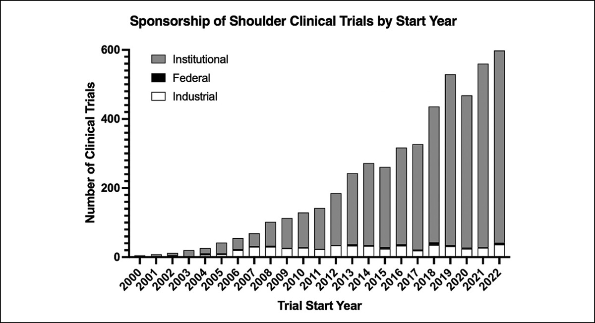
Clinical CourseAn 11-year-old girl with no notable medical history presented as a trauma after ejection from a moving vehicle approximately 12 hours before initial evaluation. She had diminished strength in the distal muscle groups of her bilateral lower extremities and absent sensation on the plantar surfaces of her feet. She had no rectal and perineal sensation, absent ankle wink reflex, and weak resting and volitional rectal tone. A catheter was placed for urinary retention. Her clinical picture was consistent with acute cauda equina syndrome. Imaging demonstrated an isolated H-type sacral fracture with focal kyphotic deformity through the S2 body (Figures 1 and 2). The patient and her family were counseled extensively that given her acute cauda equina syndrome and the unstable nature of the fracture, she was at high risk of complications. The family opted to pursue surgical intervention, and the patient was consented for open reduction and instrumentation of L4-ilium and percutaneous transiliac-transsacral screw fixation at the S1 level.
 Figure 1:
 AP (A), inlet (B), and outlet (C) radiographs of the pelvis demonstrating a complex sacral fracture and a nondisplaced right pubic ramus fracture.
 Figure 2:
 Axial (A), coronal (B), and sagittal (C) radiographic slices of a computed tomography scan of the pelvis demonstrating an H-type sacral fracture through the S2 body with marked shortening and focal kyphotic deformity.
Surgical TechniqueThe patient was taken to the operating room with the orthopaedic spine and orthopaedic trauma teams and induced into general endotracheal anesthesia. Gardner-Wells tongs and bilateral distal femoral traction pins were placed, and the patient was placed prone on a Jackson table with hips extended and knees flexed (Figure 3). The patient was secured to the table using distal femoral skeletal traction pins and counterweight was hung from the Gardner-Wells tongs. Closed reduction was done under fluoroscopic guidance, with a combination of traction and hyperextension.
 Figure 3:
 Photograph showing the intraoperative prone patient positioning on a Jackson frame OSI table with a traction arc and the patient in Gardner-Wells tongs and bilateral distal femoral skeletal traction.
Once adequate reduction was confirmed on fluoroscopy, a midline posterior approach to the lumbosacral spine was used, and the fracture was directly visualized. A dorsal fracture line was appreciated without any major displacement, and the reduction was acceptable. Bilateral iliac bolts and L4 and L5 pedicle screws were placed in a standard fashion. The iliac bolts were used to extend the pelvis and disimpact the anterior sacrum. Fluoroscopy was used to confirm appropriate alignment and implant position, and bilateral 5.5-mm rods were placed. The posterior elements were not decorticated, and no bone graft was placed to reduce the risk of arthrodesis. A hemilaminectomy was performed at L4-5 and L5-S1, and a catheter was used to flush hematoma and fracture debris from the canal (Figure 4). A transiliac-transsacral screw was placed percutaneously from the right side (Figure 5).
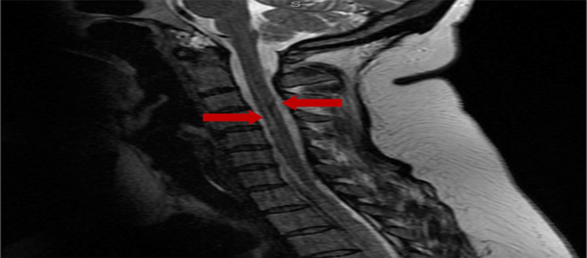
 Figure 4:
 Sagittal magnetic resonance image (MRI) demonstrating a compressive epidural hematoma from L4-S2.
 Figure 5:
 AP (A), inlet (B), and outlet (C) views of the pelvis and lateral lumbar spine (D) radiographs demonstrating L4-ilium instrumentation with S1 transiliac-transsacral screw.
Postoperative CourseThe patient was able to ambulate with therapy starting on postoperative day 1, and she was discharged home on postoperative day 5 after passing a trial of void. She had complete resolution of motor, sensory, bowel, and bladder function within 2 months. Her radiographs demonstrated bony consolidation across fracture sites with maintained fracture reduction. Her lumbopelvic parameters demonstrated appropriate postoperative alignment, with a lumbar lordosis of 52.7°, pelvic incidence of 51.5°, and sacral slope of 40.9°. A CT scan 3.5 months postoperatively showed bony healing and maintained fracture reduction (Figure 6), with no radiographic evidence of fusion at the L4-L5 or L5-S1 levels or evidence of instability because of the hemilaminectomy.
 Figure 6:
 Axial (A), coronal (B), and sagittal (C) slices of a CT scan of the pelvis demonstrating bony healing at prior H-type sacral fracture with maintained fracture reduction.
Given the patient's chronological age in the setting of bony healing, she was indicated for implant removal to reduce the risk of lumbosacral arthrodesis. The patient underwent a removal of implant 4 months postoperatively and was released to full activity without restrictions. Intraoperatively, she had maintained reduction, and there were no apparent degenerative changes or signs of bony fusion at the L4-L5 and L5-S1 levels. She reported no pain, numbness, or any sequelae of the trauma at her most recent follow-up (14 months).
ConclusionThe lumbosacral junction is particularly difficult to immobilize because of the notable forces present at the L5-S1 junction and the relatively poor bone quality in the sacrum.7 Triangular osteosynthesis constructs provide spinopelvic stability by incorporating horizontal and vertical fixations through a combination of iliosacral and lumbopelvic fixations, respectively.8 This allows for load transfer from the lumbar spine directly into the ilium, minimizing loadbearing by the injured sacrum.8,9 The purpose of this fixation was to maintain fracture reduction while allowing for early weight-bearing.7 In the setting of neurologic injury, a decompression was indicated to remove compressive hematoma and fracture debris. In our patient, a hemilaminectomy was used instead of a full laminectomy to limit the risk of iatrogenic instability. The utility of a decompression remains controversial in these types of injuries, but it is the senior author's experience that a small hemilaminectomy can mitigate some of the risks of wide decompression. This, combined with transiliac-transsacral fixation, allowed fracture union while avoiding fusion (Figure 6).
This case presentation redemonstrates the utility of this construct in the setting of an unstable lumbopelvic injury with kyphotic deformity and neurologic compromise. Furthermore, it expands on the existing literature through its application in the pediatric population. Our patient had complete functional and neurologic recovery within 12 months and was able to undergo implant removal (Figure 7). Given the patient's young age at presentation, it is unclear whether she will have any growth abnormalities or whether she will experience early degenerative changes or loss of motion at her lumbosacral junction. However, her early implant removal without signs of degenerative changes or fusion is reassuring for the long-term health and function of her lumbosacral junction. Given her surgical outcome and excellent recovery to date and the ability to perform triangular osteosynthesis with a combined orthopaedic trauma and orthopaedic spine team, we think that it is a safe and effective treatment of lumbosacral dissociation in the pediatric population suffering from acute neurologic deficits.
 Figure 7:
 AP Pelvis (A) and lateral lumbar (B) radiographs after implant removal demonstrating interval healing of prior H-type sacral fracture.
References 1. He S, Zhang H, Zhao Q, He B, Guo H, Hao D: Posterior approach in treating sacral fracture combined with lumbopelvic dissociation. Orthopedics 2014;37:e1027-e1032. 2. Sullivan MP, Smith HE, Schuster JM, Donegan D, Mehta S, Ahn J: Spondylopelvic dissociation. Orthop Clin North Am 2014;45:65-75. 3. Schildhauer TA, Josten C, Muhr G: Triangular osteosynthesis of vertically unstable sacrum fractures: A new concept allowing early weight-bearing. J Orthop Trauma 1998;12:307-314. 4. Hu X, Pei F, Wang G, He J, Kong Q, Tu C: Application triangular osteosynthesis for vertical unstable sacral fractures. Eur Spine J 2013;22:503-509. 5. Jindal R, Gupta S, Patil B, Patil A, Garg SK: Role of triangular osteosynthesis in vertically unstable transforaminal sacrum fractures: Clinical and radiological outcomes. Eur J Trauma Emerg Surg 2022;48:1369-1379. 6. Moon AS, Atesok K, Niemeier TE, Manoharan SR, Pittman JL, Theiss SM: Traumatic lumbosacral Dislocation: Current concepts in Diagnosis and management. Adv Orthop 2018;2018:6578097. 7. Esmende SM, Shah KN, Daniels AH: Spinopelvic fixation. J Am Acad Orthop Surg 2018;26:396-401. 8. Schildhauer TA, Ledoux WR, Chapman JR, Henley MB, Tencer AF, Routt ML Jr: Triangular osteosynthesis and iliosacral screw fixation for unstable sacral fractures: A cadaveric and biomechanical evaluation under cyclic loads. J Orthop Trauma 2003;17:22-31. 9. Steelman K, Bray R, Vaidya R: Technical note on placement of low-profile triangular osteosynthesis for unstable posterior pelvic ring injuries. J Orthop Trauma 2022;36:e337-e342.
留言 (0)