Patients
Patients with FAP who visited the Osaka International Cancer Institute and Ishikawa Gastrointestinal Clinic from January 1998 to September 2018 were retrospectively assessed for enrollment in this study. The diagnostic criteria for FAP in this study were (i) the presence of either ≥ 100 adenomas in the colon and rectum or 10 to 99 adenomas with a family history of FAP and (ii) a pathogenic germline variant in APC. Patients with mosaic variants in APC were excluded. Esophagogastroduodenoscopy (EGD) was generally recommended for all patients with FAP. Patients who had not undergone EGD at our institutions and patients with a history of duodenal surgery, such as gastric surgery or pancreatoduodenectomy, were not enrolled in this retrospective study because we could not ascertain the progression of the duodenal neoplasms in such patients. Most of the cases in this study were also analyzed in the study by Shimamoto et al. (Genotype-phenotype correlation for life-threatening complications in patients with familial adenomatous polyposis, accepted in Cancer Science).
Extraction of clinical data and definition of measured variables
We collected clinical data from the patients’ medical and endoscopic records. The genetic information used in this study comprised APC pathogenic variant and pedigree data aggregated at the Medical Research Support Co., Ltd. (Osaka, Japan), a data center operated by academic doctors. The genetic information was aggregated and controlled at the center in accordance with strict guidelines. All patients received genetic counseling and provided informed consent for APC genetic testing.
The observation period was defined as the duration from the day on which the first EGD was performed to the day on which the last EGD was performed according to the patient’s medical record or until immediately before the treatment intervention, if any. We generally performed annual surveillance EGD and targeted biopsies of polyps. We considered treatment interventions for patients with an advanced duodenal adenoma, namely > 10 mm or HGD, defined as lesions with severe atypia according to the previous criteria, because advanced adenoma is generally considered a precursor of invasive cancer in colorectal polyp management, and we needed to prevent the development of duodenal cancer to secure the patients’ safety.
Based on the number of colorectal adenomas, severe FAP was defined as the inability to visualize a patient’s normal mucosa macroscopically because of the profusion of colorectal adenomas, and sparse FAP was considered typical FAP (which is not severe FAP). Attenuated FAP was defined as the presence of 10 to 99 colorectal adenomas. Patients were considered to have Helicobacter pylori infection when a positive result was obtained by a serological test, urea breath test, rapid urease test, or histological examination, and when they had a history of eradication therapy for H. pylori.
Endoscopic system and settings
The endoscopic system consisted of a video processor (CV-260, CV-260SL, or CV-290; Olympus Co., Tokyo, Japan and VP-4450HD or VP-7000; Fujifilm Co., Tokyo, Japan) and a light source (CLV-260, CLV-260SL, or CLV-290; Olympus Co. and LL-4450, XL-4450, LL-7000, or BL-7000; Fujifilm Co.). A videoendoscope (GIF-H260Z, Q240Z, H290Z, PCF-Q260JI, or PCF-H290TL/I; Olympus Co. and EG-L590ZW, EG-L600ZW, or EGL600ZW7; Fujifilm Co.) was also used. Observations were performed by white light imaging and non-magnifying narrow-band imaging and/or with chromoendoscopy using indigo carmine with or without magnification.
Histological examination
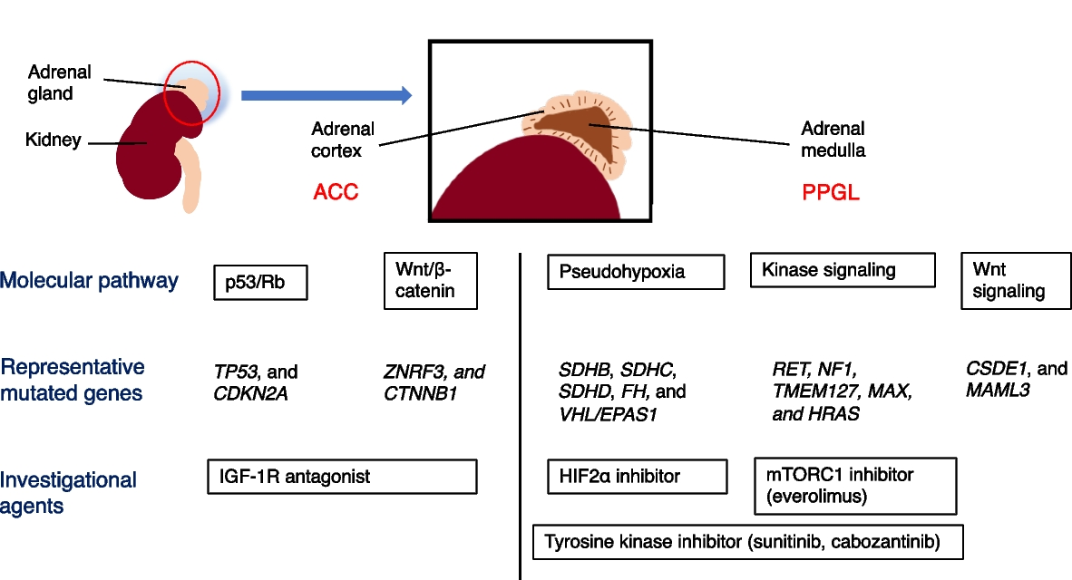
Lesions thought to be neoplastic or cancerous (well-demarcated lesions, large lesions, lesions with a depression at the center, and lesions with an irregular shape) were selected for biopsy. Biopsy tissue samples or endoscopic resection samples were collected and diagnosed. In patients who underwent intervention for duodenal neoplasms without prior biopsy, we collected information obtained by the following intervention. According to the Japanese classification of colorectal carcinoma, a histopathological examination was performed by two pathologists with expertise in gastrointestinal pathology. According to the Japanese classification, noninvasive cancer was evaluated as severe atypia by the Spigelman classification, which corresponds to Western standards. Because the grading of dysplasia according to the Vienna classification changed from mild/moderate/severe to low-grade/high-grade in 2000, our patients straddled the two eras. Our pathologists followed the mild/moderate/severe grading classification for a while, and then we adopted the original Spigelman stage (SS), using mild/moderate/severe in the analysis. We thus refer to severe atypia as “HGD” in this study. The most severe histopathological diagnosis during the observation period was adopted as the final histopathological diagnosis.
Spigelman classification
When NADAs were found, they were scored using the Spigelman classification to indicate the severity of duodenal polyposis [18]. The Spigelman classification includes the number of polyps, maximum diameter, tissue structure, and degree of atypia. Stages 0 to IV were determined by the total score obtained using the above-mentioned criteria. When a detected duodenal polyp was not histopathologically diagnosed as an adenoma, it was not considered to be a NADA.
Statistical analysis
We evaluated the prevalence of NADAs in patients with FAP, the progression of these adenomas during the observation period, the risk factors for the lifetime development of advanced adenoma, and SS IV until the end of the observation period.
The prevalence of NADAs in patients with FAP was indicated by the number and percentage of patients with NADAs. The progression of NADAs during the observation period was assessed with the SS, which includes the number of adenomas, size of adenomas, and development of severe dysplasia. Finally, we evaluated the lifetime risk of developing an advanced duodenal adenoma, and SS IV, which were reported to be risk factors for developing duodenal cancer [18, 20].
For the analysis of SS progression, patients for whom the observation period was < 5 years without SS progression were excluded because the observation period was considered too short. We then examined the progression of patients with SS 0 to III at the first EGD because there was no room for progression for patients with stage IV at the first EGD. We also examined the progression of patients with SS 0 and I at the first EGD, who were recommended to undergo follow-up at 5-year intervals according to the guidelines. Furthermore, we examined the occurrence of HGD according to the SS at the first EGD.
HGD, a tumor size ≥ 10 mm, the surveillance period, a history of colon cancer, age, the SS at the first EGD, classic FAP, and the location of several mutations in APC were evaluated as risk factors for disease progression because they were relevant to the severity of duodenal polyposis and reported to be risk factors for duodenal cancer [2,3,4,5,6, 21,22,23]. We also investigated whether APC mutations at codons 1250 to 1464, which are common in patients with severe FAP, were associated with the incidence and severity of NADAs [23]. Because we used only the protein truncation test for mutation detection in APC in patients whose first endoscopy procedure was performed before 2009, we could not obtain detailed information on the gene mutation site in some cases. We excluded those cases from the analysis of risk factors for disease progression.
Statistical analyses were performed using R software version 3.3.0 (R Foundation for Statistical Computing, Vienna, Austria; http://cran.r-project.org/). The data were analyzed using the χ2 test and Kruskal–Wallis test. Statistical significance was set at P < 0.05.


留言 (0)