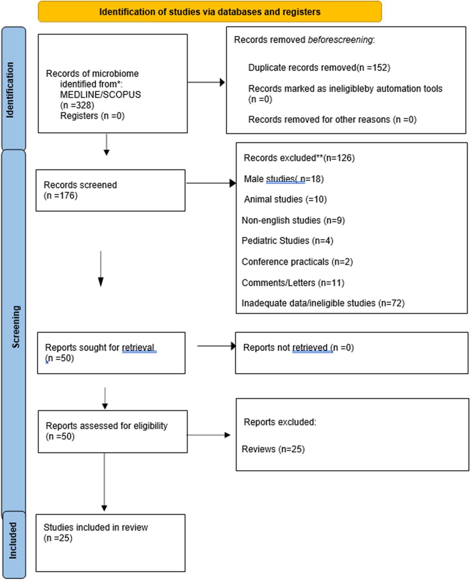
This study investigated the preoperative 3DCTA images of patients scheduled for LSC to examine IIV variants. The VW anterior to the SP was measured by comparing groups with or without the variants. The variant group had a significantly smaller VW than the other group, and the ANCOVA model was used to analyze VW adjusted for sociodemographic factors and comorbidities. There was a significant difference between the groups with and without IIV variants, excluding the effect of covariates on the VW. This study also showed that the distance from the SP to the aortic bifurcation decreased with age. Accordingly, the results were consistent with the stated hypothesis.
Previous studies have shown varying results regarding the measurement of the VW in front of the SP in sacrocolpopexy or anterior lumbar interbody fusion due to differences in race, measurement methods, and sex [11, 14, 15]. As expected, the presence of IIV variants in ALL of the SP seems common (20.2%) [5, 11]. If the left or right IIV flows into the contralateral CIV (types 3 and 5a) or if the left and right IIV merge to form a common trunk and their common trunk is the confluence of the left or right CIV or both sides (type 4), then the VW would be considered narrow [4, 11].
Furthermore, the distance from the SP to the IVC bifurcation was also significantly shorter in the variant group than that in the standard group, suggesting that the LCIV and SP were relatively close. The LCIV, the site of major vessel injury during LSC [16,17,18], requires careful ALL dissection and retraction. In this study, the ANCOVA model showed a significant difference in VW between the variant and standard groups. The VW in the variant group was narrower than that in the standard group, which should be considered during surgery. The surgeon was able to visualize the relationship between the vascular structures and the SP from all directions with 3DCTA and clearly understood the vascular structures. As a result, careful dissection could be performed intraoperatively.
The distance between the aortic bifurcation and SP decreased with age [2, 10, 19, 20]. This may be biologically plausible because the aging of the lumbar spine is accompanied by disk compression and loss of bone mineral density, resulting in decreased height [10]. Moreover, aging may result in calcium accumulation (a measure of atherosclerosis) and increased aortic diameter (a possible measure of elastin loss) [19,20,21,22], which may contribute to a downward shift of the aorta.
This study has several limitations. First, blinding was not incorporated into the analysis. The corresponding author (H.S.) and coauthors, M.K. and S.O., reviewed all the preoperative 3DCTA images, and a rigorous review of the accuracy of the imaging diagnosis was not conducted. Second, ALL dissection in the VW was limited to the area where suture fixation of the mesh could be safely performed. Therefore, the vascular structures around the ALL (e.g., the middle sacral artery and vein) were not completely confirmed intraoperatively, and there may have been cases in which the preoperative diagnosis was inaccurate owing to differences between the preoperative 3DCTA findings and the actual anatomical structures. However, the extent of the ALL dissection allowed suturing; moreover, unnecessary dissection carries the risk of vascular injury [18]. If the anatomy of the IIV could not be confirmed intraoperatively, caution may be warranted in women with a low BMI or retroperitoneal adhesions after laparotomy [12].
Third, we employed CT, and a limitation of this modality is that structures with high attenuation, such as bones, may mask the target anatomy [23]. In the present study, delineating the variant vessels was difficult in some patients, who had low retroperitoneal fat, owing to the proximity of some bones to the vessels.
A strength of this study is that all procedures were completed without vascular injury.
In summary, in this prospective cohort study we demonstrated that 1 in 5 women undergoing LSC had a vascular variant that restricts the safe VW for promontory dissection and fixation to a distance of 28 mm. Older women with a low BMI had a significantly higher risk of a vascular variant and consideration for preoperative 3DCTA may be indicated in these patients to avoid vascular injury during surgery.


Comments (0)