
記住我


We excluded 117 cases from this study. Firstly [24, 25], therapeutic doses of sulfonamides would affect serum BDG and P. jirovecii qPCR tests. Thus, 27 such cases were excluded. Secondly, 27 cases exceeding a long interval (> 3 days) that would affect the consistency between serum BDG and P. jirovecii qPCR [25] were excluded. Thirdly, co-infection with other fungi [26] would also elevate serum BDG, leaving the contribution of PJP uncertain; thus, 35 such cases were excluded. Fourthly, we found that cardiopulmonary bypass could lead to a spurious increase in BDG (data unpublished), so 13 cases were excluded. Finally, 15 cases with incomplete medical records were excluded.
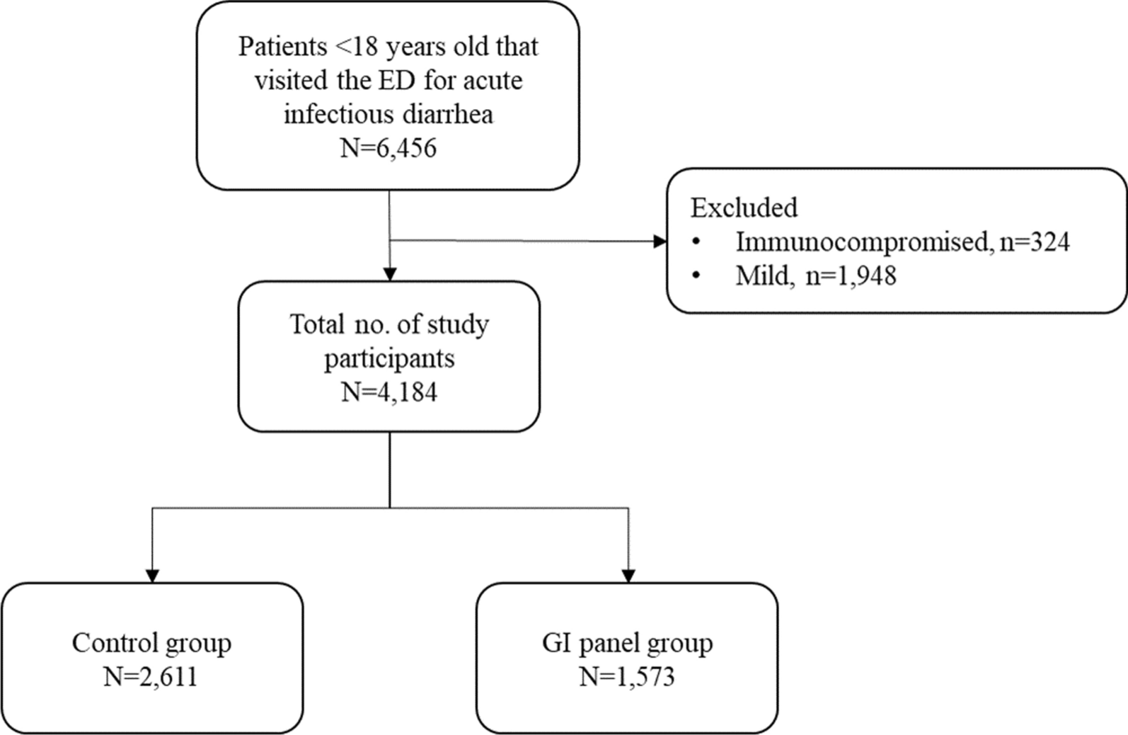
Ultimately, there were 213 patients enrolled in the study (Fig. 1), 51.6% were female, and their median age was 60 years. There were 22 (10.3%) with hematologic malignancies, 31 (14.6%) with solid cancer, 100 (46.9%) with autoimmune or inflammatory disorders, 15 (7.0%) with interstitial pneumonia due to noninfectious causes, 24 (11.3%) with nephrotic syndrome, 14 (6.6%) with infectious diseases (severe infections caused by bacteria and/or viruses), and 7 (3.3%) with other diseases (including 4 thrombotic diseases and 3 pericarditis). Based on the gold standard, there were 159 PJP patients and 54 P. jirovecii-colonized patients.
Fig. 1
Flow chart of study design. P. jirovecii qPCR positive HIV-uninfected patients (n = 330) were recruited from 2019 to 2021. After exclusion, 213 participants were included and divided into a derivation cohort (to define the cut-off value of serum BDG) and a validation cohort (to verify the defined cut-off value of serum BDG). A discharge diagnosis of PJP was taken as the gold standard to divide the participants into two groups: the PJP group and the P. jirovecii-colonized group
Differences in mortality and comorbid composition between PJP and P. jirovecii-colonized patientsBased on the gold standard, there were 159 PJP patients and 54 P. jirovecii-colonized patients. The colonization rate was 25.4%, consistent with other studies (24.5%) [8]. As indicated in Table 1, no significant differences existed in age (P = 0.1803) and gender (P = 0.0827) between the two groups. In contrast, the comorbid composition was significantly different (P = 0.0227). The in-hospital mortality was significantly higher in the PJP group than in the P. jirovecii-colonization group (13.2 vs. 3.7%, P = 0.0374), consistent with previous studies [1, 3]. Furthermore, the proportion of BALF samples in the PJP group was significantly higher than that in the P. jirovecii-colonization group (37.7% vs. 9.3%, P < 0.0001). This is because patients with PJP are more likely to meet the indication for alveolar lavage operation than patients with P. jirovecii-colonization.
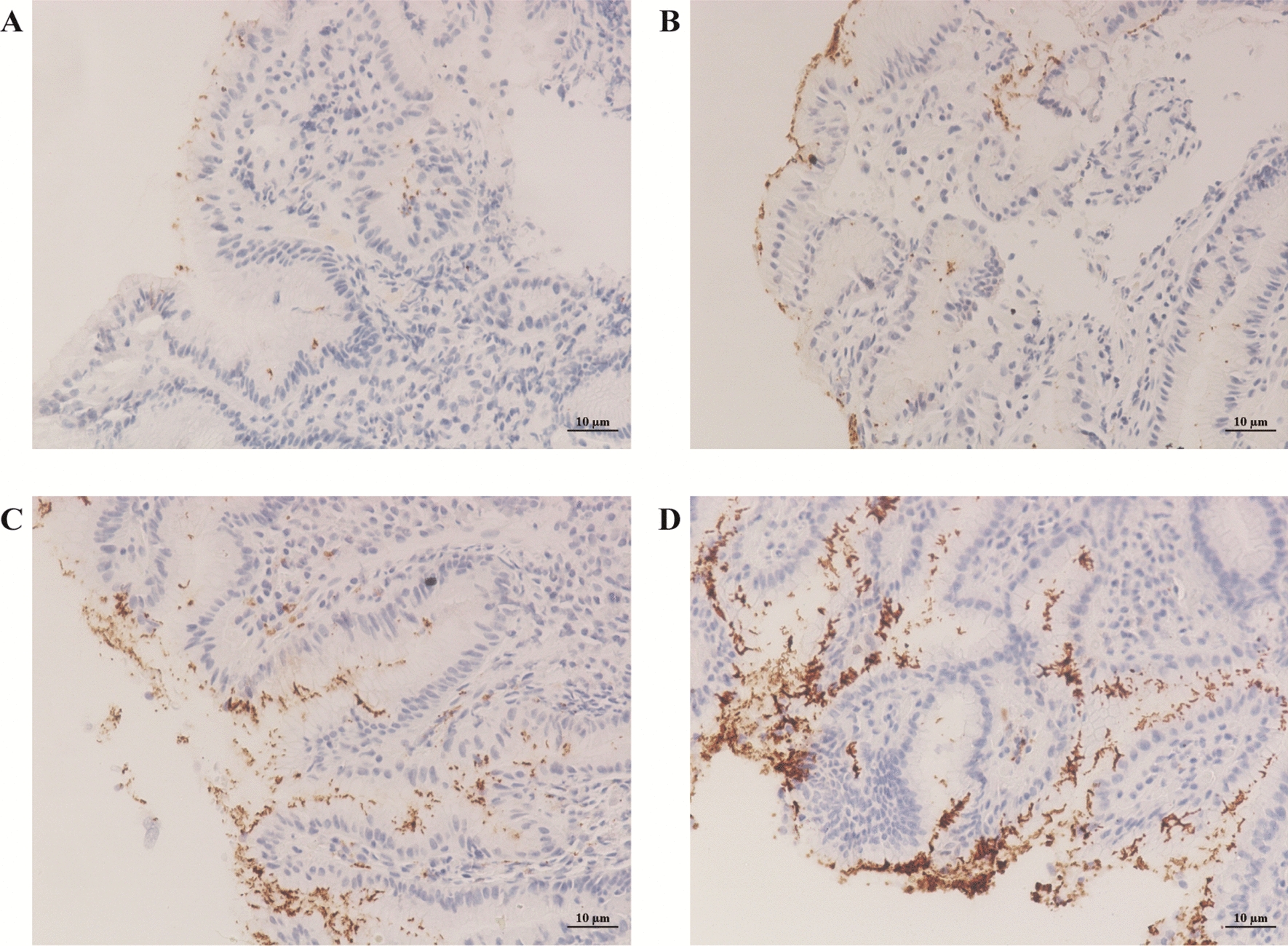
Table 1 Characteristics of the investigated populationDifferences in lymphocytes and inflammatory factors between PJP and P. jirovecii-colonized patientsAs shown in Fig. 2, both lymphocytic counts and proportions were significantly lower in PJP patients (0.46 × 10⁹/L, 6.8%) than in P. jirovecii-colonized patients (0.75 × 10⁹/L, 10.8%; P = 0.0004, P = 0.0009), being consistent with other studies [27]. Additionally, the percentage of B cells in lymphocytes was reduced in PJP patients (7.8% vs. 12.7%; P = 0.0187). In contrast, the percentage of NK cells in lymphocytes was comparable between the two groups (10.1% vs. 8.9%; P = 0.9953).
Fig. 2
Peripheral lymphocytes and inflammatory indicators were compared between PJP and P. jirovecii-colonized patients. Differences of lymphocyte counts (A), lymphocyte percentage (B), B cell percentage (C), CD4+T cell percentage (D), CD8+T cell percentage (E), CD4+T cell to CD8+T cell ratio (F), NK cell percentage (G), C reactive protein (H), and erythrocyte sedimentation rate (I) between PJP and P. jirovecii-colonized patients. The p-values were calculated using Mann-Whitney tests, and statistical significance is displayed as *P < 0.05, **P < 0.01, ***P < 0.001
Notably, CD4+ T cells and CD8+ T cells revealed an opposite trend. The percentage of CD4+ T cells in lymphocytes decreased among PJP patients (29.9% vs. 36.9%; P = 0.0265), consistent with previous studies [28, 29]. In contrast, the percentage of CD8+ T cells in lymphocytes increased among PJP patients (39.9% vs. 25.9%; P = 0.0030), inconsistent with previous studies [30]. Consequently, the ratios of CD4+ T cells to CD8+ T cells were lower among PJP patients (0.8 vs. 1.5; P = 0.0015), indicating that PJP patients have both immunodeficiency and hyperimmunity.
Moreover, two inflammatory factors, CRP and ESR, were higher in PJP patients (49.6 mg/L, 47 mm/h) than in P. jirovecii-colonized patients (27.9 mg/L, 39 mm/h). However, a significant difference was only observed in CRP (P = 0.0020), reflecting the hyperinflammatory state correlated with P. jirovecii infection.
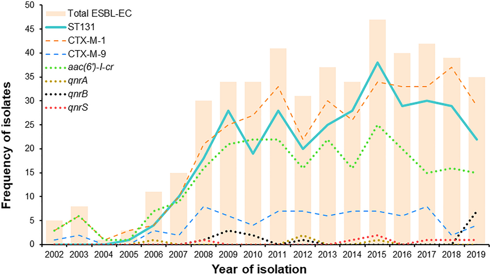
ROC curve and cut-off value of serum BDG for the diagnosis of PJPAs indicated in Table 1; Fig. 3A, the serum BDG level of PJP patients was significantly higher than those in P. jirovecii-colonized patients (P < 0.0001). The AUC of derivation cohort, validation cohort, and total subjects were 0.92, 95% CI (0.86, 0.98); 0.89, 95% CI (0.82, 0.95); and 0.91, 95% CI (0.86, 0.95); respectively (Fig. 3B).
Fig. 3
ROC curves of serum BDG in different cohorts and subgroups. (A) Comparison of serum BDG between PJP and P. jirovecii-colonized patients. (B) ROC curves of serum BDG based on the Derivation cohort, Validation cohort and the total cohort. (C) ROC curves of serum BDG based on subjects aged ≤ 60 years and > 60 years. (D) ROC curves of serum BDG based on subjects with comorbidities of infectious diseases, solid cancer, autoimmune or inflammatory disorders, hematologic malignancies, lung cancer, ILD, or nephrotic diseases. * Patients with lung cancer were excluded from the solid tumor subgroup. The p-values were calculated using Mann-Whitney test, and statistical significance is displayed as ****P < 0.0001
A serum BDG cut-off of 117.7 pg/mL was determined using the derivation cohort and then verified by the validation cohort. As shown in Table 2, with the prevalence of 74.6%, BDG ≥ 117.7 pg/mL showed outstanding specificity, LR, and PPV in the derivation (90.0%, 8.9, and 96.4%) and validation (91.7%, 9.2, and 96.3%) cohort. At the same time, the sensitivity and NPV were slightly lower in the derivation cohort (89.0% and 73.0%) and lowered in the validation cohort (76.5% and 57.9%). Notably, the median value of serum BDG in the derivation cohort was significantly higher than in the validation cohort (300.3 pg/mL vs. 170.2 pg/mL, P = 0.0253). This could be related to its better diagnostic efficacy in the derivation cohort than in the validation cohort.
Table 2 Evaluation of diagnostic performance of serum BDG (at ≥ 117.7 pg/mL)Age and comorbidities affect the diagnostic efficacy of serum BDGStratified ROC analysis was performed based on comorbidities (Table 2). As indicated in Fig. 3D, the AUC of serum BDG was 1.00 in the infectious disease subgroup, 0.90 in the solid cancer subgroup, and 0.89 in the autoimmune or inflammatory disorders subgroup, 0.89 in the hematologic malignancy subgroup, 0.83 in both the lung cancer and the ILD subgroups, and 0.70 in the nephrotic syndrome subgroup. Therefore, the diagnostic efficacy of BDG varied in patients with varied comorbidities, though without statistical difference (P = 0.3698). BDG could not significantly distinguish PJP from P. jirovecii-colonization in subgroups of lung cancer, ILD and nephrotic syndrome (P = 0.0961, P = 0.0833, and P = 0.3472). Of note, the suboptimal AUC could also be related to the limited number of P. jirovecii-colonized cases (n < 5) in the subgroups (Table 2; Fig. 3).
Moreover, stratified ROC analysis was performed by dividing the subjects into two subgroups: ≤ 60 years (n = 108) and > 60 years groups (n = 105). As represented in Fig. 3C, the AUC of serum BDG was higher in patients aged ≤ 60 years (0.96) than in patients aged > 60 years (0.86), but the difference was not statistically significant (P = 0.1196).
As shown in Table S1, PJP patients with lung cancers had the lowest median BDG levels(151.6), although the difference was not statistically significant (P = 0.1971).
Preliminary analysis of false-negative related factorsThe sensitivity and NPV of BDG were suboptimal, particularly in the validation cohort. We compared the characteristics of the true-positive group (n = 137) and the false-negative group (n = 22) to analyze the factors associated with false negatives (Table 3).
In the current study, there were 3,4,12,and 3 PJP cases not identified by BDG in the hematologic malignancie, lung cancer, autoimmune or inflammatory disorders, and ILD subgroups, respectively (Fig. 4). And the proportion of lung cancer and ILD in the false-negative group (18.2%, 13.6%) was higher than that in the true-positive group (6.3%, 9.5%), though without a statistical difference (P = 0.2815). Furthermore, there were no significant differences in peripheral lymphocyte count, peripheral lymphocyte proportion and gender composition between the true positive group and the false negative group (P = 0.4353, P = 0.1422, and P = 0.1059). Of interest, the median age of the false negative group was significantly higher than that of the true positive group (63 vs. 58 years, P = 0.0259) (Table 3). OR analysis showed that when the patient was older than 57 years old, the odds ratio of false negative to true positive were 4.307 (p = 0.007).
Fig. 4
Distribution of BDG in the PJP and P. jirovecii-colonized groups. (A) In each subgroup, PJP patients were diagnosed by BDG ≥ 117.7pg/mL, proportions of true positive and false negative cases were shown in proportion composition diagrams. (B) Scatter plot showing comparison of serum BDG between PJP and P. jirovecii-colonized patients in different subgroups. * Patients with lung cancer were excluded from the solid tumor subgroup
Table 3 Comparison of characteristics between true positive group and false negative groupOdds ratios for PJP versus P. jirovecii-colonization was correlated with immune markers, inflammatory markers, and history of glucocorticoid therapyAs shown in Fig. 5, for the whole subjects, the odds ratios for PJP versus P. jirovecii-colonization were 2.314, 2.554, 2.628, 3.244, 3.367, 3.481, 3.769, and 5.375, respectively, when the proportion of CD4+ T cells in T cells < 30%, the proportion of CD8+ T cells in T cells > 40%, lymphocyte counts < 1.0×10⁹/L, ESR > 15 mm/h, CD4+T/CD8+T < 1.4 or > 2.0, CRP > 10 mg/L, the proportion of B cells in lymphocytes < 9% and had a history of treatment with glucocorticoids within 2 weeks. For patients with autoimmune or inflammatory disorders, the odds ratios for PJP versus P. jirovecii-colonization were 6.667 and 11.846 when CRP > 10 mg/L and ESR > 15 mm/h. For participants without autoimmune or inflammatory disorders, odds ratios for PJP versus P. jirovecii-colonization were 3.048, 5.098 and 6.680 when proportion of CD8+ T cells in T cells > 40%, CD4+T/CD8+T < 1.4 or > 2.0, and had a history of treatment with glucocorticoids within 2 weeks.
Fig. 5
Odds ratios of PJP versus P. jirovecii-colonization with different characteristics in the whole cohort and Autoimmune or inflammatory disorders subgroup. (A) In the whole cohort, Odds ratios of PJP versus P. jirovecii-colonization elevated significantly when proportion of CD4+ T cells in T cells < 30%, proportion of CD8+ T cells in T cells > 40%, lymphocyte counts < 1.0×10⁹/L, ESR > 15 mm/h, CD4+T/ CD8+T < 1.4 or > 2.0, CRP > 10 mg/L, proportion of B cells in lymphocytes < 9% and treatment with glucocorticoids within 2 weeks. From top to bottom, the odds ratios are arranged from small to large. (B) In the autoimmune or inflammatory disorders subgroup, odds ratios of PJP versus P. jirovecii-colonization elevated magnificently when ESR > 15 mm/h, CRP > 10 mg/L. (C) For subjects without autoimmune or inflammatory disorders, odds ratios of PJP versus P. jirovecii-colonization elevated magnificently when CD8+ T cells in T cells > 40%, CD4+T/ CD8+T < 1.4 or > 2.0, and treatment with glucocorticoids within 2 weeks. The p-values were calculated using Chi-square test and Fisher test
留言 (0)