
記住我


See Article, page 1135
From a historical perspective, the value of provocative and analgesic spinal injections dates back to the late 1930s, when Steindler and Luck1 first demonstrated the utility of the transforaminal approach for the diagnosis of low back and radicular pain by injecting procaine hydrochloride. Since then, targeting the epidural space via transforaminal epidural steroid injections (TFESIs) has evolved into a common intervention for the treatment of radicular pain. Published literature has established the efficacy of TFESIs for short- and long-term treatment of radicular pain, in addition to avoidance of surgery.2–4
The general approach to TFESIs involves accessing the epidural space via the intervertebral foramen, the boundaries of which are defined by the superior and inferior pedicles, the posterior disk annulus, the facet joint, and the ligamentum flavum.5 The contents of the intervertebral foramen include the exiting nerve root, radicular arteries, veins, connective tissue, and epidural fat. In the thoracolumbar region, the angle of the vertebral foramen and exiting spinal nerve is posterior, and the nerve roots exit at the superior aspect of the foramen and travel inferiorly.6,7 Vascular structures in the region include singular anterior and paired posterior spinal arteries, as well as radicular arteries supplying the spinal nerve roots. The radiculomedullary arteries—the largest and most caudal of which is the artery of Adamkiewicz—typically enter through the anterior-superior aspect of the intervertebral foramina on either side (usually left) from T5 to S1.8,9
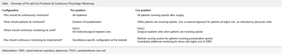
Table - Summary Table of Pros/Cons for Each Transforaminal Approach Superior (“Safe”) triangle Inferior (Kambin’s) triangle Pros Decreased risk of intradiscal injection Decreased risk of intravascular injection Decreased risk of nerve root irritation Decreased risk intrathecal injection Approach may be modified to reduce intravascular spread and paresthesia without compromising procedural efficacy Cons Increased risk of intravascular injection Arteries are still present in this region, so approach may not avoid vascular penetration entirely Increased risk of nerve root irritation Inferior triangle is highly variable and actual area may be smaller than superior triangle; may be harder to access transforaminal epidural space due to superior articular process, preventing appropriate epidural spreadFrom a technical standpoint, TFESIs consist of image-guided needle placement using either fluoroscopy or computed tomography, contrast injection to confirm epidural spread while excluding vascular and intrathecal uptake, and treatment injection of corticosteroid.10 Despite their efficacy and general safety, these procedures do carry the risk of rare, catastrophic neurological complications.11,12 The precise mechanism of neurological compromise is debated and may include vasospasm-induced infarction, direct vascular trauma, chemical injury, and/or intravascular injection leading to embolization, but it is important to recognize that all postulated mechanisms involve the vessels.8,12–14 Due to the anterior and inferior location of the vessels with respect to the cervical foramina, the accepted approach for cervical TFESI is to remain in the posterior aspect of the foramen, immediately anterior to the superior articular process.13,15 However, the optimal approach to lumbar TFESI remains debated. It is, therefore, with great interest and focus on safety that we consider the percutaneous approach to the lumbar neural foramen. In this commentary, we examine and compare the 2 most common techniques for TFESI, the superior (“safe triangle”) approach and the inferior (“Kambin’s triangle”) approach (Table). Note that in the literature, approaches to the TFESI may also be described as supraneural or infraneural, subpedicular or suprapedicular, retrodiscal, retroneural, or preganglionic. Such terminology can be inconsistent given that the location of neural structures within the foramen is highly variable and not easily identified on fluoroscopy, the most common modality for TFESI image guidance. For the sake of clarity and consistency, we will primarily use the term “superior triangle” to refer to techniques targeting the superior aspect of the intervertebral foramen, and “inferior triangle” for an approach targeting the inferior aspect of the foramen.
PRO: THE TRADITIONAL SUPERIOR (SAFE) TRIANGLE IS THE PREFERRED SITE OF NEEDLE INSERTIONUnderstanding the rationale for the superior triangle approach to lumbar TFESIs requires detailed knowledge of the foraminal anatomy. The superior triangle approach was developed with the intent to minimize risk of damage to the exiting nerve root, as well as avoid intrathecal puncture and vascular injection.16–18 Anatomically, it is a right triangle comprised by the transverse line from the “6 o’clock” position on the pedicle to the lateral pedicular line (upper border), the sagittal line from the lateral pedicle to the imagined exiting spinal nerve (lateral border), and the imagined superior border of the exiting spinal nerve root (hypotenuse) (Figure). In the traditionally described approach, the needle is advanced to the dorsal periosteum of the vertebral body, anterior to the nerve, allowing for medication administration in the anterior epidural space.19 Care is taken to avoid unintentional dural puncture by placing the needle within the bounds of the safe triangle, lateral to the midpedicular line, with contrast administration showing appropriate spread to the nerve sheath and/or medial epidural space.19
 Figure.:
 Superior and inferior triangles of the transforaminal space. Image courtesy of Imran Omar, MD, Department of Radiology, Northwestern University Feinberg School of Medicine.
Although the traditional superior triangle approach remains widely used, opponents argue that it fails to account for variability in the anatomic location of the radicular arteries and the artery of Adamkiewicz. This could increase the risk of inadvertent vascular damage, vasospasm, or intravascular injection (inclusive of particulate steroid), leading to spinal cord infarction, which is the probable etiology of paraplegia and other neurological sequelae associated with this procedure.8,12–14 Accordingly, some practitioners have advocated transforaminal injection via the inferior (Kambin’s) triangle to avoid intra-arterial injection or trauma to the radiculomedullary arteries.20 Very few studies have compared the 2 TFESI approaches directly. A trial of 75 patients comparing the superior and inferior triangle approaches for lumbar TFESI found no significant differences in terms of contrast spread to the anterior epidural space, rate of intravascular needle puncture under intermittent fluoroscopy, needle paresthesia, pain relief, or functional improvement at up to 2-month follow-up.21
Given the likely mechanism of catastrophic spinal cord injury is via the radiculomedullary arteries, the location of these vessels is of utmost importance. A retrospective review of spinal angiograms reported that among the 113 cases in which the radiculomedullary arteries could be clearly evaluated, the artery of Adamkiewicz was predominantly located in the superior aspect of the neural foramen at L3 and above.10 Dorsal-ventral location in this study could not be determined due to use of posterior-anterior projections. Cadaveric dissections of 24 individuals found that at thoracolumbar levels, the artery was almost always (92% ± 15%) anterosuperior to the nerve, while at thoracic levels, the artery was more often directly anterior to the nerve; there was no comment or report of posteriorly located vessels.22 In a different study of 94 datasets using flat panel catheter angiotomography (aka, conebeam computed tomography [CT] angiography), the radicular arteries could be visualized in 78 (83%) cases. Of those cases, the arteries were found in the anterosuperior quadrant of the neural foramen in 75 cases (96.2%), in the posterosuperior quadrant in 2 cases (2.6%), and in the anteroinferior quadrant in 1 case (1.3%), with no cases in the posteroinferior quadrant.9 Another study examined the location of the radicular spinal arteries in 256 CT angiograms of the lumbar region. Although anterior/posterior positioning was not assessed, arteries were located in the safe triangle in 129/256 cases (50.4%) and in Kambin’s triangle in 52/256 cases (20.3%); notably, an artery was found in both locations in 21 cases (8.2%).23
Superficially, if the 2 approaches have comparable efficacy, favoring the inferior triangle over the classical superior triangle might seem attractive to improve safety. One could argue that an inferior triangle approach would decrease the theoretical risk of intravascular injection since there are fewer vessels present, it reduces potential neural trauma because the nerve root is located more anteriorly lower in the foramen, and the vast majority of cases of catastrophic neurological injury that have been reported use the traditional “safe triangle” approach.24,25 However, such a view fails to appreciate the intervertebral foramen as a 3-dimensional space.
As discussed, the thoracolumbar radicular arteries are overwhelmingly in the anterior-superior quadrant of the foramen, and case reports of neurologic injury from superior triangle TFESI describe the classical approach of targeting the anterior-superior quadrant.24–29 The Spine Intervention Society guidelines for TFESI even specifically describe anterior placement of the needle.30 However, placement of the needle within posterior-superior aspect of the vertebral foramen, within the superior triangle, can also be used to access the epidural space, thus reducing the risk of intravascular trespass.18,30,31 An observational study found that the degree of pain relief with the needle placement in the posterolateral epidural space was not significantly different from the conventional approach, and elicited significantly less procedural pain.31 A comparative trial of 50 patients evaluating the conventional approach via the anterior-superior quadrant of the foramen versus a posterolateral approach via the posterior-superior quadrant found no significant differences in pain or disability outcome measures, intravascular injection, or dural puncture, as detected by real-time fluoroscopy.32 However, the conventional (anterior-superior) approach had a significantly higher incidence of nerve root irritation (26.9% vs 4.2%, P = .050) and spinal nerve pricking (23.1% vs 0%, P = .023) compared to the posterolateral approach.32 A retrospective study of 83 patients comparing anterior-superior to posterior-superior TFESI found greater ventral epidural spread with anterior injections, as well as greater reduction in pain scores, although the follow-up period was only 2 to 4 weeks.33 To our knowledge, no study has directly compared the safety and efficacy of approach via the posterior-superior aspect of the foramen to the inferior triangle approach.
Approach via the superior triangle may also have distinct advantages compared to the inferior triangle approach. The inferior triangle approach was originally developed as a technique to provide a safe corridor for surgical instrumentation to access the intervertebral disk space.34 Safety in surgical instrumentation targeting the disk should not necessarily translate to safety of needle insertion targeting the epidural space or nerve sheath. In fact, a retrospective review of 257 retrodiscal (ie, inferior approach) lumbar TFESIs found that unintentional intradiscal injections occurred in 12 (4.7%) cases.35 By contrast, the rate of intradiscal injection with TFESI via the superior approach has been reported as 0.17% to 2.3%.36,37 Although no cases of discitis were reported in any of these studies, intradiscal injection in other interventional pain procedures (eg, diskography) is known to be associated with risk of discitis.38 Intradiscal injection has also been associated with both in vivo and in vitro evidence of accelerated disk degeneration,39–41 although other studies are contradictory.42
A multi-institutional review of nearly 15,000 cervical, thoracic, and lumbar TFESIs using the traditional superior triangle approach found the rate of dural puncture under live fluoroscopy to be <0.1%.43 A different retrospective review reported that intrathecal injections occurred in 8 (3.1%) cases of retrodiscal lumbar TFESI: 3 were subarachnoid, 4 were subdural extra-arachnoid, and 1 was both.35 Notably, although the procedures were performed under fluoroscopy, the exact method of intrathecal detection was not reported. Given the increased sensitivity of real-time fluoroscopy or digital subtraction angiography to detect intravascular and intrathecal needle placement, these rates may have been underestimated. Pain interventionalists are well acquainted with the risks of inadvertent dural puncture and intrathecal injection of local anesthetic, contrast, corticosteroid, or air,44 which should be carefully considered when attempting the inferior triangle approach.
Finally, the size and shape of the inferior triangle are known to be highly variable and sensitive to adjacent spinal pathology such as facet hypertrophy, disk herniation, and loss of disk height.45–47 In some patients, the inferior triangle may be no more than a theoretical space, without any appreciable surface area between the exiting nerve root, the superior endplate of the adjacent vertebral body, and the facet joint.45 Thus, successful TFESI via the inferior triangle may impair epidural contrast spread and present additional technical challenges above and beyond the safe triangle approach, while not necessarily obviating intravascular injection. Although the risk of arterial puncture is presumably decreased compared to the superior triangle approach, 1 of the studies discussed above reported a considerable intravascular injection rate of 6.6% with the inferior approach.35
No procedure is without risks, and the approach to any procedure must account for individual patient anatomy, technical considerations (eg, imaging modality), and physician experience. There is insufficient evidence to demand a single, one-size-fits-all approach, and no area of the intervertebral foramen should truly be considered absolutely “safe.” Accordingly, most professional societies do not firmly endorse one approach over the other.48,49 Based on reported cases of catastrophic neurological consequences following TFESI and their putative mechanism of injury, it is tempting to proclaim the inferior triangle definitively as the gold standard approach. Yet we must carefully consider that there are alternative strategies to minimize injury via the superior triangle, including use of real-time fluoroscopy/digital subtraction angiography; posterior needle placement; slow, small-volume injection; and possibly nonparticulate over particulate steroid, although the last suggestion is highly controversial.48–50 We must also recognize potential harms that may be associated with more widespread use of the inferior triangle approach, including theoretical increased risk of intradiscal injection or dural puncture, without necessarily avoiding intravascular injection altogether. In their 2019 epidural recommendations, the World Institute of Pain Benelux Work Group observed that superior safety and efficacy of the inferior triangle approach has not been established, it carries a higher risk of intradiscal injection, and lack of evidence confirming appropriate epidural contrast flow via this approach.50 The Benelux Work Group recommended TFESI approach via the superior or “‘safe triangle,’ with a clear preference to keep the needle tip posterior in the neuroforamen.”50 We agree with this recommendation and advocate for the superior triangle approach to TFESI to remain the default technique, with consideration of the inferior triangle approach in suitable patients based on anatomy and spinal pathology, for practitioners who feel comfortable with the approach and remain cognizant of its associated risks (Table).
CON: THE INFERIOR (KAMBIN’S) TRIANGLE IS THE PREFERRED SITE OF NEEDLE INSERTIONTransforaminal epidural steroid injections (TFESIs) provide therapeutic relief of radicular pain generated from the nerve root, such as cases of nerve irritation due to ipsilateral retrodiscal pathology.51–54 TFESIs may also be preferred over interlaminar epidural steroid injections in the case of selective nerve root involvement for lumbar radicular pain or if prior instrumentation prevents interlaminar access. Complications from TFESI are rare but potentially devastating, and the approach to the foramen can impact risk of adverse events.8,55
The inferior triangle approach, first described in 2011,56 is safer than the traditional superior triangle approach and equally efficacious.34,51,56–59 Anatomically, it is a triangle superior to the dorsolateral disk that is bordered by the superior endplate of the inferior vertebral body (base of the triangle), the superior articulating facet (height of the triangle), and the imagined exiting superior nerve root (hypotenuse of the triangle) (Figure). The method for performing a TFESI via the inferior triangle is with an oblique fluoroscopic view and a needle entry point slightly lateral to the superior articular process of the vertebra caudal to the target level. The needle is then advanced into the inferior triangle of the foramen using the coaxial (tunnel view) technique, along the axis of the fluoroscopic beam.51,60
Catastrophic neurological injury can occur during a TFESI due to occlusion of the anterior spinal artery from either particulate steroid-induced thrombosis/vasospasm or direct needle puncture of the artery or its supplying branches. Occlusion of the anterior spinal artery can result in spinal cord injury leading to paralysis and death.9,24,25,55,61,62 A multitude of case reports has revealed catastrophic neurological injury after TFESI using the superior triangle approach.8,29,55,63–67 Following a left-sided T12-L1 TFESI using the “safe” triangle technique, a patient suffered permanent paraplegia due to vascular injury.55 In another case series, 3 patients experienced sudden lasting paraplegia or paraparesis after TFESI with spinal cord edema on postprocedure magnetic resonance imaging.8 One of the patients underwent TFESI at L3-4 on the left, and 1 at L3-4 on the right.8 In each of the cases reported, a superior triangle approach was utilized, but an inferior triangle approach would potentially have circumvented the arterial vasculature (Figure).
Avoiding arterial injury is of high concern for patient safety. Therefore, the safest approach for performing a TFESI requires that the proceduralist possess robust understanding of the location of the anterior radiculomedullary artery and the radicular arteries within the neural foramen. Specifically, in the review of 248 thoracic and lumbar spinal angiograms previously discussed in the superior triangle section, the artery of Adamkiewicz was located in the superior one-half of the foramen in 97% of cases, with 80% in the upper third, 9% in the middle third, and only 2% of cases in the lower third; it was never observed in the inferior one-fifth of the foramen. The cadaveric dissections also previously discussed found that of the 39 anterior medullary arteries identified, 29 (74%) were in the upper third of the foramen, 9 (23%) were located in the middle third, and only 1 (3%) in the lower third.10 Therefore, the safest needle placement for a TFESI, particularly at L3 and above, is likely the inferior and slightly posterior position within the foramen relative to the nerve.
The inferior triangle approach is favorable for reasons other than avoidance of the artery of Adamkiewicz. It can also minimize the risk of accidental injection into the radiculomedullary artery.56 As noted in the superior triangle discussion, an in vivo study of flat panel cathether angiotomography datasets referenced in the Pro section above revealed that thoracolumbar radicular branches providing an anterior radiculomedullary artery were overwhelmingly located in the superior quadrants, while no cases were found in the posteroinferior quandrant.9 These findings support that needle placement during a TFESI should avoid the superior half of the neural foramen, especially the anterior-superior quadrant. The inferior triangle approach avoids the superior quadrant, which minimizes risk of arterial injury and subsequent devastating patient complications. There are also several reports concluding that the area of the inferior triangle increases as the spinal level descends from levels L1 to L5, with the maximum triangular area being at the L4-5 level.68–71 Thus, in addition to being the most prudent approach in upper lumbar levels, the inferior triangle approach may also be the safest needle placement for a TFESI at L3-5.
The superior triangle approach may cause irritation of the spinal nerve root during injection if the patient has severe spinal stenosis, epidural fibrosis, or intervertebral disk degeneration.57 One study reported fewer complications when the inferior approach was used for TFESI compared to the superior approach.56 In the inferior approach group, there were no incidents of nerve root pricking during injection, whereas 5 cases were reported with the superior triangle approach. The study also reported that the inferior triangle approach was as efficacious as the superior triangle approach for short-term pain relief. Another study of over 1200 injections had similar findings, concluding that needle tip position does not impact patient pain relief in lumbar selective nerve blocks.72 Finally, in some patients, the epidural space cannot be accessed through the superior triangle, making the inferior approach a favorable alternative to provide pain relief.73
Spread of contrast media is important to evaluate both safety and efficacy, given that contrast spread will ensure the absence of vascular uptake of contrast media. This avoids inadvertent injection of agents into the radiculomedullary artery, which could lead to vascular injury and neurological complications. The spread of contrast media also verifies correct needle position and ensures delivery of medication to the target site. A retrospective study of fluoroscopic images determined positioning the needle tip medial to the pedicle aids in the spread of the contrast media into the epidural space during TFESI when using the inferior approach.60 Factors other than the needle tip position, including patient magnetic resonance imaging (MRI) findings and history of surgery, did not significantly affect the contrast media dispersion pattern.
Our specialty is long overdue to follow the evidence on patient safety and begin using the inferior triangle approach. No longer should we rely on the conventional “safe triangle” as the evidence for all safety metrics demonstrates the advantages of the inferior triangle. We suggest wide adoption of a new safe triangle for all transforaminal procedures to reduce occurrence of rare but catastrophic neurologic injuries.
DISCLOSURESName: Tina L. Doshi, MD, MHS.
Contribution: This author was lead author of the Pro section of the manuscript, wrote portions of the Con section, designed the summary table, contributed to the creation of the manuscript, provided review and commentary, and made revisions based on group feedback.
Name: Alyson M. Engle, MD.
Contribution: This author was lead author of the Con section of the manuscript, wrote portions of the Pro section, contributed to the creation of the manuscript, provided review and commentary, and made revisions based on group feedback.
Name: Aaron J. Przybysz, MD, PhD.
Contribution: This author was lead author of the introduction section of the manuscript, contributed primarily to the writing of the Pro section and portions of the Con section, contributed to the creation of the manuscript, provided review and commentary, and made revisions based on group feedback.
Name: Ariana M. Nelson, MD.
Contribution: This author contributed primarily to the writing of the Con section and portions of the Pro section of the manuscript, contributed to the creation of the manuscript, provided review and commentary, and made revisions based on group feedback.
This manuscript was handled by: Honorio T. Benzon, MD.
REFERENCES 1. Steindler A, Luck J. Differential diagnosis of pain in low in the back. J Am Med Assoc. 1938;110:106. 2. Roberts ST, Willick SE, Rho ME, Rittenberg JD. Efficacy of lumbosacral transforaminal epidural steroid injections: a systematic review. PM&R. 2009;1:657–668. 3. Buenaventura RM, Datta S, Abdi S, Smith HS. Systematic review of therapeutic lumbar transforaminal epidural steroid injections. Pain Physician. 2009;12:233–251. 4. Riew KD, Park J-B, Cho Y-S, et al. Nerve root blocks in the treatment of lumbar radicular pain. J Bone Jt Surg. 2006;88:1722–1725. 5. Raj PP, Heavner JE, Lou L, et al. Interventional Pain Management: Image-Guided Procedures. Saunders/Elsevier; 2008. Accessed September 10, 2022. https://books.google.com/books/about/Interventional_Pain_Management_Image_Gui.html?id=f6UwFyyvVkEC 6. Bicket MC, Benzon HT, Cohen SP. Chapter 63—transforaminal epidural steroid injections and selective nerve root blocks. In: Benzon HT, Raja SN, Liu SS, Fishman SM, eds. Essentials of Pain Medicine. 4th ed. Elsevier, 2018:573–584.e2. 7. Demondion X, Lefebvre G, Fisch O, et al. Radiographic anatomy of the intervertebral cervical and lumbar foramina (vessels and variants). Diagn Interv Imaging. 2012;93:690–697. 8. Houten JK, Errico TJ. Paraplegia after lumbosacral nerve root block. Spine J. 2002;2:70–75. 9. Gregg L, Sorte DE, Gailloud P. Intraforaminal location of thoracolumbar radicular arteries providing an anterior radiculomedullary artery using flat panel catheter angiotomography. Am J Neuroradiol. 2017;38:1054–1060. 10. Murthy NS, Maus TP, Behrns CL. Intraforaminal location of the great anterior radiculomedullary artery (artery of adamkiewicz): a retrospective review. Pain Med. 2010;11:1756–1764. 11. Manchikanti L, Pampati V, Falco FJ, Hirsch JA. An updated assessment of utilization of interventional pain management techniques in the medicare population: 2000–2013. Pain Physician. 2015;18:E115–E127. 12. Bicket MC, Chakravarthy K, Chang D, Cohen SP. Epidural steroid injections: an updated review on recent trends in safety and complications. Pain Manag. 2015;5:129–146. 13. Hoeft MA, Rathmell JP, Monsey RD, Fonda BJ. Cervical transforaminal injection and the radicular artery: variation in anatomical location within the cervical intervertebral foramina. Reg Anesth Pain Med. 2006;31:270–274. 14. Baker R, Dreyfuss P, Mercer S, Bogduk N. Cervical transforaminal injection of corticosteroids into a radicular artery: a possible mechanism for spinal cord injury. Pain. 2003;103:211–215. 15. Kim RE, Cohen I, Furman MB. Cervical transforaminal epidural steroid injection. In: Furman MB, Berkwits L, Cohen I, et al, eds. Atlas of Image-Guided Spinal Procedures. 2nd ed. Elsevier, 2018:417–425. 16. Bogduk N, Aprill C, Derby R. Selective nerve root blocks. In: Wilson D, ed. Interventional Radiology of the Musculoskeletal System. Edward Arnold, 1995:121–132. 17. Bogduk N, Aprill C, Derby R. Epidural steroid injections. In: White AH, ed. Spine Care, Volume One: Diagnosis and Conservative Treatment. Mosby, 1995:322–343. 18. Berkwits L, Davidoff SJ, Buttaci CJ, Furman MB. Lumbar transforaminal epidural steroid injection, supraneural (traditional) approach. In: Furman MB, Berkwits L, Cohen I, et al, eds. Atlas of Image-Guided Spinal Procedures. 2nd ed. Elsevier, 2018:205–215. 19. Mandell JC, Czuczman GJ, Gaviola GC, Ghazikhanian V, Cho CH. The lumbar neural foramen and transforaminal epidural steroid injections: an anatomic review with key safety considerations in planning the percutaneous approach. Am J Roentgenol. 2017;209:W26–W35. 20. Petrolla JJ, Furman MB. Lumbar transforaminal epidural steroid injection, infraneural approach. In: Furman MB, Berkwits L, Cohen I, et al, eds. Atlas of Image-Guided Spinal Procedures. Elsevier, 2018:227–234. 21. Ghai B, Gupta AK, Makkar JK, Dhatt SS. Contrast medium volume needed to reach anterior epidural space via the Kambin triangle or subpedicular approach for transforaminal epidural injection. Pain Physician. 2020;23:383–391. 22. Kroszczynski AC, Kohan K, Kurowski M, Olson TR, Downie SA. Intraforaminal location of thoracolumbar anterior medullary arteries. Pain Med (United States). 2013;14:808–812. 23. Simon JI, McAuliffe M, Smoger D. Location of radicular spinal arteries in the lumbar spine from analysis of CT angiograms of the abdomen and pelvis. Pain Med. 2015;17:46–51. 24. Atluri S, Glaser SE, Shah R, Sudarshan G. Needle position analysis in cases of paralysis from transforaminal epidurals: consider alternative approaches to traditional technique. Pain Physician. 2013;16:321–334. 25. Glaser SE, Shah RV. Root cause analysis of paraplegia following transforaminal epidural steroid injections: the “unsafe” triangle. Pain Physician. 2010;13:237–244. Accessed September 10, 2022. www.painphysicianjournal.com. 26. Kumar N. Spinal cord injury complicating a thoracolumbar selective nerve root block in a deformed spine: neurological and functional outcome. Spinal Cord. 2015;53:S3–S5. 27. Jeon SH, Jang W, Kim S-H, Cho Y-H, Lee HS, Ko HC. Paraplegia after transforaminal epidural steroid injection in a patient with severe lumbar disc herniation—a case report. Anesth Pain Med. 2021;16:96–102. 28. Kim SI, Lee DH, Kim SH, Cho YH. Spinal epidural hematoma occurring at a distance from the transforaminal epidural injection site: a case report. Medicine (Baltimore). 2019;98:e16654. 29. Kennedy DJ, Dreyfuss P, Aprill CN, Bogduk N. Paraplegia following image-guided transforaminal lumbar spine epidural steroid injection: two case reports. Pain Med. 2009;10:1389–1394. 30. Bogduk N, ed. Practice Guidelines for Spinal Diagnostic and Treatment Procedures. International Spine Intervention Society; 2013. 31. Lee IS, Kim SH, Lee JW, et al. Comparison of the temporary diagnostic relief of transforaminal epidural injection approaches: conventional versus posterolateral technique. Am J Neuroradiol. 2007;28:204–208. Accessed September 10, 2022. www.ajnr.org. 32. Park JW, Nam H-S, Park Y. Usefulness of posterolateral transforaminal approach in lumbar radicular pain. Ann Rehabil Med. 2011;35:395. 33. Desai MJ, Shah B, Sayal PK. Epidural contrast flow patterns of transforaminal epidural steroid injections stratified by commonly used final needle-tip position. Pain Med. 2011;12:864–870. 34. Kambin P, Sampson S. Posterolateral percutaneous suction-excision of herniated lumbar intervertebral discs. Report of interim results. Clin Orthop Relat Res. 1986;207:37–43. 35. Levi D, Horn S, Corcoran S. The incidence of intradiscal, intrathecal, and intravascular flow during the performance of retrodiscal (infraneural) approach for lumbar transforaminal epidural steroid injections. Pain Med. 2016;17:1416–1422. 36. Hong JH, Kim SY, Huh B, Shin HH. Analysis of inadvertent intradiscal and intravascular injection during lumbar transforaminal epidural steroid injections. Reg Anesth Pain Med. 2013;38:520–525. 37. Plastaras CT, Casey E, Goodman BS, Chou L, Roth D, Rittenberg J. Inadvertent intradiscal contrast flow during lumbar transforaminal epidural steroid injections: a case series examining the prevalence of intradiscal injection as well as potential associated factors and adverse events. Pain Med. 2010;11:1765–1773. 38. Sharma SK, Jones JO, Zeballos PP, Irwin SA, Martin TW. The prevention of discitis during discography. Spine J. 2009;9:936–943. 39. Cuellar JM, Stauff MP, Herzog RJ, et al. Does provocative discography cause clinically important injury to the lumbar intervertebral disc? A 10-year matched cohort study. Spine J. 2016;16:273–280. 40. Gruber HE, Rhyne AL, Hansen KJ, et al. Deleterious effects of discography radiocontrast solution on human annulus cell in vitro: changes in cell viability, proliferation, and apoptosis in exposed cells. Spine J. 2012;12:329–335. 41. Carragee EJ, Don AS, Hurwitz EL, et al. 2009 ISSLS prize winner: does discography cause accelerated progression of degeneration changes in the lumbar disc: a ten-year matched cohort study. Spine (Phila Pa 1976). 2009;34:2338–2345. 42. McCormick ZL, Lehman VT, Plastaras CT, et al. Low-Pressure lumbar provocation discography according to Spine Intervention Society/International Association for the Study of Pain Standards does not cause acceleration of disc degeneration in patients with symptomatic low back pain: a 7-year matched cohort study. Spine (Phila Pa 1976). 2019;44:E1161–E1168. 43. El-Yahchouchi CA, Plastaras CT, Maus TP, et al. Adverse event rates associated with transforaminal and interlaminar epidural steroid injections: a multi-institutional study. Pain Med (United States). 2016;17:239–247. 44. Goodman BS, Lyle AE, Posecion WF, et al. Complications and pitfalls of lumbar interlaminar and transforaminal epidural injections. Curr Rev Musculoskelet Med. 2008;1:212–222. 45. Ozer AF, Suzer T, Can H, et al. Anatomic assessment of variations in Kambin’s triangle: a surgical and cadaver study. World Neurosurg. 2017;100:498–503. 46. König A, Joseph F, Janse van Rensburg C, Myburgh J, Keough N. Kambin’s triangle and the position of the dorsal nerve root in the lumbar neural foramen. Clin Anat. 2020;33:1204–1213. 47. Fanous AA, Tumialán LM, Wang MY. Kambin’s triangle: definition and new classification schema. 2019;32:390–398. 48. Manchikanti L, Knezevic NN, Navani A, et al. Epidural interventions in the management of chronic spinal pain: American Society of Interventional Pain Physicians (ASIPP) comprehensive evidence-based guidelines. Pain Physician. 2021;24:S27–S208. 49. Rathmell JP, Benzon HT, Dreyfuss P, et al. Safeguards to prevent neurologic complications after epidural steroid injections: consensus opinions from a multidisciplinary working group and national organizations. Anesthesiology. 2015;122:974–984. 50. Van Boxem K, Rijsdijk M, Hans G, et al. Safe use of epidural corticosteroid injections: recommendations of the WIP Benelux Work Group. Pain Pract. 2019;19:61–92. 51. Seo D-K, Lee S, Lee G, et al. Retrodiscal epidural balloon adhesiolysis through Kambin’s triangle in chronic lumbar spinal stenosis. Medicine (Baltimore). 2018;97:e12791. 52. Vad VB, Bhat AL, Lutz GE, Cammisa F. Transforaminal epidural steroid injections in lumbosacral radiculopathy. Spine (Phila Pa 1976). 2002;27:11–15. 53. Manchikanti L, Cash KA, Pampati V, Damron KS, McManus CD. Evaluation of lumbar transforaminal epidural injections with needle placement and contrast flow patterns: a prospective, descriptive report. Pain Physician. 2004;7:217–223. 54. Botwin KP, Gruber RD, Bouchlas CG, et al. Fluoroscopically guided lumbar transformational epidural steroid injections in degenerative lumbar stenosis. Am J Phys Med Rehabil. 2002;81:898–905. 55. Glaser SE, Falco F. Paraplegia following a thoracolumbar transforaminal epidural steroid injection. Pain Physician. 2005;8:309–314. 56. Park JW, Nam HS, Cho SK, et al. Kambin’s triangle approach of lumbar transforaminal epidural injection with spinal stenosis. Ann Rehabil Med. 2011;35:833. 57. Gil HY, Jeong S, Cho H, Choi E, Nahm FS, Lee P-B. Kambin’s triangle approach versus traditional safe triangle approach for percutaneous transforaminal epidural adhesiolysis using an inflatable balloon catheter: a pilot study. J Clin Med. 2019;8:1996. 58. Waguia R, Gupta N, Gamel KL, Ukachukwu A. Current and future applications of the Kambin’s triangle in lumbar spine surgery. Cureus. 2022;14:e25686. 59. Wang TY, Mehta VA, Gabr M, et al. Percutaneous lumbar interbody fusion with an expandable titanium cage through Kambin’s triangle: a case series with initial clinical and radiographic results. Int J Spine Surg. 2021;15:1133–1141. 60. Lee J, Jo D, Song S, et al. Effect of needle tip position on contrast media dispersion pattern in transforaminal epidural injection using Kambin’s triangle approach. J Pain Res. 2020;13:2869–2878. 61. Gharibo C, Koo C, Chung J, Moroz A. Epidural steroid injections: an update on mechanisms of injury and safety. Tech Reg Anesth Pain Manag. 2009;13:266–271. 62. Bogduk N, Dreyfuss P, Baker R, et al. Complications of spinal diagnostic and treatment procedures. Pain Med. 2008;9(suppl 1):S11–S34. 63. Huntoon M, Martin D. Paralysis after transforaminal epidural injection and previous spinal surgery. Reg Anesth Pain Med. 2004;29:494–495. 64. Lyders EM, Morris PP. A case of spinal cord infarction following lumbar transforaminal epidural steroid injection: MR imaging and angiographic findings. Am J Neuroradiol. 2009;30:1691–1693. 65. Somayaji HS, Saifuddin A, Casey ATH, Briggs TWR. Spinal cord infarction following therapeutic computed tomography-guided left l2 nerve root injection. Spine (Phila Pa 1976). 2005;30:E106–E108.
留言 (0)