
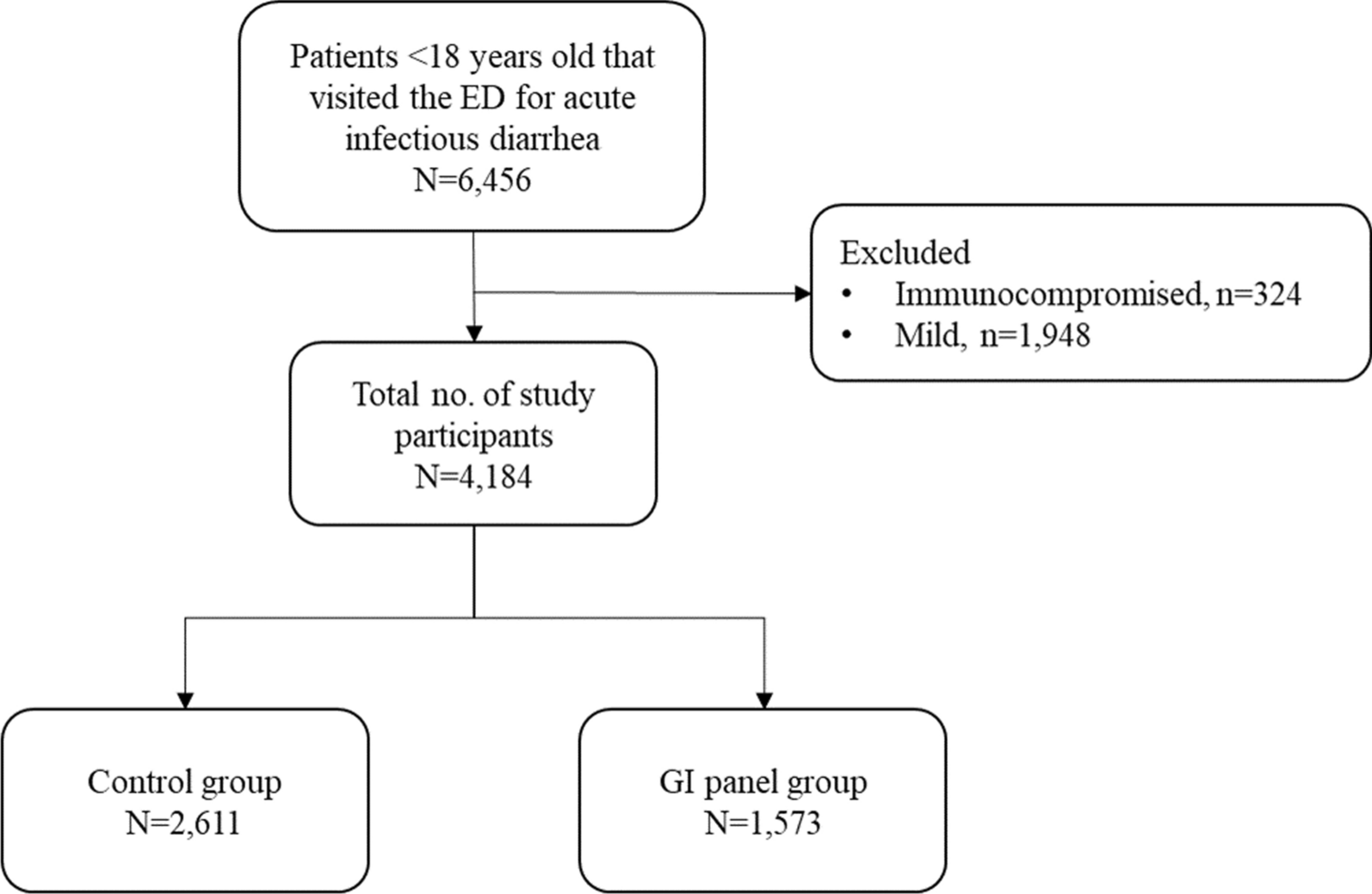
記住我


To investigate the characteristic microbial profiles in the GI tract, we conducted the 16S rRNA sequencing and compared the bacterial diversity in different GI locations first. The alpha diversity analysis revealed that richness and chao1 indices were gradually higher in fecal samples than in salivary and buccal samples (Fig. 1B, Additional file 5: Table S1), indicating a higher microbial community richness. In addition, the Simpson index was lower, and the Shannon index was higher in salivary and fecal samples than in buccal samples (Fig. 1B, Additional file 5: Table S1), which suggested higher evenness of microbial community composition in salivary and fecal samples. Furthermore, beta diversity analysis with constrained principal coordinates analysis (CPCoA) showed an adjacency between salivary and buccal samples while a separation between fecal and oral (salivary and buccal) samples in Bray–Curtis distance. Nonetheless, there were significant differences among the three groups (Adonis P < 0.05, Fig. 1C, Additional file 5: Table S2). These data suggested that salivary and buccal samples share similar microbial community composition to a certain degree, whereas fecal samples did not.
Further analysis showed that thirty-eight bacterial families were shared in three different niches and oral samples shared seven other families (Fig. 1D and Additional file 5: Table S3). In addition, there were twenty-nine special bacterial families in fecal samples (e.g., Acholeplasmataceae, Acidaminococcaceae, and Brucellaceae), four in buccal samples (Hicobacteraceae, Nocardiaceae, Nocardiodaceae, and Staphylococcaceae) and one unique bacterial family in salivary samples (Bacillaceae_1) (Fig. 1D and Additional file 5: Table S3). At the genus level, data showed that forty-two bacterial genera were shared among the three sites, and seventy-three in oral samples. As to unique genera, there were seventy-five bacteria specific in fecal samples (e.g., Akkermansia, Bilophila, and Citrobacter, etc.), seven in buccal samples (e.g., Aeromicrobium, Helicobacter, and Nocardia, etc.), while four in salivary samples (including Alloscardovia, Bacillus, Mobiluncus, and Mogibacterium) (Fig. 1E and Additional file 5: Table S3). These data indicated a higher bacterial richness in the feces, and some bacterial species were shared in oral and fecal samples.
We further analyzed the twenty most abundant bacterial families and genera and found that Streptococcaceae and Neisseriaceae mainly colonize the oral cavity at the family level, while Lachnospiraceae, Bacteroidaceae, Ruminococcaceae, and Enterobacteriaceae colonized with a high richness in feces (Fig. 1F and Additional file 5: Table S5). At the genus level, Streptococcus, Neisseria, Haemophilus, and Rothia occupied the oral cavity with high abundance; while Faecalibacterium, Bacteroides, Bifidobacterium, Escherichia/Shigella, and Lachnospiraceae_incertae_sedis were highly abundant in feces (Fig. 1G, H and Additional file 5: Table S5-6).
These results indicate that the bacteria in salivary and buccal samples are highly similar at the family and genus levels; they share many common and similar dominant bacteria but fewer unique bacteria. In contrast, there are many specific bacterial families and genera in feces but not in oral samples, indicating that it is usually difficult for most bacteria in the oral cavity to colonize in the gut. Thus, we speculate that this may be related to the different microenvironments in the oral cavity and gut.
The salivary bacterial microbes in UC patients complicated by oral ulcersTo uncover the microbial features in salivary samples of UC_OU patients, we further profiled the salivary bacterial community in all subjects. Compared with the CON group, the richness increased in the UC group, and the diversity and evenness elevated in the UC_OU group (Fig. 2A and Additional file 5: Table S1). Moreover, the beta diversity analysis revealed that the salivary microbial community composition was significantly different between the groups of patients and the CON (Adonis P = 0.046 for OU, Adonis P = 0.002 for UC, P = 0.004 for UC_OU vs. CON, respectively, Fig. 2B, S1B, and Additional file 5: Table S2). Furthermore, the analysis of the twenty most abundant bacterial families and genera showed that there were remarkable differences in abundance between the groups of patients and the CON at the family and genus level (Fig. 2C and Additional file 5: Table S5-6). Further investigation of UC_OU and OU manifested that some bacteria decreased (e.g., Blautia, Clostridium_XIII, and Faecalibacterium), and some increased (e.g., Abiotrophia) (Fig. 2D and Additional file 5: Table S7). Surprisingly, there were up to fifty different genera between the UC_OU and UC groups, including the increasing Klebsiella, Arthrobacter, Barnesiella, and the decreasing Blautia, Clostridium_XIII, and Faecalibacterium (Fig. 2E and Additional file 5: Table S7). We also compared the groups of patients with the CON respectively, and found that some genera (e.g., Arthrobacter, Barnesiella, Alistipes) increased and some (e.g., Mogibacterium) decreased consistently (Additional file 1: Figure S1C-E and Additional file 5: Table S7).
Fig. 2
The bacterial profile in salivary samples of UC patients with or without oral ulcers. A Alpha diversity indices of the microbiota, including the richness, Simpson’s, Shannon’s, and Chao1 indices. Horizontal bars within boxes represent medians. The tops and bottoms of the boxes represent the 75th and 25th percentiles, respectively. The upper and lower whiskers cover 1.5 × the interquartile range from the upper and lower edges of the box, respectively. P-values were obtained using the one-way ANOVA test (comparisons among four groups). B The constrained principal coordinate analysis based on the Bray–Curtis distance. The R software (v 4.0.1) with the vegan (v 2.5–7) package were used, and P-values were obtained using permutational multivariate analysis of variance (PERMANOVA). C Relative abundance of the top 20 bacterial families (the left panel) and genera (the right panel). Visualization was performed using Circos (http://circos.ca/). The right circle in the outer part shows the groups and relative proportions of bacterial species. The left outer circle and inner bands show the relative proportions (%) of bacterial genera in the different groups. The left inner circle represents the relative abundances of all bacteria. D and E Comparative analysis of bacterial genus abundance between two groups (D UC_OU vs. OU; E UC_OU vs. UC). The EdgeR package was used for comparative analysis. The difference between the two groups is shown as a Manhattan diagram. Point shape indicates the genus enriched, depleted, or not significant in the former group compared with the latter. Point size indicates the counts of a specific genus. CPM, count per million. F and G Comparative analysis of bacterial function between two groups (F UC_OU vs. OU; G UC_OU vs. UC). Phylogenetic Investigation of Communities annotated the pathway information by Reconstruction of Unobserved States (PICRUSt2) software by referring to the Kyoto Encyclopedia of Genes and Genomes (KEGG) database. The STAMP software was used for data visualization. CON, healthy controls; OU, patients with only oral ulcers; UC, UC patients without oral ulcers; UC_OU, UC patients with oral ulcers; ns, not significant; *P-value < 0.05; **P-value < 0.01
Furthermore, the PICRUSt2 analysis showed that in the group of UC_OU, the immune-related signaling pathways (including antigen processing and presentation, IL-17 signaling, and Th17 cell differentiation, etc.) were active compared with the OU group. The anti-inflammatory pathway (e.g., thiamine metabolism [30]) was restrained compared with the UC group (Fig. 2F, G and Additional file 5: Table S8), indicating a higher immune activation and inflammatory states in UC_OU patients.
In general, the increment of Arthrobacte and Barnesiella and the reduction of Blautia, Clostridium_XIII, and Faecalibacterium in UC_OU patients may be essential factors in mediating the occurrence and development of the disease by regulating immune responses and inflammatory pathways.
The buccal bacterial community in UC patients complicated by oral ulcersThere were no significant differences in alpha diversity among the buccal samples of the CON, OU, UC, and UC_OU. The beta diversity analysis showed that there were significant differences when the OU/UC, but not the UC_OU group, compared with the CON group (Adonis P = 0.008 for OU, P = 0.001 for UC and P = 0.102 for UC_OU vs. CON, respectively, Fig. 3B, S2A, and Additional file 5: Table S2).
Fig. 3
The bacterial profile in buccal samples of UC patients with or without oral ulcers. A Alpha diversity indices of the microbiota, including the richness, Simpson’s, Shannon’s, and Chao1 indices. Horizontal bars within boxes represent medians. The tops and bottoms of the boxes represent the 75th and 25th percentiles, respectively. The upper and lower whiskers cover 1.5 × the interquartile range from the upper and lower edges of the box, respectively. P-values were obtained using the one-way ANOVA test (comparisons among four groups). B The constrained principal coordinate analysis based on the Bray–Curtis distance. The R software (v 4.0.1) with the vegan (v 2.5–7) package were used, and P-values were obtained using permutational multivariate analysis of variance (PERMANOVA). C Relative abundance of the top 20 bacterial families (the left panel) and genera (the right panel). Visualization was performed using Circos (http://circos.ca/). The right circle in the outer part shows the groups and relative proportions of bacterial species. The left outer circle and inner bands show the relative proportions (%) of bacterial genera in the different groups. The left inner circle represents the relative abundances of all bacteria. D and E, Comparative analysis of bacterial genus abundance between two groups (D UC_OU vs. OU; E UC_OU vs. UC). The EdgeR package was used for comparative analysis. The difference between the two groups is shown as a Manhattan diagram. Point shape indicates the genus enriched, depleted, or not significant in the former group compared with the latter. Point size indicates the counts of a specific genus. CPM, count per million. F and G, Comparative analysis of bacterial function between two groups (F UC_OU vs. OU; G UC_OU vs. UC). Phylogenetic Investigation of Communities annotated the pathway information by Reconstruction of Unobserved States (PICRUSt2) software by referring to the Kyoto Encyclopedia of Genes and Genomes (KEGG) database. The STAMP software was used for data visualization. CON, healthy controls; OU, patients with only oral ulcers; UC, UC patients without oral ulcers; UC_OU, UC patients with oral ulcers; ns, not significant; *P-value < 0.05; **P-value < 0.01
We further investigated the buccal bacterial composition in different groups of subjects. At the family level, the top twenty bacteria in abundance levels differed not so markedly among all groups of subjects (Fig. 3C and Additional file 5: Table S5). While at the genus level, the abundance of some bacteria, such as Neisseria and Rothia, declined slightly in the UC_OU group. As for Neisseria, a previous study found a falling abundance of which at the inflamed site of UC patients compared with the corresponding area of non-IBD controls [31]; in addition, the abundance of Actinobacillus and Fusobacterium varied primarily among these groups (Fig. 3C and Additional file 5: Table S6).
Using further differential analysis, we found that, compared with the CON group, the variation of bacteria was considerably conspicuous among OU, UC, and UC_OU group at the genus level; for instance, the abundance of Barnesiella, Alistipes, and Rhodopseudomonas ascended, while Actinobacillus descended (Additional file 2: Figure S2B-D and Additional file 5: Table S9). Then, we compared oral bacterial richness between UC_OU and OU groups and found very few differential bacteria, which manifested the increase of Abiotrophia, Cardiobacterium, and Klebsiella (Fig. 3D and Additional file 5: Table S9). Interestingly, Abiotrophia defectiva was related to pro-inflammatory response in the oral cavity [32]; moreover, Klebsiella pneumoniae was reported to aggravate chronic intestinal inflammation by destructing the intestinal epithelial barrier [33]. In contrast with UC patients, there was a marked increase in the abundance of Abiotrophia, Cardiobacterium, and Klebsiella in the UC_OU patients; on the contrary, the abundance of Actinobacillus decreased notably (Fig. 3E and Additional file 5: Table S9). Hence, we inferred that these bacteria probably contributed to oral ulcers in the UC_OU patients.
A previous study found that decreased sphingolipids correlated with gut inflammation in IBD subjects [34]. Our PICRUSt2 analysis also observed suppressed sphingolipid signaling pathway in the UC_OU patients compared with OU patients. In addition, there was an enrichment of protein processing in the endoplasmic reticulum in the UC_OU patients (vs. OU, Fig. 3F and Additional file 5: Table S10). It was found multiple immune cells could activate that unfolded protein response at distinct levels [35]. Furthermore, the differences in signal pathways between UC and UC_OU patients showed that the expression level of some signal pathways declined, such as dorso-ventral axis formation and glycosphingolipid biosynthesis (Fig. 3G and Additional file 5: Table S10); among which glycosphingolipid was reported to correlate with regulating immune signaling with facilitating bacterial entering host cells [36]. The comparison results between OU and CON patients unveiled that some pathways diminished in the OU patients, such as pyrimidine metabolism, RNA polymerase, protein phosphatases and associated proteins, and secondary bile acid biosynthesis (Additional file 2: Figure S2E and Additional file 5: Table S10), which were all associated with immunity [37,38,39]. Moreover, there was an enriched apoptosis pathway in the UC group compared with the CON one (Additional file 2: Figure S2F and Additional file 5: Table S10), which was paralleled with a previous review that showed the pathogenic function of caspase-mediated intestinal epithelial cell apoptosis in the IBD [40].
In brief, buccal bacterial features in UC_OU subjects differed from UC alone or OU. and the abundance of some bacteria (such as Abiotrophia, and Klebsiella) in the buccal mucosa may help to distinguish between UC_OU subjects and UC alone or OU. Furthermore, the alterations of some signaling pathways related to immune cells or processes implied that immune factors might participate in the occurrence and development of oral ulcers in UC_OU patients.
The fecal bacterial microbiota in UC_OU patientsWe further analyzed the fecal bacterial composition of all subjects. We found that, compared with the CON, the alpha diversity of fecal bacteria in UC_OU patients significantly decreased, while the OU or UC patients did not (Fig. 4A and Additional file 5: Table S1); beta diversity analysis showed a marked difference in the fecal microbiota composition between patients and CON (Adonis P = 0.011 for OU, 0.002 for UC, and 0.003 for UC_OU vs. CON, respectively; Fig. 4B, S3A and Additional file 5: Table S2). In the top 20 bacterial families, Enterobacteriaceae increased in the groups of patients, while the abundances of Prevotellaceae in the UC and Veillonellaceae in the OU were decreased than in the CON group (Fig. 4C and Additional file 5: Table S5). At the genus level, compared with the CON, Prevotella and Roseburia declined in the UC group, while Escherichia/Shigella raised in the UC and UC_OU group (Fig. 4C and Additional file 5: Table S6).
Fig. 4
The bacterial profile in fecal samples of UC patients with or without oral ulcers. A Alpha diversity indices of the microbiota, including the richness, Simpson’s, Shannon’s, and Chao1 indices. Horizontal bars within boxes represent medians. The tops and bottoms of the boxes represent the 75th and 25th percentiles, respectively. The upper and lower whiskers cover 1.5 × the interquartile range from the upper and lower edges of the box, respectively. P-values were obtained using the one-way ANOVA test (comparisons among four groups). B The constrained principal coordinate analysis based on the Bray–Curtis distance. The R software (v 4.0.1) with the vegan (v 2.5–7) package were used, and P-values were obtained using permutational multivariate analysis of variance (PERMANOVA). C, Relative abundance of the top 20 bacterial families (the left panel) and genera (the right panel). Visualization was performed using Circos (http://circos.ca/). The right circle in the outer part shows the groups and relative proportions of bacterial species. The left outer circle and inner bands show the relative proportions (%) of bacterial genera in the different groups. The left inner circle represents the relative abundances of all bacteria. D and E, Comparative analysis of bacterial genus abundance between two groups (D UC_OU vs. OU; E UC_OU vs. UC). The EdgeR package was used for comparative analysis. The difference between the two groups is shown as a Manhattan diagram. Point shape indicates the genus enriched, depleted, or not significant in the former group compared with the latter. Point size indicates the counts of a specific genus. CPM, count per million. F and G, Comparative analysis of bacterial function between two groups [F UC_OU vs. OU; G UC_OU vs. UC]. Phylogenetic Investigation of Communities annotated the pathway information by Reconstruction of Unobserved States (PICRUSt2) software by referring to the Kyoto Encyclopedia of Genes and Genomes (KEGG) database. The STAMP software was used for data visualization. CON, healthy controls; OU, patients with only oral ulcers; UC, UC patients without oral ulcers; UC_OU, UC patients with oral ulcers; ns, not significant; *P-value < 0.05; **P-value < 0.01
We further performed a comparative analysis at the genus level. The results showed that, compared with the CON group, some bacteria (e.g., Arthrobacter, Knoellia, Bacillus, Peptostreptococcus, etc.) increased in the groups of patients (OU/UC/UC_OU) and some decreased (e.g., Lautropia) (Additional file 3: Figure S3B-D and Additional file 5: Table S11). Interestingly, Bacillus and Peptostreptococcus have been reported to correlate with severe infections [41] and IBD progression [42]. In addition, no differential genera could be found between UC_OU and OU groups (Fig. 4D); however, Alloprevotella, Granulicatella, Lactobacillus, and Holdemanella ascended strikingly in the UC_OU group compared with the UC group (Fig. 4E and Additional file 5: Table S11).
We then had the PICRUSt2 analysis. The results showed that some pathways, such as aminobenzoate degradation, xylene degradation, and bile secretion, lessened in the UC_OU patients compared with OU patients (Fig. 4F and Additional file 5: Table S12), among which xylene can exacerbate allergic inflammation [43]. Furthermore, the comparison between UC_OU patients and UC patients showed that metabolism-related pathways (including aminobenzoate degradation, platinum drug resistance, styrene degradation, etc.) were repressed (Fig. 4G and Additional file 5: Table S12). In addition, nucleotide metabolism and naphthalene degradation pathways in OU patients, and amino acid metabolism and fatty acid degradation in UC patients were activated compared with the CON (Additional file 3: Figure S3E-G and Additional file 5: Table S12).
To conclude, the fecal flora characteristics in the UC_OU group were distinguished from UC alone, and Alloprevotella, Granulicatella, Lactobacillus, and Holdemanella may contribute to differentiating between UC_OU and UC alone. In addition, signal pathways related to metabolization and immunity can involve the pathogenic process in the UC_OU.
The correlation of GI spatial microbiome with clinical, immunological parametersTo further assess whether the disease activities and clinical parameters correlated with the spatial microbiome alteration, we collected twenty clinical parameters responding to inflammatory and immunological statuses, including C-reaction protein (CRP), ESR, the percentage of monocytes (MO_P), etc. We performed a redundancy analysis (RDA) between bacterial beta diversity and clinical parameters and Spearman’s correlation between alpha diversity and clinical parameters (Fig. 5A–F and Additional file 5: Table S13). The inflammatory indices were statistically correlated with the salivary microbiome in alpha diversity and beta diversity (Fig. 5A). Of note, the salivary microbial alpha diversity correlated with the lymphocyte’s (rho = 0.281, FDR = 0.022 for chao1, and rho = 0.282, FDR = 0.022 for richness, respectively), and neutrophil’s percentage (rho = − 0.372, FDR = 0.002 for chao1, and rho = − 0.372, FDR = 0.002 for richness, respectively; Fig. 5B and Additional file 5: Table S13). The buccal bacterial Shannon index correlated with subjects’ inflammatory markers, such as C4 (rho = 0.567, FDR = 0.002) and CRP (rho = 0.399, FDR = 0.039) (Fig. 5C and Additional file 5: Table S13). Like the salivary microbiome, buccal bacterial beta diversity showed negative results correlating with clinical parameters (Figs. 5C, D and Additional file 5: Table S13). The fecal microbiota showed a significant correlation between beta diversity and immunological indices, such as the monocytes’ (pseudo F = 2.218, P = 0.039) and basophils’ percentage (pseudo F = 1.984, P = 0.035), rather than inflammatory markers (Fig. 5E, F, and Additional file 5: Table S13). Based on these results, we inferred that the fecal bacteria play a more important role in shaping the host immune system.
Fig. 5
The correlation between clinical parameters (inflammatory and immunological) and bacterial diversity (alpha and beta). A and B The correlation between inflammatory (A) or immunological (B) indices and salivary bacterial diversity. The length of the black bar shows the R square value of the indices-beta diversity correlation. The results were acquired by the redundancy analysis (RDA) using the R software (v 4.0.1) with the vegan (v 2.5–7) package. The color box shows the rho value of indices-alpha diversity correlation. The results were analyzed from Spearman’s correlation using the R software (v 4.0.1) with the vegan (v 2.5–7) package. The star symbols behind the left words show the P-values acquired from the RDA. The box’s star symbols show the adjusted P-values (false discovery rate, FDR) received from the Spearman’s correlation. (same methodology as in C–F). C and D The correlation between inflammatory (C) or immunological (D) indices and buccal bacterial diversity. E and F The correlation between inflammatory (E) or immunological (F) indices and buccal bacterial diversity
Treatment responses in UC patientsWe then analyzed the treatment response for UC patients with a six-month following-up. Thirty-two participants were followed-up, including twenty-two UC and ten UC_OU patients, for which we documented the Mayo clinic score to differentiate patients with or without treatment response. There were no significant difference of some clinical parameters (Mayo score, ESR, CRP, IgA, IgG, IgM, C3, and C4) before and after treatments in UC and UC_DU groups (Additional file 4: Figure S4). Interestingly, only one out of ten UC_OU patients responded to 5-ASA routine treatment, significantly lower than that in UC patients (Chi-squared test, X2 = 19.09, P < 0.001, Fig. 6A and Additional file 5: Table S14). We further analyzed the GI spatial bacterial profiles in UC patients with or without treatment responses. Only one subject had responded to the treatment in UC_OU patients, so this group of patients was excluded from this part of the investigation. Notably, there were eighteen genera with a remarkable difference in these two subgroups of patients in the salivary bacteria, including Prevotella, Alloprevotella, Fusobacteria, Oribacterium, Campylobacter, and Rothia, etc. (Fig. 6B and Additional file 5: Table S14). Compared with salivary samples, the buccal biopsies showed fewer bacterial contents, with a significant difference between UC patients with and without treatment responses (Fig. 6C and Additional file 5: Table S14). It could be found that there were three same genera also enriched in the non-responding UC patients’ oral mucosae, which were Fusobacterium, Oribacterium, and Campylobacter, respectively. Additionally, only one content, i.e., Blautia, was represented in fecal microbiota in non-responding patients (Fig. 6D and Additional file 5: Table S14). Based on these data, we deduced that the richness of Fusobacterium, Oribacterium, and Campylobacter might be involved in non-response; the salivary microbiome also had a potential for indicating treatment response in UC patients.
Fig. 6
Microbial differentiation between UC patients with and without treatment response. A The treatment response rate in UC patients with or without oral ulcers. The Chi-squared test was performed to test the response difference between the two groups. UC, UC patients without oral ulcers; UC_OU, UC patients with oral ulcers. B–D The comparative analysis of bacterial genus abundance of the samples from salivary (B), buccal (C), and fecal (D) niches. The STAMP software was used for data visualization. E The graphic summary
留言 (0)