
Remember me


All solvents used were of analytical grade [American Chemical Society (ACS), International Organization for Standardization (ISO), Reag. Ph. Eur.], consisting of methanol (Merck, Hamburg, Germany), n-hexane (Th. Geyer, Renningen, Germany), acetic acid (Carl Roth, Karlsruhe, Germany) and ethyl acetate (VWR, Darmstadt, Germany). Standard ibuprofen was obtained from Caesar & Loretz GmbH (Hilden, Germany). Analysed ibuprofen tablets (Sanofi Aventis, Frankfurt, Germany) were commercially available, claiming 400 mg API. Used UV lamp (Gebr. Rettberg GmbH, Göttingen, Germany) was equipped with a short and long wavelength. Filter paper (MN 615) and 20 × 20 cm silica gel plates (Alugram® Xtra Sil G/UV254) with a 0.2 mm layer were supplied by Macherey–Nagel (Düren, Germany).
2.2 Preparation of ibuprofen solutionsReference solution of ibuprofen was prepared by weighing out 100 mg of ibuprofen and dissolving it in 10 mL of methanol. Appropriate volumes were then transferred to a 20 mL snap-cap vial and diluted to 5, 4 and 3 mg/mL.
A 400 mg ibuprofen tablet was wrapped in aluminium foil and pulverised to prevent a loss of mass during transfer from mortar to flask. The tablet was then dissolved in 40 mL methanol for 15 min and filtered using medium-porosity cellulose filter paper (MN 615). An appropriate volume was transferred to a 20 mL snap-cap vial and diluted to 5 mg/mL for the sample solution.
2.3 Chromatographic conditionsSilica gel plates were cut to 10 × 20 cm to allow the development of several plates simultaneously. A spotting line was marked leaving 1 cm space from the bottom and edges, additionally pre-marking the solvent front line 7 cm from the origin line. Local calibration spots for the TLC plates were then applied in a row using 2 μL capillary tubes. Three spots of the reference solution (5, 4 and 3 mg/mL) were applied, followed by two spots (5 mg/mL) of the sample solution. The mobile phase was prepared following recommendations by the Ph. Eur. and consisted of acetic acid, ethyl acetate and n-hexane (5:24:71, V/V), saturating the chamber for 20 min before use. After development, the plate was dried at room temperature and visualised under shortwave UV light (254 nm).
2.4 Image settingsSeveral images of the plates were taken with a smartphone (Samsung Galaxy S22, Android 13; Suwon, South Korea) using different image settings and file formats. The camera array of the smartphone was equipped with a triple camera system with different image sensor sizes (50 MP, 12 MP and 10 MP). Images were taken by hand, resting the smartphone above the UV lamp and aligning the viewfinder grid to the spotting lines. First, two images were taken using the main camera app and the 12 MP sensor without any further adjustments (images 1 and 2). Then, images were taken using the smartphone’s 50 MP sensor. Lastly (image 3), an image using the Pro mode within the camera app (version 13.1.01.9) was taken, with the ISO set to the lowest possible setting (ISO = 50), thus limiting the light sensitivity. In addition, shutter speed and exposition were locked to prevent changes in brightness and shutter speed during image acquisition. All images of ibuprofen plates were taken in a dark room, visualising under UV light at a wavelength of 254 nm. The images were not subjected to any standardisation, resulting in images with an approximate, even illumination using an automatic focus. For analysis image sets sharing the same image settings (ISO = 50; exposure bias = 0.0 eV; f-stop = f/1.8; exposure time = 1/30 s; focal length = 5 mm; 35 mm focal length = 23 mm) were then selected.
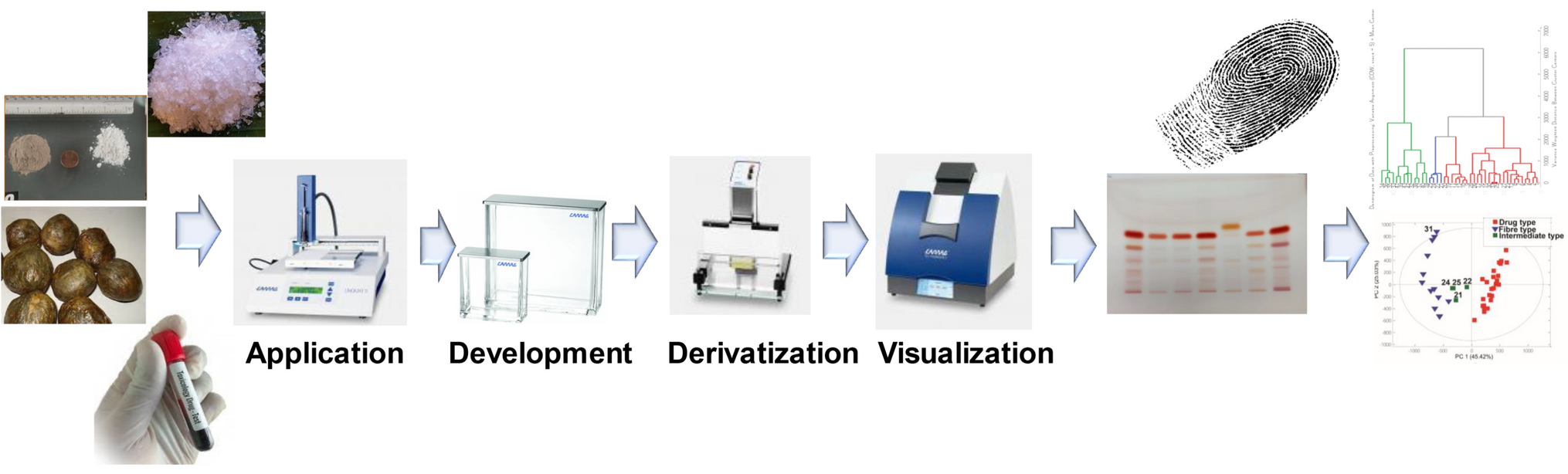
2.5 Image analysisFollowing the scheme shown in Fig. 1, the selected images were imported to Fiji (File > Open) and cropped using the rectangle tool according to the spotting pattern. Splitting the colour channels (Image > Color > Split Channels) results in three image variants of the corresponding channel (red, green, blue) in 8-bit format. The green colour channel was selected for further analysis and then cropped to fit the band area. Then, a median blur of 5 px (Process > Filters > Median) and smoothing (Process > Smooth) were applied. Additionally, a bandpass filter (Process > FFT > Bandpass filter) was used, filtering larger structures down to 100 px and smaller structures up to 0 px. For better visibility, the look-up table (LUT) was inverted (Image > Color > Invert LUTs). Visual bands were then selected using the rectangular selection tool and a plot profile was drawn using the gel analyser (Analyze > Gels > Plot Lanes). The corresponding peak areas were fitted using the line selection tool and then measured using the wand tool. For more information of using on using the gel analyser, see the ImageJ user guide [15]. Finally, the percentage was displayed using Analyze > Gels > Label Peaks. Using the 5 mg/mL reference solution as the standard, the relative density of each band was fitted and plotted against the known concentration, using open source software Jamovi and the ggplot2 package in R Studio [16,17,18].
Fig. 1
Method proposal for ibuprofen quantification using digital image processing software (Fiji)
2.6 Data analysis2.6.1 LinearityGood linearity could be found within the subjected images of plate 2 (R2 = 0.996, 0.984 and 0.994) as shown in Fig. 2. Applying the same method to the images in TIFF and RAW image format resulted in a non-ideal linearity (R2 < 0.95) when analysed with a concentration range from 3 to 5 mg/mL, thus the usage of these image files was deemed as unsuitable for the same method.
Fig. 2
Linearity plots of plate 2: three images were subjected to analysis across three measurements (n = 3)
2.6.2 AccuracyAccuracy was measured as the mean relative density of each image across the various measurements used to calculate the mean recovery of known concentrations. This resulted in accuracies of 100 ± 1.43%, 99.9 ± 3.24% and 100 ± 1.94% for the reference solution as presented in Table 1. By comparison, the analysed sample solutions resulted in accuracies of 98.1 ± 2.91%, 98.0 ± 1.86% and 98.8 ± 1.74%.
Table 1 Linearity parameters of plate 2 over three measurements of each image (n = 3)2.6.3 LimitationLimit of detection (LOD) and limit of quantification (LOQ) were calculated following recommendations by the International Council for Harmonisation (ICH), where \(LOD= 3.3\times (\sigma /slope)\) and \(LOQ=10\times (\sigma /slope)\) [19]. The use of a small sample size and linear range for the calibration curve can limit the significance of LOD and LOQ. While the detection and quantification limits matched the applied samples of image 1 and 3, the LOQ of image 2 was higher than the spotted concentrations.
2.6.4 RepeatabilitySeveral images of the plates were taken and selected for analysis. User error can occur whilst cropping the peak areas. Thus, selected images were subjected to repeated measurements to ensure repeatability throughout one image. The procedure of selecting band area, application of filters and manually cropping peak areas was repeated three times for these images. Relative standard deviation (RSD) was measured for repeatability, with an RSD between images of 1.43%, 3.24% and 1.94%.
Comments (0)