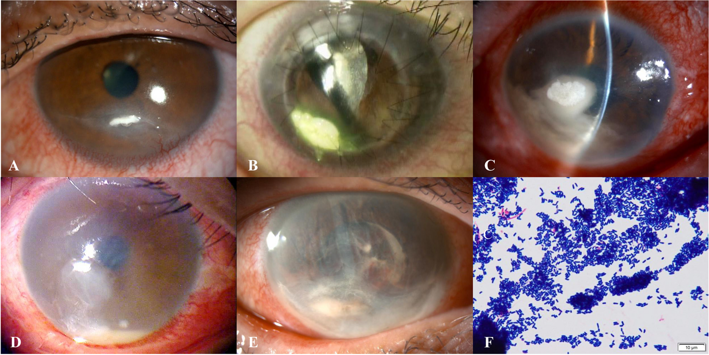
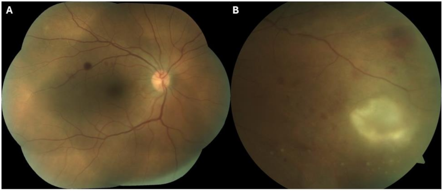
S. maltophilia is an opportunistic infection that is a rare cause of endophthalmitis and cases of S. maltophilia-related endophthalmitis have often been reported following cataract surgery, trauma or intravitreal injections [11,12,13]. We present a case of S. maltophilia endophthalmitis after keratoplasty, which is a rare entity in the literature.
There are some cases in the literature who underwent intravitreal injection for various reasons and subsequently developed S. maltophilia endophthalmitis. Boeke et al. reported S. maltophilia endophthalmitis developing 1 month later in a 70-year-old female patient who underwent intravitreal aflibercept for diabetic macular edema. Due to the suspicion of endophthalmitis a tap procedure was performed and then intravitreal vancomycin, ceftazidime and dexamethasone were injected. Topical prednisolone acetate 1% every hour, topical moxifloxacin 4 times daily and cyclopentolate 3 times daily were started. Then S. maltophilia growth was observed in the patient's aqueous humor, and his clinical improvement was observed with the continuation of the topical treatment and the need for vitrectomy did not arise [13]. Our patient did not have severe pain as stated in this case. Although hypopyon was also observed in our patient, a fibrinous reaction occurred in the anterior chamber first, and then it was gradually replaced by hypopyon. Since condensation in the vitreous and phacomorphic glaucoma developed in the follow-up of our patient, combined phaco-vitrectomy surgery was performed.
Karakurt et al. reported 6 cases who developed S. maltophilia endophthalmitis between 1 and 19 days after cataract surgery [14]. In addition, Chang et al. reported 8 cases of S. maltophilia endophthalmitis that occurred following cataract surgery [11]. Vitrectomy was required in 3 of these 8 patients. Similarly, Ji et al. published 14 cases of S. maltophilia endophthalmitis that occurred between 1 and 56 days postoperatively after cataract surgery [15]. As can be seen, S. maltophilia endophthalmitis cases in the literature were generally seen after cataract surgery. In our patient, we encountered an endophthalmitis that gradually appeared after keratoplasty.
In addition to all these, there are also cases of S. maltophilia endophthalmitis reported after ocular traumas. Lai et al. reported a case of S. maltophilia endophthalmitis after penetrating injury by a wooden splinter [12]. Patton et al. brought to the literature a case of S. maltophilia endophthalmitis in a patient with intraocular metallic foreign body after trauma [16]. Also, Kherani et al. published a S. maltophilia endophthalmitis case following penetrating corneal injury [17]. As it is known, due to impaired sterility in intraocular penetrating injuries, we frequently encounter endophthalmitis as in these cases. However, we usually see rapidly progressive endophthalmitis in these patients. In our patient, the fibrin reaction that occurred after keratoplasty was followed for a while, then it was replaced by a hypopyon, but the size of this hypopyon remained more stable than in classical endophthalmitis.
In a case published by Díez-Álvarez et al., a case of S. maltophilia-associated keratitis-endophthalmitis has been reported. In this case, an 84-year-old female patient developed a persistent epithelial defect and a dense stromal infiltrate after descemet stripping automated endothelial keratoplasty (DSAEK) surgery and S. maltophilia growth has been reported in the corneal scraping sample taken. Complete recovery was achieved in 3 weeks after oral and topical trimethoprim-sulfamethoxazole (TMP/SMX) treatment. This case emphasized the importance of keeping in mind that S. maltophilia may also be a factor in keratitis after corneal transplantation [18]. In our case, there was a patient who developed S. maltophilia-related endophthalmitis after penetrating keratoplasty, presented with progressively increasing inflammation, fibrin reaction and vitreous condensation, and was more resistant to heal.
There is one case report describing S. maltophilia keratitis that developed after penetrating keratoplasty surgery [19]. In this case, a 70-year-old patient complained of decreased vision 5.5 months after the surgery and S. maltophilia was isolated in corneal scraping samples. In this patient, as in our patient, the treatment plan was shaped according to the antibiotic susceptibility test and it was observed that the keratitis was completely resolved in 2.5 months with topical 0.3% ciprofloxacin hydrochloride treatment. In our patient, unlike this patient, S. maltophilia-related endophthalmitis was observed after keratoplasty, and the time of occurrence in our case was seen earlier after keratoplasty.


Comments (0)