
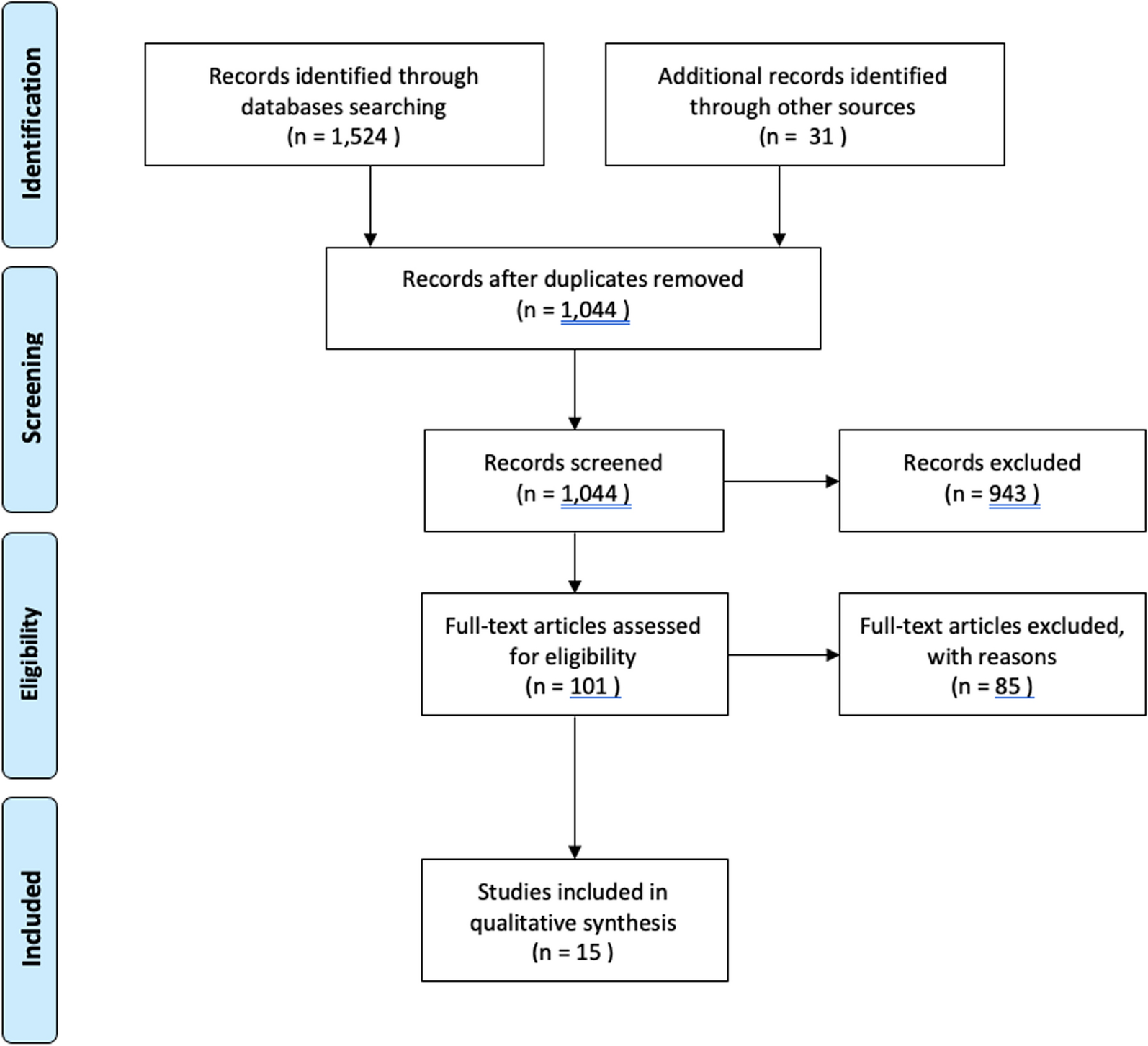
Remember me


After approval from the institutional research ethical committee, patients were retrospectively identified for this study from the electronic patient records in a regional trauma center which is also a certified center of shoulder surgery.
Study populationAll procedures between January 2017 and October 2020 adhering to the following inclusion criteria were identified:
A four-fragment PHF treated with a CFR-PEEKPowerTM Humeral Fracture Plate (PEEKPowerTM Humeral Fracture Plate (HFP), Arthrex®, Naples, United States of America)
Performed by a single surgeon (L.L.)
Isolated fracture of the proximal humerus
Satisfactory reduction and refixation of the fracture in postoperative radiographs
Surgery was performed within 14 days after trauma
Minimum follow-up of 1 year
A signed consent form.
Patients who could not attend the follow-up examination for medical reasons, who did not want a follow-up examination for personal reasons and who could not be contacted were excluded. A declaration of informed consent was signed at the follow-up examination. The data were only collected after a signed declaration of consent.
Surgical treatmentThe specific treatment decision was based on fracture morphology, bone quality and patient-specific criteria (e.g. age, physical activity, comorbidities). The risk of osteosynthesis failure of the proximal humerus was estimated following the criteria defined by Hertel [9]. If the surgeon considered the risk of complications to be high, a primary endoprosthetic joint replacement was performed instead of an osteosynthesis. In patients with preoperative symptomatic osteoarthritis, implantation of a reverse total shoulder arthroplasty (RSA) was preferred. None of the cases were intraoperatively converted to endoprosthetic replacement. The surgery was performed in the beach-chair position under general and/or regional anaesthesia using an interscalene plexus block. A deltopectoral approach was used in all cases. A three- or five-hole CFR-PEEK plate (PEEKPowerTM Humeral Fracture Plate (HFP), Arthrex®) was used. A titanium calcar screw (soft bone locking screw, 4-mm Arthrex®) was placed as close as possible to the calcar. The CFR-PEEK plate allows an angular deviation of the locking screws of up to 12° in all directions. The lengths of the humeral head screws were selected so that their tips extended to the subchondral surface of the humeral head without penetration of the articular surface. Depending on the fracture morphology, additional screws for the lesser tuberosity and a suture cerclage of the tuberosities (FiberWire®, Arthrex®) were applied, and a tenotomy or tenodesis of the long head of the biceps tendon (LBT) was performed. Depending on the fracture, the follow-up treatment included early functional therapy or a restrictive protocol of immobilization in a shoulder abduction splint for 3 weeks with subsequent passive mobilization of the shoulder. Active rehabilitation of the operated shoulder was started after 6 weeks.
Evaluation of the functional results and revision surgeryGeneral information (gender, dominant hand, diabetes, current smoking, height, weight) was gathered. Active and passive range of motion of the shoulder (abduction, flexion, external rotation, internal rotation) were assessed by the senior author (M.K.) at the follow-up examination after at least 12 months postoperatively and the Constant–Murley Score (CS) was completed. Isometric force measurement to determine the CS was performed with the patient seated with the shoulder in 90° abduction, 0° anteversion, and the elbow in extension [29]. The strap of the electrical force measurement device was applied to the distal forearm (IsoForceControl® EVO2, Herkules Kunststoff AG, Oberburg, Switzerland). Patient-reported outcome measures (PROMs) were collected, included a Visual Analog Scale for pain (VAS), the Subjective Shoulder Value (SSV) and the Quick Disabilities of the Arm, Shoulder and Hand Score (QDASH). Complications such as adhesive capsulitis, implant loosening or breakage, refracture, secondary fracture dislocation, avascular necrosis of the humeral head, secondary osteoarthritis, mal-/non-union, hematoma and iatrogenic nerve lesions as well as revision surgery (indication, performed surgery) were recorded.
Radiological analysisPreoperative radiographs (true anterior posterior (AP) view, lateral (Y) view) and computed tomography (CT) scans were analysed. Both radiographs were also evaluated 2 days after surgery and at the final follow-up. The PHFs were classified according to Codman’s four-fragment theory [4] by analysing CT scans (M.K., V.R.). Furthermore, the presence of a head split component and preexisting glenohumeral osteoarthritis (classified according to Samilson-Prieto [22]) was analysed.
To evaluate the positioning of the calcar screw, the distance between the calcar and calcar screw (Fig. 1) was measured in postoperatively performed AP radiographs. Fracture reduction was assessed on AP radiographs by measuring the neck shaft angulation (NSA) (Fig. 2a), neck shaft distance (Fig. 2b) and reduction of the greater tuberosity. A satisfactory NSA was defined as being between 110° and 150° [26]. The neck shaft distance (NSD) was measured to quantify the reduction at the medial hinge of the proximal humerus. A satisfactory reduction was defined at a distance of less than 5 mm.
Fig. 1
Radiological measurement of the distance from the calcar screw to the calcar in an anterior posterior radiograph: a calcar screw distance < 12 mm (group I), b calcar screw distance ≥ 12 mm (group II)
Fig. 2
Radiological measurements in an anterior posterior radiograph: a neck shaft angulation, b neck shaft distance
At the final follow-up examination, the following radiographic parameters were assessed: screw or plate breakage or dislocation, non-union, and osteonecrosis of the humeral head. In addition, the integrity and position of the tuberosities (resorption, dislocation ≥ 5 mm) were assessed. Secondary varus dislocation was defined as an angular deviation of more than 110° in AP radiographs. The radiological images were assessed by two orthopaedic surgeons (M.K., V.R.). In the case of disagreement, the final assessment was determined through discussion and consensus.
Statistical analysisTwo groups were created based on the calcar screw placement: group I: < 12 mm, group II: ≥ 12 mm [18]. Statistical analyses were performed using SPSS® software (version 28.0; IBM®, Armonk, United States of America). The nominal variables were summarized as percentages. The arithmetic mean and its standard deviation were used for descriptive statistics in the case of a normal distribution. The Shapiro–Wilk test was used to test the normality of the variables. The Wilcoxon–Mann–Whitney test (U test) was used for normally distributed quantitative variables. Pearson’s chi-squared test was calculated to test the association of two ordinal variables. The level of significance was set at p < 0.05.
Comments (0)