
記住我
For over half a century, interest in biomarkers for autoimmune inflammatory myopathies (AIM) has continuously expanded and there is little sign that this trend is declining. Arguably, AIM was rather ‘late to the biomarker party’ when compared to its closest relatives, systemic lupus erythematosus (SLE), systemic sclerosis (SSc), mixed connective tissue disease (MCTD), Sjögren syndrome (SjS), and rheumatoid arthritis (RA). AIMs comprise clinical subsets that include antisynthetase syndrome (ASyS), dermatomyositis (DM), immune mediated necrotizing myopathy (IMNM), sporadic inclusion body myositis (sIBM), drug-mediated myositis and overlap syndromes (OS). The accurate diagnosis of AIM can be compromised because of ‘mimics’ or doppelgangers that include metabolic myopathies, genetic myopathies, neurological diseases (e.g., amyotrophic lateral sclerosis, chronic inflammatory demyelinating polyneuropathy), acquired diseases (e.g., acute and chronic infections), vitamin D deficiency, endocrinopathies (e.g., hyper- and hypo-thyroid diseases, acromegaly, Cushing syndrome, Addison disease), and exposure to drugs, toxins and other environmental agents [1].
It is now widely appreciated that circulating autoantibodies and other proteomic biomarkers in AIM can help make an accurate diagnosis, but also stratify patients into clinically relevant and actionable subsets [2]. In this manner, they can serve as valuable predictive and prognostic aids and provide important criteria for enrollment of AIM subsets in clinical trials. At a time when it was hoped that biomarkers might replace moderately invasive diagnostic approaches such as muscle biopsy, recent findings indicate that the muscle biopsy remains a key to understanding the roles of biomarkers in the pathogenesis and classification of AIM [3,4▪,5–9].
In general, the autoantibody biomarkers in AIM can be regarded as AIM-specific (AIM-S) or AIM-related (AIM-R). Although several novel AIM-S and AIM-R are discussed in this overview, a significant serological gap persists (no detectable autoantibodies or seronegative AIM) which poses a diagnostic challenge because a delayed or equivocal diagnosis may forestall evidence-based therapy and be attended by poorer clinical outcomes and increased healthcare expenditures. In addition, as we understand more about the various presentations and pathological features of AIM, the spectrum of AIM continues to expand. For example, sIBM was once relegated to a disease category of its own. However, with the discovery of an autoantibody biomarker directed to NT5c1A (C1A, Mup44) by Greenwood and his colleagues [10,11], involvement of autoinvasive T cells [12], a type 2 interferon mediated pathogenesis [13] and an appreciation that sIBM overlaps with SjS [14–16], it is now more or less comfortably classified as an AIM along with ASyS, DM, and IMNM [17,18,19▪▪]. However, the notion that sIBM is not a classical autoimmune disease is the appreciation that conventional immunosuppressive agents have yet to show remarkable benefit (reviewed in [12]). More recently but not discussed in detail here, there has been interest in myopathies that are seen as an adverse immune response to checkpoint inhibitor treatments [20] as well as myopathies seen in the context of severe acute respiratory syndrome coronavirus 2 infection, coronavirus disease-2019 (COVID-19), and the cognate vaccines [21,22].
This overview will focus on some overlooked aspects of AIM and newer published evidence encompassing the past ∼two years. Accordingly, topics of discussion will include laboratory methods used to detect AIM autoantibodies and related biomarkers, newer biomarkers and their proposed clinical value, an exploration of some of the newer OS associated with AIM, along with unmet needs and challenges in AIM research and clinical applications.
 Box 1:
 no caption available
LABORATORY DETECTION OF AUTOANTIBODIES IN AUTOIMMUNE INFLAMMATORY MYOPATHIESThe discovery of autoantibodies in AIM dates to the early 1980s at a time that coincides with the ‘golden age’ of cell biology when techniques such as western immunoblotting, immunoprecipitation (IP), and enzyme linked immunosorbent assays (ELISA) were replacing immunodiffusion, counter-immunoelectrophoresis, and hemagglutination [23]. Since then, some of these assays are being replaced by newer and more robust technologies such as dot immunoblot assays (DIA) and line immunoassays (LIA), addressable laser bead immunoassays (ALBIA), particle-based multianalyte technology (PMAT), mass spectrometry and immunoprecipitation mass spectrometry (IP-MS) [23–26]. However, some of these novel assays that had gained favor as high throughput, multiplexed and relatively economical technologies, are faced by technical and performance challenges [25,27–33]. A narrative review the methods (IP, ELISA, LIA, ALBIA, DIA) used to detect AIM-A and AIM-S autoantibodies in AIM has recently been published [34▪▪].
IMMUNOPRECIPITATION AND IMMUNOPRECIPITATION-MASS SPECTROSCOPYIn the past several decades, IP of radiolabeled cell extracts has been regarded the ‘gold standard’ immunoassay for AIM-S (Fig. 1), but with the broadening spectrum of autoantibodies being reported, challenges have emerged [29,35]. For example, not all AIM-S or AIM-R autoantibodies are easily detected by IP and there is no standardized inter-laboratory protocol or commutability studies for IP. In addition, the IP assays are unique to several (research) laboratories and have not achieved regulatory approval as in vitro diagnostic devices and hence are labelled laboratory developed tests and/or designated as research use only (RUO).
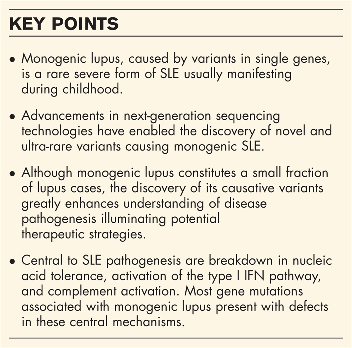
 FIGURE 1:
 35S-methionine/cysteine labeled K562 cell extract was immunoprecipitated by reference sera and fractionated on 8% (panel a) or 13% (Panel b) SDS-polyacrylamide electrophoresis (SDS-PAGE). Images were obtained by autoradiography. Components of the target autoantibodies are shown by black dots or black lines. Immunoprecipitation analysis of anti-OJ sera reveals complexity of the OJ system (left lane, panel a). Glutamyl – aspartyl, all are aminoacyl tRNA synthetases; AIMP, aminoacyl tRNA synthetase complex interacting multifunctional protein. Positions of the molecular weight marker is shown on the right. Panel a: 8% SDS-PAGE. MDA5, melanoma differentiation associated gene 5; MW, molecular weight; NXP-2, nuclear matrix protein 2; NHS, normal human serum; SAE, small ubiquitin-like molecule activating enzyme; SRP, signal recognition particle; SMN, survival of motor neuron; TIF, transcriptional intermediary factor. Panel b: 13% SDS-PAGE. MW molecular weight; NHS, normal human serum; RNP, ribonucleoprotein; SMN, survival of motor neuron; SRP, signal recognition particle.
IP-MS represents a powerful tool for the discovery novel autoantibodies and as an approach to closing the seronegative gap in AIM [24,36–38]. However, the significance of such findings is highly dependent on the selection of an appropriate reference or comparator group [39] and newly identified autoantibodies would be most clinically useful if they can be ported to more conventional in vitro diagnostic platforms such as ELISA [40], LIA [28], ALBIA or PMAT [26,41,42]. In addition, although IP-MS method holds promise for the discovery of novel biomarkers, due to the current lack of standardization and harmonization, it will likely remain a valuable research tool, but a challenge for it to meet In Vitro Diagnostic Regulations (IVDR) requirements [43]. It should be noted that ELISA has been routinely used in many countries for decades and some ELISA AIM-S autoantibody kits have been validated by IP with very good agreement [44▪]. In addition, newer platforms such as the use of LIA is limited and PMAT (discussed below) are not currently available in some countries. In summary, IP, once regarded the ‘gold standard’ immunoassay to detect AIM autoantibody targets (reviewed in [29]), may face IVDR and other challenges that challenge its survival as the ‘go to’ immunoassay for AIM.
LINE IMMUNOASSAYS AND ENZYME LINKED IMMUNOSORBENT ASSAYSLIA and related technologies (e.g., dot blots) to detect AIM-S and AIM-R autoantibodies have become widely available and used in clinical diagnostic laboratories. Their convenience, ease of use, and low capital equipment costs are notable assets. Each “line” on a LIA strip is ‘printed’ with the target antigen of interest. By placing 10 to 20 analytes (e.g., target antigens) on a single strip allows a multiplexed approach to detection of autoantibodies in AIM and other systemic autoimmune rheumatic diseases (SARD). One of the limitations of LIA is variable sensitivity and specificity of individual analytes on a multiplex strip leading to quantitative and qualitative variability of the different autoantibodies. Establishing site-specific reference ranges is of vital importance to limit false positive and false negative results.
A recent study reported the comparison of LIA to IP for the detection of anti-Mi-2 (nucleosome remodelling complex) antibodies in DM [45]. In their cohort of 432 consecutive DM patients, the frequency of anti-Mi-2β antibody by LIA was highest (75.0%), followed by anti-Mi-2 by IP (35.0%) (IP detects the antigenic complex) and anti-Mi-2α by LIA (20.0%), respectively. Mi-2 detected by IP had the best agreement for DM (95.0%) compared to 70.0% and 25.0% for the LIA Mi-2α and Mi-2β, respectively. Of note, anti-Mi-2β detected by LIA was significantly associated with a non-DM diagnosis.
In another study of anti-small ubiquitin-like modifier activating enzyme (SAE) in suspected inflammatory myositis [46], a higher cut-off on LIA >=36 units) yielded better agreement with IP. Similar limitations and approaches to LIA testing of anti-melanoma differentiation-associated protein (MDA5), anti-NXP2 (nuclear matrix protein) and anti-TIF1-γ (transcriptional intermediary factor) were reported by others [47,48,49▪,50,51]. A study of anti-HMGCR (3-hydroxy-3-methylglutaryl-coenzyme A reductase) antibodies showed that detection of anti-HMGCR autoantibodies using LIA had high estimated clinical sensitivity and good concordance with an in-house laboratory developed ELISA [52]. However, the diagnostic specificity of LIA was 88.5% leading to the suggestion that ‘dual positivity’ by another anti-HMGCR immunoassay should be used to improve specificity should be considered.
PARTICLE-BASED MULTI-ANALYTE TECHNOLOGYPMAT represents a newer solid-phase diagnostic platform that is anticipated to address some of the current limitations relating to precision and accuracy of autoantibody testing in AIM [53]. One of these limitations includes the lack of analyte specific controls and proper calibration as well as the temperature control of the test. A strategy to address this is to include quality controls for every analyte included in the array. This means that each test run is based on a specific calibration curve. Several studies on patients with AIM were carried out using PMAT. While some studies leveraged a RUO multianalyte assay [26,32,42], one other study specifically focussed on Mi-2α/Mi2β [41]. A more recent study that compared IP and LIA to a beta (RUO) version [26] of a PMAT AIM kit [26] reported remarkable variations among all methods. The PMAT assay containing Jo-1, MDA-5, NXP2, SRP, Mi-2, TIF-1γ, and EJ analytes showed slightly better correlation with IP than LIA, although the kappa agreement was strongly dependent on the antibody tested. When the data obtained from IP were used as the reference for a receiver operating characteristic analysis, good discrimination, and a high area under the curve (AUC) values were found for PMAT (AUC = 0.83, 95% confidence interval, CI 0.70–0.95) which was significantly higher (P =0.0332) than the LIA method (AUC = 0.70, 95% CI 0.56–0.84).
In another study of 264 AIM patients using PMAT, 80 (30.3%) tested positive for at least one of the AIM-S as compared to 12/200 (6.0%) in the control group, the majority of which had antibodies levels close to the upper range of normal [32]. Of note, 6/264 (2.3%) AIM were positive for more than one antibody. The overall sensitivity and specificity were 68.2% and 94.0%, respectively, leading to an odds ratio (OR) of 33.8. Additional studies based on larger cohorts using regulatory body approved (i.e., Food and Drug Administration USA; European Union CE marked) PMAT kits are needed to fully assess the ‘postmarketing’ performance of this novel system for the detection of autoantibodies in AIM. The PMAT used to detect a spectrum of AIM-S autoantibodies represents a potential high throughput and more standardized alternative to IP and other diagnostic assays.
NEWER AUTOANTIBODIES: CLOSING THE SERONEGATIVE GAP IN AUTOIMMUNE INFLAMMATORY MYOPATHIESAlthough several novel AIM autoantibodies have recently been reported, a serological gap persists (autoantibody ‘negative’ AIM), posing a diagnostic challenge. This review will primarily focus on newer autoantibodies as the spectrum of AIM-S and AIM-R autoantibodies continues to widen (Table 1).
Table 1 - Autoimmune inflammatory myopathy autoantibodies with emphasis on recent findings. AIM Subtype Clinical Features/Comments Index and Recent References ASyS Myositis, polyarthritis, ILD, mechanic's hands. Most, if not all, cytoplasmic ARS may be target antigens. New ARS may fill AIM seronegative gap [54,55▪▪] Jo-1/histidyl ∗ Most common ASyS autoantibody; reported in 8.5% of DM; associated with mild-moderate muscle disease (necrotizing myopathy∗ ); ILD (up to 90%); arthritis including erosive disease [56–58] PL-7/threonyl ∗ Second most common ASyS autoantibody; associated with severe interstitial lung disease; reported overlap with anti-MDA5 [59,60] PL-12/alanyl ∗ May present with ILD only; myopathy responsive to treatment; case report of anti-PL-12 +ve AIM who developed ALS [61,62] OJ/isoleucyl ∗ OJ is a macromolecular complex making detection of these autoantibodies more challenging. Compared to other anti-ASyS antibodies, it is associated with more severe myositis. IARS epitope peptide promising new analyte [25,63,64] EJ/glycyl ∗ Nonspecific interstitial pneumonia (NSIP) with overlapping anti-Ro52/TRIM21 common; report of PHT [65,66] KS/asparaginyl ∗ Fill seronegative gap in ASyS. Strong association with ILD. May identify an AIM subset with sicca symptoms, CADM, chronic ILD and a relatively favorable outcome. [67–70] Zo/phenylalanyl ∗ Fever, myopathy, ILD, arthritis, mechanic's hands, HLA 8.1 ancestral haplotype.. Reported in <1% ASyS [71,72] Ha/YRS/tyrosyl ∗ Fill seronegative gap in ASyS. Reported in <1% ASyS; 2% in ILD; rash and arthritis [73,74] CARS1/Ly/cysteinyl ∗ Fill seronegative gap in ASyS [24,75] VARS/varyl ∗ Fill seronegative gap in ASyS [75] DM Myositis, distinctive skin rash, nailfold capillary enlargement Mi-2α Mi-2β Mild to moderate DM, cancer risk low. Favorable prognosis. Interpret LIA results with caution. PMAT assay becoming available. [45,48,49▪,76] SAE Classical DM cutaneous rash may be presenting feature; Moderate muscle; cancer in up to 15% [46,77] MDA5 Severe cutaneous/clinical amyopathic DM; rapidly progressive ILD with variable but progressive HRCT findings; lymphopenia and co-existent anti-Ro52/TRIM21 associated with poorer outcome; high activated type I interferon score; [78▪,79–82,83▪,84,85] NXP2 Higher risk of malignancy, dysphagia, mild-moderate skin; type I interferonopathy; ILD (NSIP/OP) with good prognosis; may present with isolated seronegative polyarthritis ILD; interpret LIA with caution. [33,47,86–89] TIF1-γ/TRIM33 Higher risk of malignancy; decreased risk if coincident other AIM-S antibodies such as anti-CCAR1 and anti-Sp4 (see below); interpret LIA with caution; possible molecular mimicry with TRIMs and viruses [50,90,91] Sp4 transcription factor Associated with DQA1∗04 and DRB1∗08. In JDM, frequent Raynaud's phenomenon and less pronounced muscle involvement. Reported overlap with anti-TIF1-γ decreased cancer risk [92▪,93] CCAR1 When overlap with anti-TIF1-γ, associated with decreased cancer risk [94▪▪,95] FHL1 Fills seronegative AIM gap; may not be AIM-S; Juvenile DM; associated with anti-Ro52/TRIM21 but milder disease [96,97] IMNM Severe myopathy, high CK levels SRP Severe muscle, dysphagia, cardiac involvement; skin not typically involved; use of PLEX and anticomplement therapies advocated [98,99] HMGCR Severe muscle (IMNM); statin exposure a factor but not as frequent as previously reported [100,101] SMN Severe muscle disease [102,103] PM Diagnosis of Exclusion FHL1 Severe myositis. Seen in PM, sIBM, juvenile AIM. Up to 14% of AIM, fills seronegative gap [96,97,104,105] sIBM Older >50; distal upper limbs and quadriceps, slow progression. Poor response to conventional AIM therapies; Associated with SjS Mup44/cN1A/NT5c1A Sensitivity 50%; Specificity 90%. Identifies distinct subset of AIM [106–108] VCP Sensitivity 26%; Specificity 87%; helps fill seronegative gap [109] FHL1 Also seen in PM; ∼11% juvenile AIM; fills seronegative gap. [96] (see above) NXP2 Detected by LIA but not confirmed by IP. Dysphagia a significant association. [47] OS PM/Scl PM/DM overlap with SSc: myositis, cutaneous DM, calcinosis, ILD with good functional outcome; low risk of malignancy; hand joint contractures [110,111] U1RNP MCTD, pulmonary hypertension Absence of AIM-S antibodies, absence of muscle inflammation or typical dermatomyositis skin rash, less disturbed pulmonary function tests, presence of puffy hands, Raynaud's phenomenon. Those with myositis: severe necrotizing myositis associated with frequent extra-muscular manifestations; anti-RNP+ myositis a separate entity with features of IMNM but different from other subgroups of myositis (dermatomyositis, ASyS). Only a minority of anti-RNP+ patients had AIM-S antibodies: anti-SRP; anti-OJ antibodies. [112] SMN Scleromyositis; poor outcome; more studies needed. [102,113] RuvBL1/2 Scleromyositis; PM/SSc overlap [114] Ku Lupomyostits; Axial myopathy, mild muscle disease. AIM/SSc: synovitis, joint contractures, myositis; negatively associated with vascular manifestation of SSc. Anti-Ku cohort: high association with ILD, arthritis, Raynaud's phenomenon; moderate association with antidsDNA and nephritis [115–117] U3RNP/fibrillarin Diffuse SSc with myositis, scleromyositis. More common in African American patients [118,119,120▪] OTHER Ro52/TRIM21 Accompanies other AIM-S and AIM-R antibodies; most commonly anti-Jo-1, anti-MDA-5. More severe and rapidly progressive disease and ILD (except juvenile AIM; aberrant cytokine circuit; isolated anti-Ro52/TRIM21 may identify a distinct clinical disease. [82,97,121▪▪,122▪▪,123–125] Cortactin Not AIM subset specific; rapidly progressive ILD; overlap with anti-Ro52/TRIM21, Mi-2, NXP2 [82,126,127] Mitochondria AMA detected in up to 10% of AIM. Associated with severe arrhythmia and slowly progressive proximal muscle weakness and lordotic posture. [128,129,130▪▪]∗Respective amino acyl transfer RNA synthetase.AIM, autoimmune inflammatory myopathy; AIM-R, AIM-related; AIM-S, AIM-specific; ALS, amyotrophic lateral sclerosis; ARS, aminoacyl tRNA synthetases; ASyS, antisynthetase syndrome; CADM, clinically amyopathic dermatomyositis; CARS1, cysteinyl tRNA synthetase; CCAR1, cell division cycle and apoptosis regulator protein 1; cN1A, cytosolic 5’ nucleotidase 1A; DM, dermatomyositis; FHL1, four and a half LIM domains 1; HMGCR, 3-hydroxy-3-methylglutaryl-coenzyme A reductase; HRCT, high resolution computed tomography; IARS, isoleucyl-tRNA synthetase; ICI, immune checkpoint inhibitor; IFN, interferon; ILD, interstitial lung disease; IMNM, immune-mediated necrotizing myopathy; Ku, regulatory subunit of DNA-dependent protein kinase; LIA, line immunoassay; MCTD, mixed connective tissue disease; MDA, melanoma differentiation-associated protein; Mi-2, nucleosome remodelling complex; NSIP, nonspecific interstitial pneumonia; NXP2, nuclear matrix protein 2; OP, organizing pneumonia; OS, overlap syndromes; PM, polymyositis; PM/Scl, exosome protein complex; PMAT, particle based multianalyte technology; RNP, ribonucleoprotein; SAE, small ubiquitin-like modifier activating enzyme; sIBM, sporadic inclusion body myositis; SjS, Sjögren syndrome; SMN, survival of motor neuron; SP4, SP4 transcription factor; SRP, signal recognition particle; SSc, systemic sclerosis; TIF, transcriptional intermediary factor; TRIM21, tripartite motif containing 21; TRIMs, tripartite motifs; VCP, valosin containing protein.
The anti-OJ autoantigen system is very complex and hence it is among the most difficult AIM-S to detect accurately [63,64] (Fig. 1). According to an international survey, as discussed above despite concern about its accuracy, LIA is commonly used for the detection of AIM-S including anti-OJ [28]. Recently, a summary of studies analyzing the performance of LIA for the detection of anti-OJ antibodies concluded poor performance of LIA for the detection of anti-OJ [25]. Poor performance was also observed in a recent study by Preger et al.[55▪▪] as well as by Vulsteke et al.[24] who detected anti-OJ antibodies via IP-MS in a serum that was negative by LIA.
When Mimori et al. developed an antiaminoacyl tRNA synthetase ELISA (mixture of Jo-1, EJ, PL-7, PL-12, KS), they originally included the isoleucyl-tRNA synthetase (IARS) epitope/peptide but disregarded it because of lack of concordance with IP results [40]. Similarly, the original studies using the PMAT AIM-S assay excluded anti-OJ from the analysis due to the lack of a reliable assay. A more recent PMAT study included OJ as an AIM-S analyte based on isoleucyl tRNA synthetase (IARS) and lysyl tRNA synthetase (KARS), two components of the OJ complex [25]. This and another recent study, concluded that IARS and KARS represent promising antigens for anti-OJ detection [64]. Comparison with IP indicated that anti-KARS might show higher correlation with IP than anti-IARS antibodies. Since the commercial LIA uses IARS [25], which was also successfully used in ELISA [64], it is most likely that the protein construct or the immobilization of the analyte in the LIA are responsible for the remarkable difference in performance between LIA and PMAT. Another reason might be the origin of the protein used by Muro et al.[64] which was derived from a cell free system allowing for other proteins of the OJ complex to be present in the cell—free derived antigen.
Interestingly, Vulstake et al. [24] showed that the amount of immunoprecipitated individual components of the OJ complex varied between different sera, suggesting heterogeneity in the reactivity of anti-OJ antibodies. High correlation between samples for components of macromolecular complexes is expected since the entire complex is immunoprecipitated. However, different reactivity patterns in IP are frequently observed for anti-OJ [63] (Fig. 1, panel a).
CYTOPLASMIC CYSTEINYL-tRNA-SYNTHETASE AND VARYL-tRNA SYNTHETASEVulsteke et al. utilized untargeted protein IP combined with gel-free liquid chromatography-tandem mass spectrometry (IP-MS) as an approach to close the seronegative gap in AIM [24]. As part of that effort, a novel autoantibody to cytoplasmic cysteinyl-tRNA-synthetase (CARS1, anti-Ly), a new member of the ASyS group was identified and reported (Table 1). Other than filling the seronegative gap for AIM, the clinical significance of these new autoantibodies is unknown and needs to be elucidated. Muro et al. also detected one serum each for CARS and varyl tRNA synthetase by ELISA and IP using in vitro translated proteins [75].
SURVIVAL OF MOTOR NEURON COMPLEXAutoantibodies to the survival of motor neuron (SMN) complex, at one time relegated to autoantibody ‘Death Valley’ [131], has been reclaimed with renewed interest in autoantibodies associated with the nuclear dots HEp-2 IFA staining pattern (ICAP AC-7). Anti-SMN complex antibodies was described in three Caucasian females with PM [132], followed by more recent reports employing ALBIA described anti-SMN in a patient with severe IMNM and cardiovascular collapse [102] and in scleromyositis [113] (discussed in more detail below). While anti-SMN can be associated with the AC-7 HEp-2 IFA staining pattern (Fig. 2), it is important to appreciate that this pattern can be obscured by other staining patterns such as the AC-5 HEp2 IFA pattern typically seen in anti-U1RNP sera and MCTD. Hence, it is interesting to postulate that sera with anti-U1RNP may harbor anti-SMN antibodies and when present may be associated with different clinical features than anti-U1RNP alone in MCTD, SSc, and SLE. Of interest and relevance to the findings mentioned above, a previous study reported that 20% of patients with anti-U1-RNP as detected by RNA IP techniques had histological evidence of IMNM [133], but because RNA IP was used to detect autoantibodies, anti-SMN might not have been detected in that study. IP can be used to confirm the presence of anti-SMN (Fig. 1, panel b).
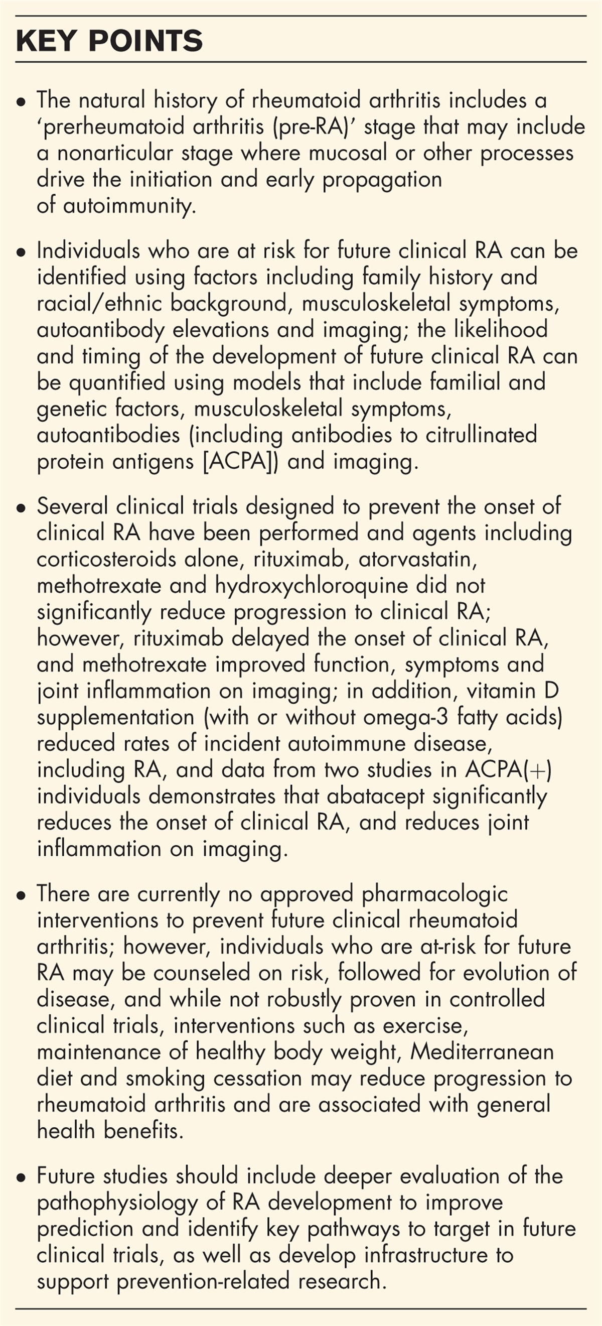
 FIGURE 2:
 Anti-survival of motor neuron (SMN) antibodies associated with the AC-7 HEp-2 indirect immunofluorescence asssay (IFA) staining pattern where intense staining of nuclear Cajal bodies (arrows) is a characteristic feature in a serum with monospecific anti-SMN (a) and a serum with anti-U1-RNP and anti-SMN. SMN is localized to coarse granules characteristic of AC-5 and anti-U1-RNP sera. IFA, immunofluorescence assay; SMN, survival of motor neuron.
CELL DIVISION CYCLE AND APOPTOSIS REGULATOR 1Using a proteomic approach, Fiorentino et al. identified ten additional autoantibodies in DM patients bearing anti-TIF1-γ autoantibodies, a known risk factor for malignancy [94▪▪,95]. Of the ten novel targets, autoantibodies directed against the cell division cycle and apoptosis regulator protein 1 (CCAR1) were the most common and were negatively associated with contemporaneous cancer (discovery cohort OR 0.27 [95% CI 0.7–1.00], P = 0.050; validation cohort OR 0.13 [95% CI 0.03–0.59], P = 0.008). Of note, when cancer eventually appeared in some patients, it occurred significantly later in anti-CCAR1-positive individuals (median time from DM onset 4.3 vs. 0.85 years, respectively; P = 0.006) and the malignancies were more likely to be localized (89% of anti-CCAR1-positive malignancies presenting at stage 0 or 1 compared with 42% of patients without anti-CCAR1 antibodies, P = 0.02). In addition, when the number of additional autoantibodies increased in anti-TIF1-γ-positive DM, the frequency of cancer decreased (P < 0.001). Hence, it appears that as the diversity of B cell responses in anti-TIF1-γ DM patients increases, the likelihood of malignancy decreases. Importantly, these findings indicate that more detailed autoantibody detection at diagnosis might better predict cancer risk. Unfortunately, as is the case for many of the newer autoantibodies associated with AIM [29], there was no evidence presented that anti-CCAR1 is associated with a specific IFA pattern on HEp-2 substrates. Clearly, these intricacies of autoantibody profiles lend support to the use of multiplexed autoantibody arrays in the diagnosis and staging of DM and other AIM.
SP4 TRANSCRIPTION FACTORIn a similar approach, a phage IP sequencing technology was used to identify transcription factor Sp4 is a novel autoantigen in sera from DM patients [93]. In an ELISA (using a full-length human recombinant protein) testing 371 AIM (255 DM, 28 ASyS, 40 IMNM, 29 sIBM and 19 PM), 80 SARD controls and 200 healthy comparators, anti-Sp4 autoantibodies were detected in 10.5% DM patients and in a single RA patient but in none of the other comparator cohorts. Sp4 is a probable transcriptional activator that binds to GT and GC box promoter elements. Remarkably, there was ∼96% overlap of anti-SP4 with anti-TIF1-γ positive patients. Among these anti-TIF1-γ-positive patients, none of those bearing anti-Sp4 had a malignancy. In contrast, among 35 anti-TIF1-γ-positive patients without anti-Sp4 autoantibodies, 14% (P = 0.04) had cancer. Similar findings were derived from a validation cohort from another center. Hence, anti-SP4 joins anti-CCAR1 as a biomarker that appears to help rule out malignancy in DM patients with anti-TIF1-γ antibodies.
CORTACTINAutoantibodies directed to cortactin, a member of the actin-binding protein family important in cell movement involving the cytoskeleton, were detected in 7/34 (20%) PM, 9/117 (7.6%) DM, 2/7 (26%) IMNM, but none of the 4 sIBM [126]. However, there was no apparent association with specific clinical features. Anticortactin antibodies were more frequently positive in patients with PM and IMNM than in DM or sIBM. Of note, it was the only myositis autoantibody found in sera of three patients suggesting anticortactin may help close the seronegative gap in AIM. In a more recent study of 670 adult AIM and 343 juvenile AIM using an ELISA [127] anticortactin autoantibodies were more common in adult DM patients (15%; P = 0.005), particularly those with coexisting anti-Mi-2 autoantibodies (24%; P = 0.03), anti-NXP-2 autoantibodies (23%; P = 0.04), anti-Ro52/TRIM21 autoantibodies (47% vs. 26%; P = 0.001), or anti-NT5c1a autoantibodies (59% vs. 33%; P = 0.001). Notably, the titers of anticortactin antibodies were higher in patients with interstitial lung disease (ILD) (0.15 vs. 0.12 arbitrary units; P = 0.03). The prevalence of these autoantibodies was not different in juvenile myositis patients (2%) as compared to juvenile healthy controls (4%).
ANTI-MITOCHONDRIAL ANTIBODIESThere is considerable interest in the malfunction of mitochondria in AIM and other rheumatic diseases [134▪,135] suggesting that the presence of antimitochondrial antibodies (AMA) found in up to 10% of AIM [130▪▪] may add important light to the pathophysiology and diagnosis of AIM. AMA-associated myopathies are reported as a homogeneous disease entity with severe arrhythmia and slowly progressive proximal muscle weakness with lordotic posture, features which are irrespective of the presence of primary biliary cholangitis (PBC) [129]. Albayda et al. reported a small cohort of seven AIM (DM and PM) with conventional AMA (M2 EP, MIT3) detected by ELISA [128]. Aberrations pointing to mitochondrial dysfunction were seen in 2/7 patients and co-existing PBC, autoimmune hepatitis, psoriasis, and Hashimoto's thyroiditis were seen in 5/7 individuals. Of note, in this study AMA was associated with a distinct inflammatory myopathy phenotype that was frequently associated with chronic skeletal muscle disease and severe cardiac involvement. A recent study concluded that mitochondria are central to skeletal muscle involvement and calcinosis of juvenile dermatomyositis (JDM) [135]. Last, Kainaga et al. reported a 48-year-old female with myositis associated with PBC but without classical AMAs [129]. This and related observations summarized above suggest the importance at determining AMA in a more systematic approach as recently reported in SLE [136].
CONCLUSIONThe techniques employed for the discovery and detection AIM-S and AIM-R autoantibodies have undergone significant advancements, aligning with our growing understanding of AIM disease pathogenesis. Technologies such as IP-MS has expanded our repertoire of new diagnostic and prognostic biomarkers available for AIM, while high-throughput methods including ELISA, LIA, ALBIA, and PMAT, have helped the adoption of these newer biomarkers into clinical practice. However, there continues to be a need to fill the seronegative gap for AIM and identify markers of disease severity, poor outcome (e.g., ILD, malignancy), and novel targets for therapy. Several promising candidate biomarkers have emerged recently, and the expectation is that this growth and success will continue in AIM, despite being a late bloomer in the field of biomarkers.
AcknowledgementsWe thank Ms. Tomoko Hasegawa for her excellent technical assistance.
Statement of ethics and consent: This study was performed in accordance with the Helsinki Declaration of 1975 as revised in 2013.
Financial support and sponsorshipNone.
Conflicts of interestM.J.F. is the Director and M.Y.C. is the Associate Director of Mitogen Diagnostics Corporation. M.J.F. is a consultant for and received speaking honoraria from Werfen International (San Diego, CA; Barcelona, Spain). M.S. has no disclosures to declare. M.Y.C. has received consulting fees from AstraZeneca, Mallinckrodt Pharmaceuticals, Werfen, and MitogenDx. M.Y.C. has received speaking honoraria from AstraZeneca and Organon.
REFERENCES AND RECOMMENDED READINGPapers of particular interest, published within the annual period of review, have been highlighted as:
▪ of special interest
▪▪ of outstanding interest
REFERENCES 1. Chinoy H, Lilleker JB. Pitfalls in the diagnosis of myositis. Best Pract Res Clin Rheumatol 2020; 34:101486. 2. Mariampillai K, Granger B, Amelin D, et al. Development of a new classification system for idiopathic inflammatory myopathies based on clinical manifestations and myositis-specific autoantibodies. JAMA Neurol 2018; 75:1528–1537. 3. Kabeya Y, Okubo M, Yonezawa S, et al. Deep convolutional neural network-based algorithm for muscle biopsy diagnosis. Lab Invest 2022; 102:220–226. 4▪. Tanboon J, Inoue M, Saito Y, et al. Dermatomyositis: muscle pathology according to antibody
留言 (0)