
Remember me


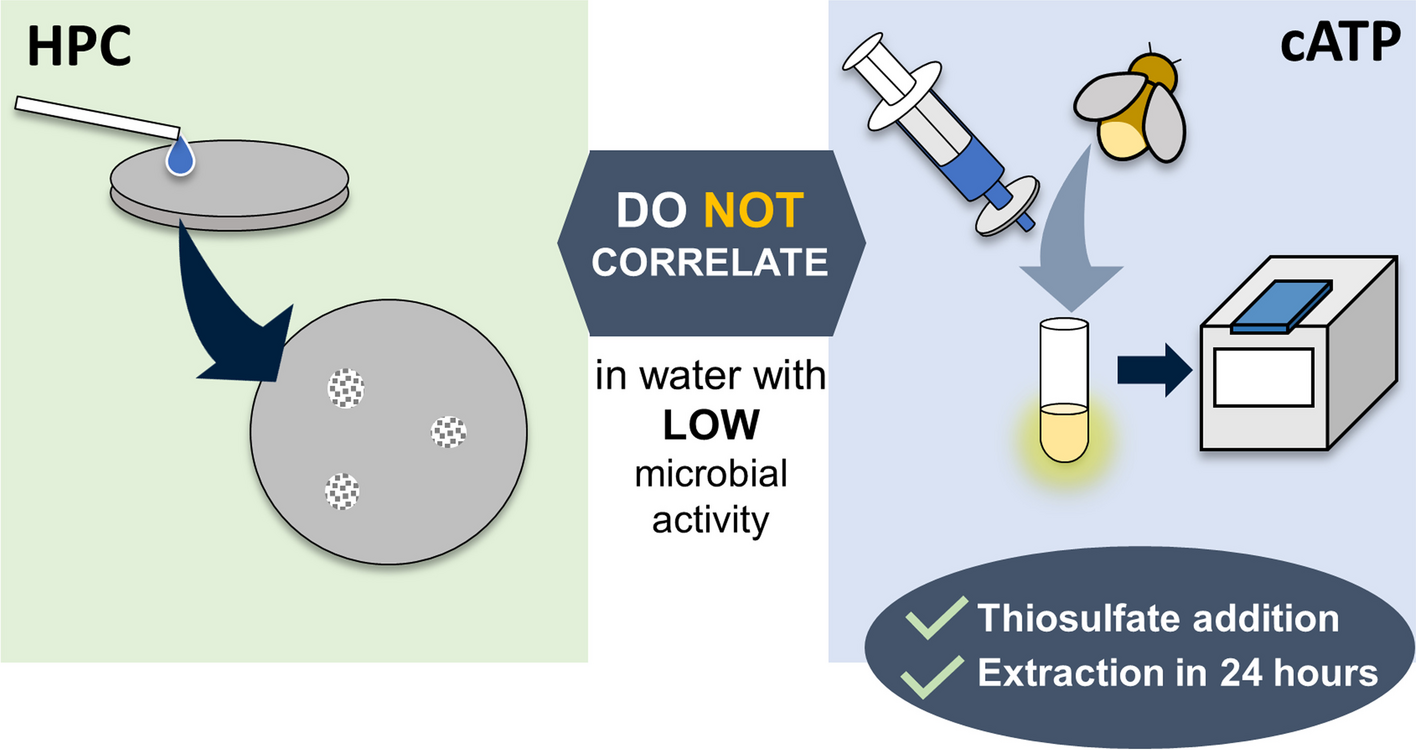
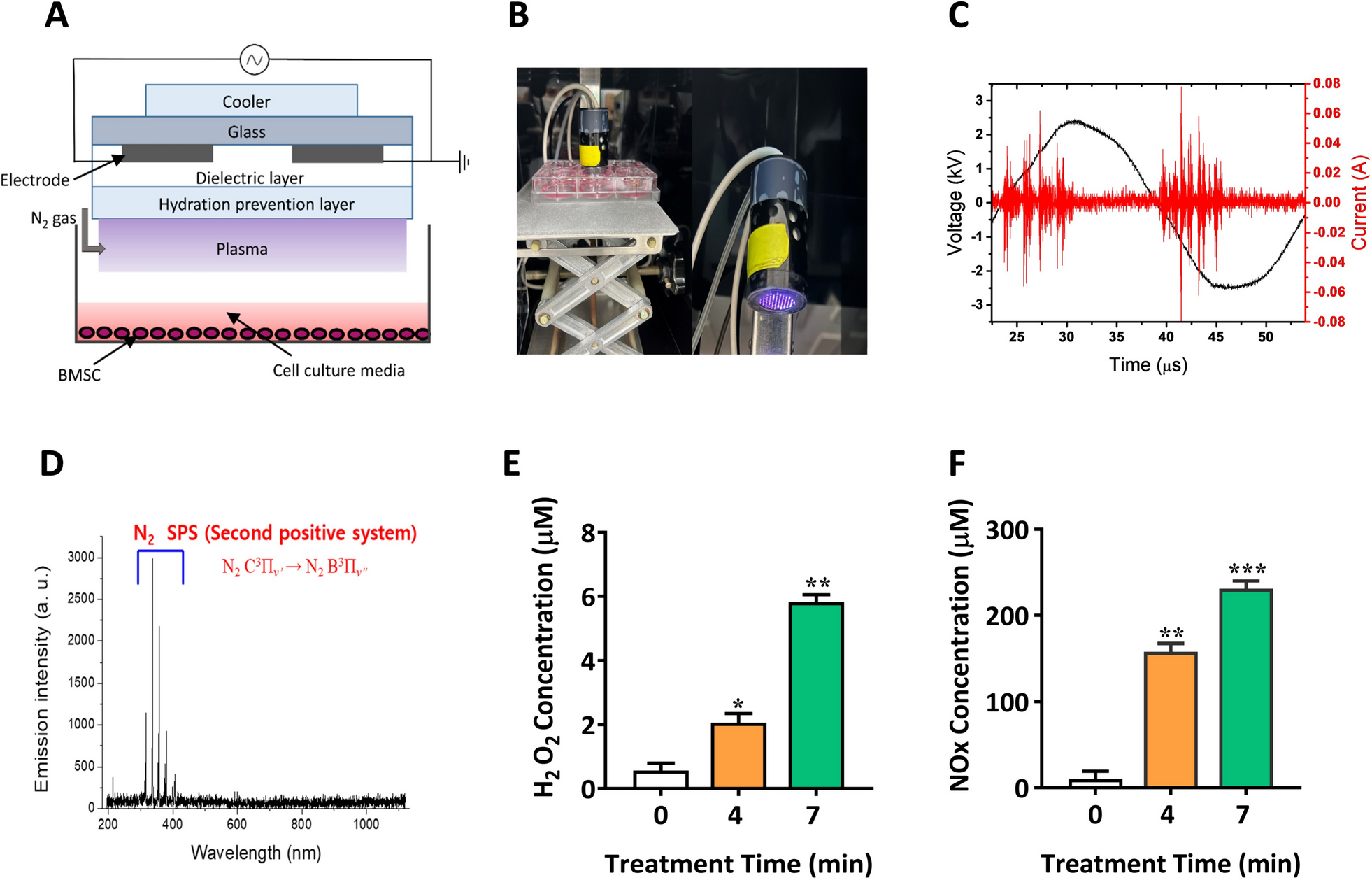


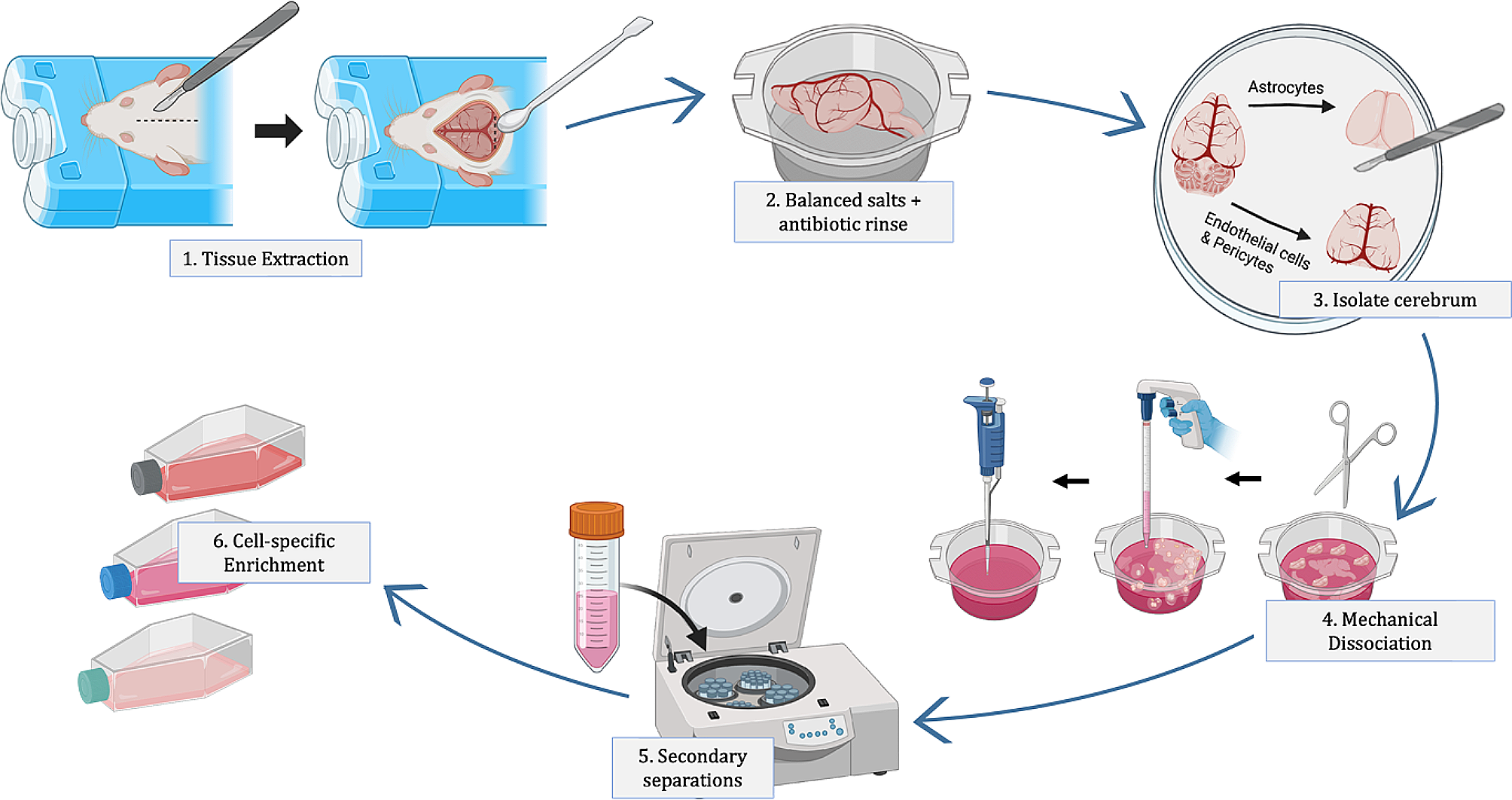
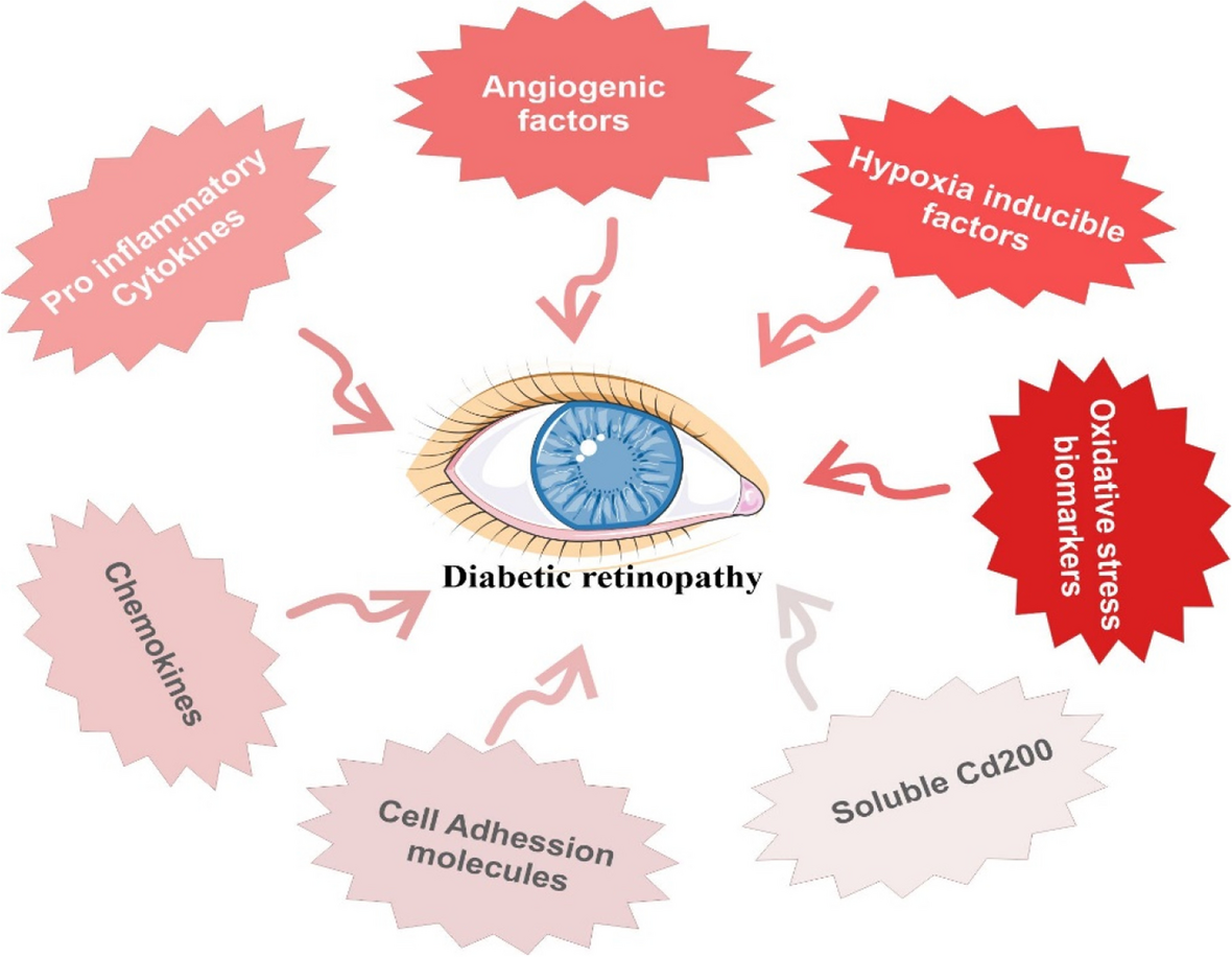
Figure 1a and b show the XRD pattern of NiCoFe2O4 nanoflowers before and after the calcination, respectively. The diffraction signals at 2θ = 11.75°, 23.33°, 34.44°, 38.9°, 46.6° 59.91°, and 61.20° in the non-calcined NiCoFe2O4 nanoflowers are attributed to the cubic spinel structure with space group of Fd-3−m NiCoFe2O4NPs (Fig. 1a). These findings are in agreement with previous reposts [26,27,28]. In the calcinated nanoflower the diffraction signals are appeared at 18.2°, 43.8°, 50.9°, and 74.8° which are correspond to the (111), (400), (102), and (622) of cubic spinel structure (FCC) of NiCoFe2O4 NPs (Fig. 1b). Figure 1b shows a broadening and sharp diffraction signal confirming, the formation of polycrystalline NiCoFe2O4 NPs (space group Fd-3 m) [29,30,31,32]. Notably, during the synthesis of these multimetallic NPs, some metal elements segregated from the final product. These elements existed in metallic or oxidized forms as by-products. As is apparent from Fig. 1b, the calcination temperature caused the NiCoFe2O4 nanoparticles to exhibit a two-phase spinel structure consisting of NiFe2O4 and CoFe2O4. The diffraction signals appear at 37.2° and 63°, which correspond to the (222) and (440) planes of the cubic spinel structure (FCC) of NiFe2O4 nanoparticles [33]. Additionally, the diffraction signals appear at 35.8° and 30.1°, which correspond to the (311) and (220) planes of the cubic spinel structure (FCC) of CoFe2O4 nanoparticles [34, 35]. Furthermore, the calcination of the nanoparticles (NPs) reduced the presence of these metallic elements, resulting in peak displacement and sharpening. In this case, the peak originally located at 34.44° shifted to the 35° region (see Fig. 1b). According to the literature, during the synthesis of ferrite nanoparticles, the dissolution of nickel and cobalt precursors into the iron phase may increase over time. The increasing dissolution of precursors within the iron phase results in observable shifts in the peaks. These shifts towards larger and smaller 2θ values indicate the dissolution of impurities, specifically nickel and cobalt precursors, within the iron phase. Simultaneously, the dissolution of these additional metal elements contributes to their incorporation into the structure of the final product, consequently altering the elemental weight ratio of the resultant material. Notably, cobalt atoms tend to integrate into the iron particle structure more rapidly than nickel. Consequently, nickel ions, possessing a smaller ionic radius, can more easily replace cobalt ions, which have a larger ionic radius, in the structure [36]. In the ternary synthesis of NiCoFe2O4, varying quantities of nickel and cobalt precursors induced subtle peak shifts. The presence of the 30.1° peak exclusively in Fig. 1b signifies the incorporation of numerous iron ions (Fe3+) into the tetrahedral site, attributable to the increased presence of nickel (Ni2+) and cobalt ions (Co2+) in the calcined nanoflower. The shift of the diffraction peak from 61.20° to 63° results from the substitution of several nickel ions (Ni2+) into the octahedral site in lieu of iron ions. Notably, in the production of multimetallic NPs, the absence of a precursor led to alterations in both the type and structure of the final product [27, 29, 31, 32, 36,37,38,39]. Rosemary has been identified as effective in mitigating pollution caused by metal elements in the environment, owing to its capacity for absorption. It is established that rosemary leaves and stems encompass diverse metallic elements [40]. In this study, rosemary extract served as both a reducing and stabilizing agent for the synthesis of NiCoFe2O4 nanoflowers. The resultant nanoflowers, prior to calcination, contained ionic elements and carbonaceous materials derived from rosemary's secondary metabolites. Subsequent to the synthesis, calcination was employed to ensure product stability and eliminate surplus materials from the non-calcined nanoflowers.
Fig. 1
XRD patterns of (a) the non-calcined NiCoFe2O4 nanoflowers and (b) the calcined nanoflowers at 400 °C
To study the surface characteristics of the nanoflowers, the XPS analysis was performed. The XPS survey spectrum has substantiated the presence of Fe 2p, Ni 2p, Co 2p, O 1 s, and C 1 s, as depicted in Fig. 2a. The significant presence of the C element, characterized by a binding energy of 284.56 eV, serves as confirmation of the existence of an organic precursor originating from rosemary extract. This precursor was evidently present surrounding the metal ions, functioning as a coating agent [41]. In Fig. 2, the Co 2p, Ni 2p, and Fe 2p spectra exhibited two sets of spin-orbital characteristics: Co 2p3/2, Co 2p1/2, Ni 2p3/2, Ni 2p1/2, Fe 2p3/2, and Fe 2p1/2, respectively. These characteristic peaks, as indicated in Fig. 2, align with the findings reported in the literature [42,43,44]. Notably, the nickel spectrum displayed four primary peaks and eight satellite peaks (depicted as the yellow band) (Fig. 2b). The satellite peaks were detected at binding energy 856.38 eV, 864.26 eV, 873.87 eV and 881.75 eV, which proved the presence of Ni2+, oxidized Ni, Ni0, and Ni2+ respectively [45,46,47,48]. Cobalt spectrum showed 4 main peaks and 9 satellite peaks (yellow band) (Fig. 2e). The peaks in the region of 780.20, 781.87, 786.19, and 789.12 eV indicated the Co2p3/2 orbitals. Peaks in the region of 795.94, 797.32, 801.39, and 804.32 indicated the Co2p1/2 orbitals. The main sharp peak at binding energy 780.20 eV proved the presence of Co (II) ion in the octahedral position. The main sharp peak at binding energy 795.94 eV proved the presence of Co (II) ion in CoFe2O4 structure. Satellite peaks at 781.87, and 797.32 eV region proved the presence of Co(II) ion in CoFe2O4 structure [49, 50]. Satellite peaks at 783.42 eV regions confirmed the presence of iron in CoFe2O4 structure. Three satellite peaks could be detected in the Co2p1/2 orbital spectrum (797.32, 801.39 eV, and 804.32 eV). Other peaks exhibiting strong binding energies correspond to the oxidation state of Co(II) [37]. The satellite peaks were detected at binding energy 786.19 eV, and 789.12 eV proved the presence of Co2+ and oxidized Co species on surface, respectively [51, 52]. Iron spectrum shows 3 main peaks and 9 satellite peaks (yellow band) (Fig. 2d). The peaks in the region of 708.91, 710.75, 713.72, 717.39 eV indicated the Fe2p3/2 orbitals. The peaks in the region of 722.51, 724.35, 727.32, and 730.99 showed the Fe2p1/2 orbitals. Two main peaks at binding energy 710.75 and 724.35 eV confirmed the oxidation state of Fe3+ in octahedral and tetrahedral sites. As a result, Fe3+ ions were equally placed in these two places, and the tendency of double positive Ni2+ and Co2+ ions with inverted spinel structure was more towards the octahedral place [21, 53]. Satellite peaks were detected in 717.39 eV, showing the oxidation state of Fe3+ in octahedral and tetrahedral sites [54,55,56]. The satellite peak at 713.72 eV confirmed the presence of Fe3+ ion. Satellite weak peak at binding energy 708.91 eV confirmed the presence of Fe2+ ions [12]. Two satellite peaks at binding energy 727.32 and 730.99 eV confirmed the presence Fe3+ ion and Fe3+ in octahedral and tetrahedral sites, respectively [57, 58]. The satellite peak at binding energy 722.51 eV confirmed the presence of Fe3+ ions. The oxygen spectrum showed one symmetric main peak and 4 satellite peaks (Fig. 2c). Sharp peaks within the 530.84 eV region were distinctly observed in the O1s orbital. This particular peak serves as confirmation of the presence of oxygen ions within the cobalt ferrite/nickel ferrite structure [59]. The peak at 529.40 eV corresponds to the ionic state of O2− in oxygen-metal bonding. Peaks within the 532.98 eV region provide confirmation of oxygen bonding with carbon. Notably, the highest satellite peak at 531.64 eV affirms the absence of O2− ions, signifying an oxygen vacancy, within the trimetallic structure. The co-existence of Fe, Co, and Ni in the synthetic trimetallic nanomaterials resulted in the formation of 26.6% oxygen vacancy regions, potentially enhancing their catalytic properties. It is noteworthy that this phenomenon led to the surface magnetic properties of the trimetallic nanomaterials being greater than those of their internal structure, as previously reported in studies [60, 61]. Given that the calcination of NPs altered the percentage of ionic elements, this analysis was conducted to validate the experimental formula of the NPs.
Fig. 2
XPS spectra of (a) non-calcined NiCoFe2O4 nanoflowers and deconvolution of (b) Ni 2p region, (c) O 1 s region, (d) Fe 2p region, and (e) Co 2p region
The components of the non-calcined nanoflowers are depicted in Fig. 3a based on the EDS analysis spectrum. The non-calcined biogenic NiCoFe2O4 NPs were found to comprise elements such as iron, nickel, cobalt, oxygen, and carbon, with respective weight percentages of 21.44%, 11.36%, 11.68%, 42.02%, and 13.50%. The presence of the carbon element was attributed to the existence of plant precursors, serving as both reducing and stabilizing agents, within the structure of the nanoparticles. This observation aligns with the findings obtained through XPS analysis. Figure 3b displays the SEM micrograph of non-calcined biogenic NiCoFe2O4 NPs. The non-calcined NPs exhibit both spherical and rod-like morphologies, with varying sizes of 11.26 nm, 14 nm, 17 nm, and 24.67 nm, arranged in a sequential fashion, resembling a rosary bead configuration (see Fig. 3c). Additionally, the SEM images reveal the presence of spherical particles distributed uniformly, forming thin, convex plates (Fig. 3d). These convex plates exhibit diverse orientations reminiscent of flower petals, ultimately forming rose-like nanoflowers. Notably, the spherical and rod-like particles are sequentially organized within the light-colored petals, which are convex in shape and possess a dark hue. The components of the calcined nanoflowers are illustrated in Fig. 3e. According to the EDS analysis spectrum, the calcined NiCoFe2O4 NPS were found to encompass elements such as Fe, Ni, Co, O, and C, with respective weight percentages of 31.8, 20, 18.1, 19.7, and 10.4 wt%. These EDS results highlight that the calcination process of NiCoFe2O4 NPs resulted in a reduction in the weight percentages of O and C elements. Conversely, there was an increase in the weight percentages of Fe, Ni, and Co. It is noteworthy that O and C are abundant elements found in rosemary extract, which contains polyphenolics, with a particularly high concentration of polyphenols in the aqueous extract of rosemary. Therefore, the calcination of NPs effectively eliminated organic components (plant extracts) and metal impurities [62, 63]. The calcined NPs display a morphology characterized by spherical and rod-like structures arranged in a linear fashion, resembling the configuration of rosary beads. In this calcined state, the convexity of the petals is accentuated, and the petals are closely juxtaposed, giving rise to the appearance of flower buds. In contrast, the non-calcined NPs exhibit an open, flower-like structure. Furthermore, the micrograph reveals the presence of clusters with diameters measuring 405.1 nm and 517.5 nm. These clusters can be attributed to the fusion and accumulation of NPs brought about by the calcination process and its associated temperature conditions [62].
Fig. 3
FE-SEM images of NiCoFe2O4 nanostructures (a), EDX spectrum of non-calcined NiCoFe2O4 nanoflowers (b-d), and SEM micrographs of the non-calcined nanoflowers, (e), EDX spectrum of the calcined nanoflowers at 400 °C (f), and SEM micrographs of the calcined nanoflowers at 400 °C
Figure 4 shows HR-TEM bright-field micrographs of the nanoflowers at 200 and 5 nm scales. NiCoFe2O4 individual rose-like nanostructures with uniform and distinct size (~ 200 nm) are documented in Fig. 4a. In this micrograph, the needle-shaped plates were flower petals with different orientation. These needle-shaped plates densely penetrated each other. As a result, the center of each nanoflower was darker, where was overlapping the highest number of petals [60, 64]. The petals of the flower micrograph are shown in higher magnification in the Fig. 4b. In this micrograph, the petals were plate-shaped with curvature and darker contrast at the edge suggesting its higher thickness, which is consistent with SEM image in Fig. 3d. In some regions, these needle-like ends of petals (petal edge) consisted of 3 to 5 layers, which were ~ 5 nm. In other places, due to the accumulation of NPs and the formation of rosary clusters (corresponding to the FESEM micrograph), the edges of the petals were folded towards the inside of the clusters, and the number of the end layers of the petals could not be recognized. As a result of these folds along with crystallization, empty spaces could be formed in the structure of the petals and their random growth [64, 65]. The spindle-like shapes in Fig. 4c showing the lattice fringes with a distance of 0.209 and 0.211 nm respectively confirmed the (111) and (311) different crystal planes of the cubic spinel structure of NiCoFe2O4 NPs [43]. The lattice fringes of each petal have grown densely in different directions, and in some areas, they were broken due to the change of petal direction. This could cause the surface of the petals to be smooth in some areas and rough in some areas. The rough areas acted as the functional sites and the porous space of NiCoFe2O4 NPs. As a result, each petal plate of an individual rose-like nanostructure contained a large number of NiCoFe2O4 single crystal. The SAED pattern in Fig. 4d shows clear and discrete point electron diffraction rings. These results confirm the polycrystalline nature of NiCoFe2O4 nanoflowers. The presence of a ring indexed to the d111 plane further substantiates the spinal cubic structure of NiCoFe2O4 [66, 67]. This diffraction pattern confirmed the different orientations of the face-centered-cubic crystal structure of NPs, which was in consistent with the XRD results [23, 43, 60]. The EDS analysis results of these regions confirmed the co-existence of carbon, iron, nickel, cobalt, and oxygen elements, as shown in Fig. 4d. Nickel atoms overlapped with cobalt, while carbon overlapped with oxygen. The composition also contained a small amount of oxygen, which was consistent with the XPS results.
Fig. 4
HR-TEM images of the non-calcined nanostructures. a Individuals NiCoFe2O4 nanoflowers and (b) a detail of petals structure (c) SAED patterns
Figure 5 a and b present STEM bright-field (STEM-BF) and STEM dark-field (STEM-DF) images of the non-calcined multi-petal NiCoFe2O4 nanoflowers, respectively, and Fig. 5 c and d shows STEM tomography visualization of surface and internal volume for the morphology of the same multi-petal NiCoFe2O4 nanoflower imaged in Fig. 5 a and b. The different orientations and fractures of the petals can be clearly identified in Fig. 5a. Also, two identical individual nanoflowers can be identified in Fig. 5b. The brighter contrast in fracture areas and some areas of needle-shaped plates is due to the presence of hollow spaces (mesoporous nature) of nanoflowers (Fig. 5b) [68]. In Fig. 5 c and d, the folds of the petal edges and the holes in the nanoflower structure are clearly visible from different angles. Video S1 (in Supporting Information) shows the internal 3D visualization of NiCoFe2O4 nanoflower obtained through sectioning in all three spatial directions. Observation of the NiCoFe2O4 nanoflower using video imaging from various angles revealed the absence of a central core in its structure. These analyses confirmed the existence of non-calcined NiCoFe2O4 in the form of a multi-petal nanoflower structure with individual petals arranged in various orientations. The visualization displayed a rose-shaped structure with irregular sheets of petals situated closely to each other. The petals of the rose were curved and intertwined, and their thickness remained uniform throughout the nanoflower, approximately 5 nm. Furthermore, compacted petals were identified at the center of the NiCoFe2O4 nanoflower.
Fig. 5
STEM images of the non-calcined nanostructure used for 3D reconstruction (the round particles are Au beads used for image alignment): (a) STEM-BF image, b STEM-DF image, c STEM tomography visualization of Side views NiCoFe2O4 nanoflower and (d) STEM tomography visualization of a cross-section of the nanoflower revealing the petal structure in 3D
The results obtained from the BET-BJH analysis of NiCoFe2O4 nanoflowers are shown in Fig. 6. Based on the obtained data, specific surface area (Fig. 6b, BET plot), total pore volume (p/p0 = 0.990), and mean pore diameter (Fig. 6c, BJH plot) of the non-calcined NPs were 193.34 cm2 g–1, 0.8279 cm3 g–1 and 127.17 nm, respectively. Also, the specific surface area (Fig. 6e, BET plot), total pore volume (p/p0 = 0.990) and mean pore diameter (Fig. 6f, BJH plot) of the calcined NPs were 145.12 m2g–1, 0.9541 cm3 g–1, and 298.26 nm, respectively. Adsorption desorption plot of the non-calcined NPs type IV isotherms with H2 hysteresis (IUPAC) in relative pressure (P/P0) 0.4 to 1 is shown in Fig. 6a. Also, the adsorption desorption plot of the calcined NPs at 400 °C type IV isotherms with H3 hysteresis (IUPAC classification) in relative pressure (P/P0) 0.6 to 1 is shown in Fig. 6d. Based on the type hysteresis, the non-calcined NPs had the irregular cavities. Additionally, based on the type hysteresis, the calcined NPs had slit (plate and cut) cavities [69, 70]. Based on the BJH curve of the non-calcined NPs, the particle diameters were uniform, and the mesopore size was in the range of 1.85 to 61.31 nm. Dominant pore size was in the range of 3.52 to 12.24 nm (Fig. 6c). Also, according to the BJH curve of the calcined NPs, the particle diameters were not uniform, and the pore size was in the range of 1.85 nm to 71.89 nm. Dominant pore size was in the range of 1.85 to 12.24 nm (Fig. 6f). The resulting NiCoFe2O4 nanoflowers (the calcined at 400 °C and non-calcined) were mesoporous in nature. Remarkably, the non-calcined NiCoFe2O4 nanoflowers had the higher surface area from the calcined nanoflowers at 400 °C.
Fig. 6
a Nitrogen adsorption–desorption isotherms, b the BET surface areas, and c BJH of the non-calcined. d Nitrogen adsorption–desorption isotherms, e the BET surface areas, and f BJH of the calcined nanoflowers at 400 °C
Figure 7 depicts the magnetic behavior of the non-calcined nanoparticles at ambient temperature within a magnetic field range of -20,000 ≤ H (Oe) ≤ 20,000. The nearly linear curve suggests that the nanoflowers exhibit paramagnetic or antiferromagnetic properties, characterized by a magnetization saturation (Ms) value of 20.98 (emu/g). Furthermore, magnetic parameters such as coercivity (Hc) and remanence (Mr) were not detected in this analysis [71,72,73]. In three-metallic iron NPs, the Fe ions underwent a transformation from iron (III) to iron (II) state due to several factors including the presence of oxygen, particle size, alterations in crystal structure, and ion replacement within the Fe positions, resulting in diminished ferromagnetic behavior [74]. Figure 7b illustrates the magnetic behavior of the calcined nanoflowers at 400 °C, at room temperature, subjected to a magnetic field range of -20,000 ≤ H (Oe) ≤ 20,000. The curve indicates that the nanoflowers exhibit ferromagnetic characteristics, with a magnetization saturation (Ms) value of 50.10 (emu/g). It is worth noting that the magnetic behavior of iron nanoparticles is influenced by parameters such as size, synthesis method, shape, and calcination process [75]. The calcination of NiCoFe2O4 nanoflowers resulted in an increase in both magnetization and coercivity values, leading to a transition in magnetic behavior from paramagnetic to ferromagnetic. The enhancement of magnetic behavior and the increase in magnetization saturation (Ms) value in the calcined nanoflowers were achieved by placing Fe3+ metal cations in the octahedral site, and Co2+ and Ni2+ metal cations in the tetrahedral site within the spinel ferrite structures [76]. According to the existing literature, the magnetic moment of cobalt, nickel, and iron ions is 3 µB, 2 µB, and 5 µB, respectively [77]. Consequently, the calcined nanoflowers at 400 °C had more magnetic properties than non-calcined nanoflowers. The increase in magnetization saturation (Ms) of the calcined nanoflowers can be attributed to the cumulative state and the increase in the cluster diameter of NPs. The available surface area of the clusters was smaller than that of small spherical particles, and this surface area was inversely re
Comments (0)