
記住我


The massive worldwide vaccination for coronavirus disease 19 (COVID-19) has effectively weakened the COVID-19 pandemic and saved countless human lives. Although neurological complications of COVID-19, including peripheral nervous system diseases and neuropathic pain, affect a significant proportion of COVID-19 patients,2,22,24 serious neurological adverse events following COVID-19 vaccination have involved only a very limited proportion of the vaccinated population.15
Increasing evidence now indicates that COVID-19 vaccination is frequently associated with persistent sensory disturbances, including generalized sensory symptoms and pain, raising the possibility that this vaccination might trigger somatosensory nervous system damage, presumably affecting small nerve fibres.12,33 Accordingly, a recent observational study showed that a significant proportion of patients complaining of generalized sensory symptoms and pain one month after COVID-19 vaccination had objective evidence of small-fibre neuropathy.27
Due to the limited evidence in the literature, the association between COVID-19 vaccination and small fibre damage remains an open issue. A better understanding of whether COVID-19 vaccination triggers small fibre damage might improve how we manage this seldom encountered vaccine complication.
In this uncontrolled observational study, we aimed to identify small fibre damage in patients complaining of generalized sensory symptoms and pain after COVID-19 vaccination. To do so, we collected clinical data and investigated quantitative sensory testing (QST), skin biopsy with the assessment of intraepidermal, piloerector muscle, and sweat gland nerve fibre density, and cardiovascular autonomic tests in prospectively enrolled patients with generalized sensory symptoms and pain after COVID-19 vaccination.
2. Methods 2.1. Study design and patient cohortFrom January 2022 to June 2022, we prospectively screened 20 patients (all woman patients) experiencing generalized sensory symptoms and pain as long-term complications after COVID-19 vaccination. These patients were consecutively referred to our peripheral neuropathy and neuropathic pain unit at the Department of Human Neuroscience, Sapienza University of Rome. Inclusion criteria were age >18 years and sensory disturbances persisting for at least 6 months after COVID-19 vaccination. Exclusion criteria were previous chronic pain conditions, concomitant peripheral or central nervous system diseases, conditions potentially associated with peripheral nervous system diseases, including diabetes, B12 deficiency, kidney failure, autoimmune diseases, previous neurotoxic therapies, and previous diagnosis of fibromyalgia, psychiatric disorders, and the inability to provide informed consent.
Within a single clinical session, patients underwent a detailed neurological examination with sensory profiling, the Neuropathic Pain Symptom Inventory (NPSI) for assessing neuropathic pain symptoms, the Composite Autonomic Symptom Score (COMPASS)-31 questionnaire for autonomic symptoms, and diagnostic tests including nerve conduction study (NCS), QST, and skin biopsy. Those patients who reported orthostatic intolerance on the COMPASS-31 questionnaire (“in the past year, have you ever felt faint, dizzy, goofy, or had difficulty thinking soon after standing up from a sitting or lying position?”) underwent cardiovascular tests a week later in a separate clinical session.
In line with the widely accepted Besta criteria, the diagnosis of small-fibre neuropathy was based on the combination of at least 2 out of 3 of the following criteria: (1) distally distributed sensory signs (decreased thermal pain sensation and/or hyperalgesia and/or allodynia) as assessed with bedside clinical examination; (2) abnormal cold and/or warm detection threshold as assessed by QST; and (3) intraepidermal nerve fibre density reduction at skin biopsy from the distal calf.7
The institutional review board approved the study. The informed consent for the procedures was obtained from participants.
2.2. Clinical examination and questionnairesAll patients underwent a neurological examination using bedside tools, with precise sensory profiling. We investigated the negative sensory signs (tactile, pinprick, and thermal hypoesthesia) and positive symptoms and signs (constant pain, paroxysmal pain, allodynia, and pinprick hyperalgesia). Patients were asked to provide a complete description of pain characteristics and to mark the pain distribution area on a somatic map. The pain distribution was then analysed using a specifically designed software program that allowed the pain distribution areas to be drawn over a 3-dimensional body model consisting of 1500 surface elements.11
The pain areas were analysed to calculate the widespread pain index (WPI). In addition, we calculated the symptom severity scale (SSS) score for each patient, taking into account the presence of fatigue, waking unrefreshed, and cognitive symptoms.34
All patients completed the NPSI,5 a validated questionnaire commonly used for scoring the different types of pain. We calculated NPSI subscores for the different pain qualities, including ongoing pain (burning and pressing pain), paroxysmal pain, provoked pain, and abnormal sensation (paraesthesia, dysesthesia). The NPSI questionnaire was administered for distally distributed pain (the feet in 13 patients and the hands in 2 patients).
Autonomic symptoms were investigated with COMPASS-31, a structured interview including questions related to orthostatic intolerance, genitourinary disturbances, gastrointestinal disturbances (diarrhoea or constipation), sicca syndrome (dry mouth or dry eyes), abnormal sweating, and pupillomotor symptoms.
2.3. Nerve conduction studyAll patients underwent NCS by surface recording electrodes with standard placement. Nerve conduction study included sensory nerve action potential amplitude and conduction velocity recorded from sural, ulnar, and superficial radial nerves, and compound motor action potential amplitude and conduction velocity of peroneal, tibial, and ulnar nerves. Recording methods adhered to the recommendations of the International Federation of Clinical Neurophysiology.18 Skin temperature was maintained between 34°C and 36°C. Nerve conduction study data were compared with age-adjusted normative ranges.
2.4. Quantitative sensory testingQuantitative sensory testing was performed following the standardized protocol of the German Research Network on Neuropathic Pain.3 We selected the dorsum of the right foot as a test area in each patient (corresponding to a painful site in most patients; Fig. 1).
 Figure 1.: Pain distribution. Topographical maps showing the reported pain distribution in the 15 patients with generalized sensory symptoms and pain associated with COVID-19 vaccination. Patients 1, 2, 3, 6, and 7 had a combination of sensory symptoms and quantitative sensory testing abnormalities potentially compatible with a diagnosis of small fibre neuropathy (Table 1).
Using the log-transformed raw values for each QST variable, a z-score sensory profile was calculated (z-score = patient value − mean value of control subjects/standard deviation [SD] of control subjects). Negative z-scores indicated loss of perception, whereas positive z-scores indicated gain of perception. For individual analysis, single values were compared with published reference data. Values exceeding 0 ± 1.96 SDs were considered abnormal.4
2.5. Skin biopsyPatients underwent skin biopsy at the distal leg, 10 cm above the lateral malleolus, and from the lateral upper thigh using a 3-mm disposable circular punch after local lidocaine anaesthesia. Biopsies were performed under sterile conditions and with no suture required.
Using indirect immunofluorescence, intraepidermal and autonomic structure innervation was assessed with the pan-neuronal marker PGP9.5. Biopsies were fixed for 24 hours at 4°C in Zamboni's fixative, then cryoprotected overnight. Cut was performed at −23°C with a cryostat (MEV, SLEE medical) to obtain 50-µm-thick sections. Three nonconsecutive free-floating sections were randomly selected for immunostaining from each sample and blocked with 5% normal donkey serum for 1 hour. Sections were then incubated overnight with a rabbit anti-human PGP9.5 monoclonal antibody (Abcam, Cambridge, United Kingdom, 1:500 diluted) and a mouse antihuman collagen IV monoclonal antibody (Millipore, Milan, Italy, 1:1600). The following day, sections were incubated with antirabbit-Cy3 (Jakson, Cambridge, United Kingdom, 1:800) and antimouse-488 (Jakson, 1:400) secondary antibodies overnight.
We calculated intraepidermal nerve fibre density according to the European Federation of Neurological Societies and Peripheral Nerve Society guidelines. Epidermal linear length was measured through Image-J to obtain a linear density (number of fibres/mm). Normative values from an internationally recognized data set were used.26
We calculated piloerector muscle nerve fibre density for each patient on all piloerector muscles available in the sections. Only subjects with at least 3 distinct evaluable piloerector muscles were included. For each piloerector muscle, a photograph was taken at the level of maximum diameter, focusing on as many fibres as possible. To quantify innervation, a vertical line to the nerve fibres was drawn, and fibres intersecting the line were counted. The nerve fibre density of each piloerector muscle innervation was calculated as the ratio between the number of nerve fibres crossing the line in focus and the width of the line (nerve fibres/mm).23 For each patient, piloerector muscle nerve fibre density corresponded to the average nerve fibre density of the 3 most innervated piloerector muscles. Two blind operators (P.F., E.G.) calculated nerve fibre density through a fluorescence microscope (Leica NB, Milan, Italy) with appropriate wavelength filters. Twenty-five age-matched and sex-matched healthy subjects underwent skin biopsy as a control group for the piloerector muscle nerve fibre density calculation.
We evaluated sweat gland nerve fibre density in a semiquantitative fashion for each patient on all sweat glands available in the sections, as previously described.9 For each sweat gland, a photograph was taken at the level of the gland maximum diameter, focusing on as many fibres as possible. Images of sweat glands were scored semiquantitatively on a 0 to 4 scale. A score of 0 was assigned when no nerve fibres were identifiable, 1 to severely, 2 to moderately, and 3 to mildly reduced nerve fibre density, and 4 to normal nerve fibre density. Only patients with at least 3 evaluable sweat glands were included in the analysis. Each patient was assigned a score from 0 to 4 reflecting the average density of all analysed glands in the patient samples.
Given the lack of normative values for piloerector muscle and sweat gland nerve fibre density, we enrolled 48 age-matched and sex-matched healthy subjects as control group for these autonomic skin biopsy parameters.
2.6. Cardiovascular autonomic testsPatients who reported possible symptoms of orthostatic intolerance at COMPASS-31 questionnaire underwent cardiovascular tests. ECG and blood pressure signals were noninvasively acquired using the Task Force Monitor (CNSystem). Tests included cardiovascular reflex tests (deep breathing, Valsalva manoeuvre) and head-up tilt test. The deep breathing test was considered an index of parasympathetic function. Participants breathed maximally at a frequency of 6 breaths per minute, following the lead of an oscillating ball on a computer screen. The deep breathing test was considered normal if the heart rate variation was 15 beats/min or more, borderline if 11 to 14 beats/min, and pathological if 10 beats/min or less. The Valsalva manoeuvre was used to assess baroreflex sympathetic and parasympathetic function. Patients performed a forced expiration at 40 mm Hg for 15 seconds The Valsalva ratio was calculated, and the maximal drop of systolic and diastolic blood pressure at the different phases during Valsalva manoeuvre were computed. Tilt table testing was performed with a head-up tilt of 70° for 20 minutes after 10 minutes of supine rest, monitoring beat-to-beat changes in heart rate and blood pressure in each position. Orthostatic hypotension was defined as a sustained diastolic blood pressure drop of at least 10 mm Hg or a systolic blood pressure drop of at least 20 mm Hg. Postural orthostatic tachycardia was defined as a sustained heart rate rise of at least 30 bpm or a heart rate of at least 120 bpm in the first 10 minutes of tilt, without concomitant orthostatic hypotension.13
2.7. Statistical analysisWe used descriptive statistics to describe the main demographic, clinical, and diagnostic test variables. For continuous variables, we reported mean (SD). For discrete variables, we reported the number of observations and frequencies.
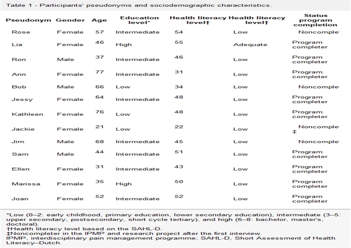
3. ResultsOf the 20 women patients experiencing generalized sensory symptoms and pain as long-term complications after COVID-19 vaccination, 4 patients were excluded due to previous chronic pain conditions, one patient was excluded because her sensory disturbances were persisting for less than 6 months after COVID-19 vaccination. We have therefore included 15 patients (mean age, 48.5 ± 11.3 years). These patients received an mRNA vaccine except one, who received an adenovector-based vaccine (Table 1).
Table 1 - Demographic and clinical characteristics of the 15 patients. Patient* Age (y) Vaccine dose† Onset of symptoms (d) NPSI score COMPASS-31 score WPI SS CDT (C°) WDT (C°) Calf IENFD (/mm) Thigh IENFD (/mm) 1 78 III 4 25 9 18 5 19.6 47.1 13.3 14.51 2 48 I 7 60 53 7 5 28.4 48.9 13.94 16.1 3 36 III 1 17 17 0 4 29.3 44.1 11.44 16.72 4 51 II 7 8 7 9 2 27.3 34.7 10.55 16.03 5 43 II 1 49 22 12 8 28.5 36.6 29.54 31.91 6 44 II 5 35 42 13 11 11.5 39.2 11.58 16.2 7 53 I 7 60 13 10 7 22.4 42.3 13.3 15.71 8 46 II 2 18 10 13 7 27.5 38.5 23.96 ‡ 9 57 II 3 60 37 19 6 24 40.5 15.65 17.23 10 32 I 3 42 10 19 5 28.5 37.8 11.92 16.72 11 41 II 5 40 2 3 5 29.2 35.3 11.77 16.28 12 44 I 1 84 56 13 8 29 36 11.86 15.83 13 39 II 30 11 31 6 8 29 34.7 16.4 15.84 14 58 I 3 65 32 11 10 29 35.5 12.15 16.36 15 57 I 30 50 17 2 5 26.4 38.8 14.85 ‡In bold the CDT and WDT whose Z-score is abnormal (0 ± 1.96 SD).
*All patients are female.
†All patients, patient 5 excluded, received mRNA-based vaccination.
‡Skin biopsy sample unavailable.
CDT, cold detection threshold; COMPASS 31, composite autonomic symptom score; IENFD, intraepidermal nerve fibre density; NPSI, neuropathic pain symptom inventory; SS, somatic symptoms; WDT, warm detection threshold; WPI, widespread index.
Of these 15 patients, 3 reported a sars-cov-2 infection more than 3 months before vaccination (patients 9, 11, 14 had a previous infection 5, 6 and 4 months before the vaccination).
In all patients, symptoms started after vaccination; all experienced generalized sensory symptoms and pain (Figure 1; Supplementary Figure 1, available at https://links.lww.com/PR9/A200), which started from 1 to 30 days after vaccination. Nine patients experienced distally distributed allodynia and/or hyperalgesia (Supplementary Table 1, available at https://links.lww.com/PR9/A200). In most patients, pain had a widespread distribution (WPI: 10.9 ± 6.5) and was commonly associated with fatigue, waking unrefreshed, and cognitive symptoms (SSS: 5.4 ± 2.3). Accordingly, 11 patients met fibromyalgia diagnostic criteria.34 The NPSI questionnaire showed that pins and needles, tingling sensation, burning pain, and electric shock–like sensations were the most frequently reported sensory disturbances (Table 2). The COMPASS-31 questionnaire showed that all patients experienced autonomic symptoms (Supplementary Table 2, available at https://links.lww.com/PR9/A200), with orthostatic intolerance being the most frequently reported (9 patients).
Table 2 - Neuropathic pain symptom inventory data. NPSI item Frequency (%) Severity 0–10 points (mean ± SD) Q1 burning pain 73 7.9 ± 1.7 Q2 squeezing pain 47 8.0 ± 2.1 Q3 pressure pain 47 8.9 ± 1.9 Q5 electric shocks 73 7.0 ± 2.1 Q6 stabbing pain 53 8.4 ± 1.3 Q8 pain provoked by brushing 60 5.8 ± 2.0 Q9 pain provoked by pressure 27 7.0 ± 2.2 Q10 pain provoked by cold 33 5.6 ± 3.4 Q11 pins and needles paraesthesia 87 6.8 ± 1.6 Q12 tingling paraesthesia 73 7.2 ± 1.9In the severity calculation, only patients reporting the specific types of pain are included. NPSI questionnaire was administered for distally distributed pain (the feet in 13 patients and the hands in 2 patients).
NPSI, neuropathic pain symptom inventory.
In all patients, NCS yielded unremarkable findings. Quantitative sensory testing investigations showed that cold detection threshold was abnormal in 3 patients and warm detection threshold was abnormal in 3 patients (1 patient had concomitant abnormalities of cold and warm detection thresholds). The mean z-scores of the different QST variables fell within the normative range of 0 ± 1.96 SDs (Fig. 2).
 Figure 2.:
 Sensory profiles as assessed with quantitative sensory testing. Scatter dot plot showing the individual values of quantitative sensory testing variables at foot and the mean values ±SD in the 15 patients with generalized sensory symptoms and pain associated with COVID-19 vaccination. CDT, cold detection threshold; CPT, cold pain threshold; HPT, heat pain threshold; MDT, mechanical detection threshold; MPS, mechanical pain sensitivity; MPT, mechanical pain threshold; PPT, pressure pain threshold; TSL, thermal sensory limen; VDT, vibration detection threshold; WDT, warm detection threshold; WUR, wind-up ratio.
In all patients, intraepidermal, piloerector muscle, and sweat gland nerve fibre density, as assessed by PGP9.5 immunostaining of the skin samples, was within normative ranges (Fig. 3).
 Figure 3.:
 Skin biopsy representative findings. PG9.5-stained intraepidermal nerve fibres (A), piloerector muscle (B), and sweat gland nerve fibres (C) in a representative patient with generalized sensory symptoms and pain associated with COVID-19 vaccination. All patients had normal skin biopsy findings. Calibration bars: 100 µm.
The 9 patients with orthostatic intolerance underwent cardiovascular autonomic tests, which did not disclose abnormal findings. Of the 15 patients enrolled, 5 had cold/warm detection threshold abnormalities at the QST associated with hyperalgesia and allodynia, potentially compatible with a diagnosis of small fibre neuropathy (Table 1 and Fig. 1, patients 1, 2, 3, 6 and 7). Nevertheless, 4 of the 5 patients with small fibre neuropathy also met the diagnostic criteria for fibromyalgia (Table 1 and Fig. 1).
4. DiscussionIn this uncontrolled observational study, we found that although 5 patients complaining of generalized sensory symptoms and pain after COVID-19 vaccination had QST abnormalities potentially compatible with a small fibre neuropathy, all patients had unremarkable findings at the skin biopsy investigations, and most of them met the diagnostic criteria for fibromyalgia, thus suggesting that COVID-19 vaccination might trigger a fibromyalgia-like syndrome.
In our study, we applied the widely agreed Besta criteria7 to identify small fibre neuropathy in patients complaining of generalized sensory symptoms and pain after COVID-19 vaccination. Due to their high sensitivity and specificity in the diagnosis of small fibre neuropathy, these criteria represent a reliable approach to identify patients with small fibre neuropathy related to COVID-19 vaccination.
The topographical distribution of pain in our 15 patients with generalized sensory symptoms and pain associated with COVID-19 vaccination resembles the distribution of pain in patients with long COVID syndrome. Previous studies indicated that pain associated with long COVID syndrome affects lower limbs or has a widespread distribution in a considerable proportion of patients.19,30
Three patients had a sars-cov-2 infection more than 3 months before vaccination. Although we cannot exclude that a previous infection may influence generalized sensory symptoms and pain associated with vaccine, these 3 patients had spared QST variables and did not differ from the other patients in clinical variables.
Of the 15 patients included in this study, we found that 5 patients had abnormal cold and/or warm detection thresholds at the QST investigation associated with hyperalgesia and allodynia, thus meeting the Besta criteria for small fibre neuropathy. However, QST is a psychophysical test that is influenced by patients' attention and cooperation, and 5% of healthy subjects are expected to present with at least one abnormal QST value.3 In addition, in our patients, skin biopsy investigations did not show clinically significant abnormalities. Skin biopsy, providing objective information on small nerve fibres, has higher sensitivity and specificity than QST in disclosing small fibre damage and is widely considered the reference standard technique in the diagnosis of small fibre neuropathy.10,14,31 The lack of clinically important small fibre damage we found at skin biopsy also agrees with the lack of abnormalities at the cardiovascular tests in our patients.
Most of our patients (including 4 of the 5 patients fulfilling criteria for small fibre neuropathy) met the diagnostic criteria for fibromyalgia. The clinical picture was characterized by a constellation of symptoms, including widespread pain, fatigue, waking unrefreshed, and cognitive symptoms. This finding, in combination with the lack of abnormalities at objective diagnostic tests (namely, skin biopsy and cardiovascular tests), suggests that COVID-19 vaccination might rather trigger a fibromyalgia-like syndrome. This interpretation is consistent with evidence that different vaccines are associated with fibromyalgia-like syndromes.1,6,20
Admittedly, multiple observations now raise the possibility that fibromyalgia and small-fibre neuropathy may manifest as a spectrum disorder.11,32 A recent study provided preclinical data showing that dorsal root ganglia damage due to circulating autoantibodies may play a role in fibromyalgia.17 Fibromyalgia-like syndromes may therefore hide small fibre damage. Mechanisms underlying small fibre damage due to COVID-19 vaccination are unclear. We may hypothesize that vaccination triggers immune-mediated damage to small fibres. Preclinical studies showed that small fibre–related sensory ganglia preferentially express ACE-2 receptors,21,29 which are the cell targets of the SARS-CoV-2 spike protein.28 Because COVID-19 vaccines are based on spike protein immunization, the spike protein may directly target small sensory neurons.
It follows that we cannot exclude that our 15 patients who complained of a similar combination of sensory and autonomic symptoms might experience immune-mediated small fibre impairment manifesting with mild or negligible signs of axonal loss. Furthermore, because the spike protein might directly target sensory neurons in the dorsal root ganglion17,29 with negligible axonal loss, sensory symptoms might develop without clinically evident somatosensory system damage. Therefore, the currently accepted criteria for small fibre neuropathy diagnosis might have poor sensitivity in detecting small fibre impairment in these patients.
Unexpectedly, all the patients we enrolled in this study were women. We may hypothesize that this women-restricted involvement may reflect peculiar susceptibility of women to develop chronic pain and fibromyalgia-like syndrome. Admittedly, we cannot exclude a sampling bias, due to the small sample of patients we enrolled; it follows that the generalizability of this finding requires further case–control studies, including large sample of patients.
In our patients, we assessed piloerector muscle and sweat gland nerve fibre density to investigate autonomic skin innervation. However, although intraepidermal nerve fibre density is a widely accepted reference standard parameter for diagnosing small fibre neuropathy, no standardized consensus exists regarding the quantitative assessment of autonomic skin innervation. Accordingly, we did not include piloerector muscle and sweat gland nerve fibre density as critical measures for the diagnosis of small fibre neuropathy. To investigate autonomic skin innervation, we performed a semiquantitative analysis of sweat gland innervation, in line with previous studies9 and calculated piloerector muscle nerve fibre density with a quantitative procedure, as recommended.23 Admittedly, we did not investigate autonomic nerve fibres with specific immunostaining, such as vasoactive intestinal peptide or dopamine-beta-hydroxylase. Recent evidence, however, suggests that PGP 9.5 per se may also function as a reliable marker for autonomic innervation quantification.16
4.1. LimitationsAdmittedly, our study has several limitations. This is an observational study of self-referred patients who were selected because they complained of generalized sensory symptoms and pain after COVID-19 vaccination. It follows that the validity of our findings is limited by referral bias. Furthermore, this is an uncontrolled study. Although there was a close temporal association between symptoms and the vaccine and we retrospectively excluded potential causes of small fibre impairment, we cannot reliably conclude that the vaccine has a causative role.
In our study, we did not assess nociceptive fibre function through specific neurophysiological testing such as laser-evoked potentials. Skin biopsy identifies axonal loss alone, and normal intraepidermal nerve fibre density could leave peripheral nerve damage undisclosed; for instance, small fibre neuropathy at onset possibly causes negligible axonal loss, and the symptoms predominantly reflect irritable nociceptor phenomenon.14 In patients with predominant irritable nociceptor phenomenon, unlike skin biopsy, laser-evoked potential recording may identify nociceptive fibre dysfunction.8 However, we believe that the use of QST, a psychophysical test assessing nociceptive system function, may partly vicariate the laser-evoked potential recording.
Although all patients complained of autonomic symptoms as assessed with the COMPASS-31, only the patients with orthostatic intolerance underwent cardiovascular tests. In contrast to previous studies investigating small fibre neuropathy associated with COVID-19 and the related vaccination,27 we did not use the quantitative sudomotor axon reflex test. However, in our skin biopsy investigation, we included the assessment of sweat gland innervation density and found that all patients had spared innervation of the sweat gland. This skin biopsy finding tends to exclude significant axonal loss affecting autonomic small fibres of the skin.
Given that in this study, we aimed to detect small fibre damage, we followed standard rules in the diagnosis of small fibre neuropathy (ie, we collected QST and skin biopsy data at the foot and at the distal leg).7,25 It follows that we cannot exclude that our patients may experience additional sensory abnormalities in untested sites. For instance, we did not test pressure pain threshold in large muscles such as low back regions and trapezius muscle. Although these muscle sites may represent ideal regions for testing hypersensitivity, we believe that the main findings of our study would be probably unchanged by testing additional body areas.
5. ConclusionsOur uncontrolled observational study showed that in our patients generalized sensory symptoms and pain associated with COVID-19 vaccination are unrelated to skin biopsy abnormalities; rather these symptoms are potentially compatible with a fibromyalgia-like syndrome. Admittedly, 5 patients out of the 15 we included in this study had QST abnormalities associated with hyperalgesia and allodynia, in line with a small fibre damage; however, the psychophysical characteristic of this diagnostic test and the lack of concomitant skin biopsy abnormalities lead us to consider that this finding is hardly compatible with a definitely diagnosed small fibre neuropathy. Further larger controlled studies are needed to reliably address the association between small fibre neuropathy and COVID-19 vaccination. Furthermore, longitudinal studies, with multiple clinical, skin biopsy and QST data collection may help understanding the time course of symptoms and signs associated with COVID-19 vaccination; in particular, multiple testing at distance from the vaccine may allow comparing those patients who still have pain and those who have no more pain.
DisclosuresA. Truini received consulting fees or payment for lectures from Angelini, Grunenthal, Viatris. The other authors have no conflicts to declare.
Appendix A. Supplemental digital contentSupplemental digital content associated with this article can be found online at https://links.lww.com/PR9/A200.
References [1]. Allen AD. Is RA27/3 rubella immunization a cause of chronic fatigue? Med Hypotheses 1988;27:217–20. [2]. Attal N, Martinez V, Bouhassira D. Potential for increased prevalence of neuropathic pain after the COVID-19 pandemic. Pain Rep 2021;6:e884. [3]. Backonja MM, Attal N, Baron R, Bouhassira D, Drangholt M, Dyck PJ, Edwards RR, Freeman R, Gracely R, Haanpaa MH, Hansson P, Hatem SM, Krumova EK, Jensen TS, Maier C, Mick G, Rice AS, Rolke R, Treede RD, Serra J, Toelle T, Tugnoli V, Walk D, Walalce MS, Ware M, Yarnitsky D, Ziegler D. Value of quantitative sensory testing in neurological and pain disorders: NeuPSIG consensus. PAIN 2013;154:1807–19. [4]. Baron R, Maier C, Attal N, Binder A, Bouhassira D, Cruccu G, Finnerup NB, Haanpää M, Hansson P, Hüllemann P, Jensen TS, Freynhagen R, Kennedy JD, Magerl W, Mainka T, Reimer M, Rice ASC, Segerdahl M, Serra J, Sindrup S, Sommer C, Tölle T, Vollert J, Treede RD. German Neuropathic Pain Research Network (DFNS), and the EUROPAIN, and NEUROPAIN consortia. Peripheral neuropathic pain: a mechanism-related organizing principle based on sensory profiles. PAIN 2017;158:261–72. [5]. Bouhassira D, Attal N, Fermanian J, Alchaar H, Gautron M, Masquelier E, Rostaing S, Lanteri-Minet M, Collin E, Grisart J, Boureau F. Development and validation of the neuropathic pain symptom inventory. PAIN 2004;108:248–57. [6]. Buskila D, Atzeni F, Sarzi-Puttini P. Etiology of fibromyalgia: the possible role of infection and vaccination. Autoimmun Rev 2008;8:41–3. [7]. Devigili G, Rinaldo S, Lombardi R, Cazzato D, Marchi M, Salvi E, Eleopra R, Lauria G. Diagnostic criteria for small fibre neuropathy in clinical practice and research. Brain 2019;142:3728–36. [8]. Di Stefano G, La Cesa S, Leone C, Pepe A, Galosi E, Fiorelli M, Valeriani M, Lacerenza M, Pergolini M, Biasiotta A, Cruccu G, Truini A. Diagnostic accuracy of laser-evoked potentials in diabetic neuropathy. PAIN 2017;158:1100–7. [9]. Donadio V, Cortelli P, Elam M, Di Stasi V, Montagna P, Holmberg B, Giannoccaro MP, Bugiardini E, Avoni P, Baruzzi A, Liguori R. Autonomic innervation in multiple system atrophy and pure autonomic failure. J Neurol Neurosurg Psychiatry 2010;81:1327–35. [10]. Egenolf N, Zu Altenschildesche CM, Kreß L, Eggermann K, Namer B, Gross F, Klitsch A, Malzacher T, Kampik D, Malik RA, Kurth I, Sommer C, Üçeyler N. Diagnosing small fiber neuropathy in clinical practice: a deep phenotyping study. Ther Adv Neurol Disord 2021;14:17562864211004318. [11]. Fasolino A, Di Stefano G, Leone C, Galosi E, Gioia C, Lucchino B, Terracciano A, Di Franco M, Cruccu G, Truini A. Small-fibre pathology has no impact on somatosensory system function in patients with fibromyalgia. PAIN 2020;161:2385–93. [12]. Finsterer J, Scorza FA, Scorza CA, Fiorini AC. Peripheral neuropathy in COVID-19 is due to immune-mechanisms, pre-existing risk factors, anti-viral drugs, or bedding in the Intensive Care Unit. Arq Neuropsiquiatr 2021;79:924–8. [13]. Freeman R, Wieling W, Axelrod FB, Benditt DG, Benarroch E, Biaggioni I, Cheshire WP, Chelimsky T, Cortelli P, Gibbons CH, Goldstein DS, Hainsworth R, Hilz MJ, Jacob G, Kaufmann H, Jordan J, Lipsitz LA, Levine BD, Low PA, Mathias C, Raj SR, Robertson D, Sandroni P, Schatz I, Schondorff R, Stewart JM, van Dijk JG. Consensus statement on the definition of orthostatic hypotension, neurally mediated syncope and the postural tachycardia syndrome. Clin Auton Res 2011;21:69–72. [14]. Galosi E, Di Pietro G, La Cesa S, Di Stefano G, Leone C, Fasolino A, Di Lionardo A, Leonetti F, Buzzetti R, Mollica C, Cruccu G, Truini A. Differential involvement of myelinated and unmyelinated nerve fibers in painful diabetic polyneuropathy. Muscle Nerve 2021;63:68–74. [15]. Garg RK, Paliwal VK. Spectrum of neurological complications following COVID-19 vaccination. Neurol Sci 2022;43:3–40. [16]. Gibbons CH, Wang N, Kim JY, Campagnolo M, Freeman R. Skin biopsy in evaluation of autonomic disorders. Continuum (Minneap Minn) 2020;26:200–12. [17]. Goebel A, Krock E, Gentry C, Israel MR, Jurczak A, Urbina CM, Sandor K, Vastani N, Maurer M, Cuhadar U, Sensi S, Nomura Y, Menezes J, Baharpoor A, Brieskorn L, Sandström A, Tour J, Kadetoff D, Haglund L, Kosek E, Bevan S, Svensson CI, Andersson DA. Passive transfer of fibromyalgia symptoms from patients to mice. J Clin Invest 2021;131:e144201. [18]. Kimura J, editor. Peripheral nerve diseases, handbook of clinical neurophysiology. Amsterdam: Elsevier, 2006. [19]. Kubota GT, Soares FHC, da Fonseca AS, Rosa TdS, da Silva VA, Gouveia GR, Faria VG, da Cunha PHM, Brunoni AR, Teixeira MJ, de Andrade DC. Pain paths among post- COVID- 19 condition subjects: a prospective cross- sectional study with in- person evaluation. Eur J Pain 2023;27:636–50. [20]. Martínez-Lavín M. HPV vaccination syndrome: a clinical mirage, or a new tragic fibromyalgia model. Reumatol Clin (Engl Ed) 2018;14:211–4. [21]. McFarland AJ, Yousuf MS, Shiers S, Price TJ. Neurobiology of SARS-CoV-2 interactions with the peripheral nervous system: implications for COVID-19 and pain. Pain Rep 2021;6:e885. [22]. Meyer-Frießem CH, Gierthmühlen J, Baron R, Sommer C, Üçeyler N, Enax-Krumova EK. Pain during and after COVID-19 in Germany and worldwide: a narrative review of current knowledge. Pain Rep 2021;6:e893. [23]. Nolano M, Provitera V, Caporaso G, Stancanelli A, Vitale DF, Santoro L. Quantification of pilomotor nerves: a new tool to evaluate autonomic involvement in diabetes. Neurology 2010;75:1089–97. [24]. Oaklander AL, Mills AJ, Kelley M, Toran LS, Smith B, Dalakas MC, Nath A. Peripheral neuropathy evaluations of patients with prolonged long COVID. Neurol Neuroimmunol Neuroinflamm 2022;9:e1146. [25]. Oaklander AL, Nolano M. Scientific advances in and clinical approaches to small-fiber polyneuropathy: a review. JAMA Neurol 2019;76:1240–51. [26]. Provitera V, Gibbons CH, Wendelschafer-Crabb G, Donadio V, Vitale DF, Stanca
留言 (0)