
記住我


Infections originating from the teeth or their supporting structures, known as odontogenic infections, are among the most common diseases in the oral and maxillofacial region. Due to the anatomical proximity of maxillary sinuses, periapical infections as well as periodontal or endodontic surgeries may lead to bacterial dislocation and maxillary sinusitis. Odontogenic etiology accounts for 10–12% of these cases [1]. Odontogenic origin is also one of the most common causes of deep neck abscess [2,3], which requires aggressive diagnostic and therapeutic management. Both conditions, if left without proper treatment, can lead to life-threatening complications.
Description of the casesHerein we present two different clinical manifestations of aggressive abscesses in the head and neck region of probable odontogenic origin.
The first case involved a maxillary sinus abscess. A 31-year-old man was admitted because of a tumor-like lesion within the right maxillary sinus revealed by a computed tomography. On admission, the patient reported 2 months of nasal obstruction and purulent unilateral nasal discharge. He was afebrile and did not report any pain. Despite antibiotic therapy administered during previous hospitalization in another center, the patient's condition had not improved. The physical examination revealed dental caries of all teeth. Inflammatory parameters were elevated [white blood cells (WBCs) 12.66 103/μl, C-reactive protein (CRP) 228 ml/l]. Computed tomography (CT) of the sinuses (Fig. 1) displayed a tumor-like lesion filling the whole right maxillary sinus and obstructing its ostium. Preliminary diagnosis of the right maxillary sinus abscess was established. However, a neoplastic process was also taken into consideration. Surgical removal of tumor was planned. The patient was given ceftriaxone 1 g i.v. as a perioperative antimicrobial prophylaxis. Following the Caldwell-Luc approach, the front wall of the sinus was fenestrated, revealing a cyst filled with a purulent collection. About 15 ml of pus was drained and the cyst sac was removed. Material collected during the surgery (Fig. 2) was sent for bacteriological analysis and histopathological examination. After the surgery, a treatment of i.v. metronidazole (500 mg t.i.d.) and clindamycin (600 mg b.i.d.) was administered for 6 days. The peri- and postoperative course was uneventful. Microbiological analysis revealed Prevotella melaninogenica sensitive to the entire panel of antibiotics. The patient underwent dental consultation with a recommendation for elective oral cavity sanitation, indicating advanced dental caries as the probable cause of the maxillary sinus abscess. During hospitalization, inflammatory parameters normalized (WBCs 7.52 103/μl, CRP 5.72 mg/l). The patient was discharged in good general condition with a recommendation to continue the antibiotic therapy (clindamycin p.o. 600 mg t.i.d.) for the next 8 days. At a follow-up visit one week after discharge, the patient remained in good general condition, without additional treatment required.
 Fig. 1:
 Case 1: computed tomography of the sinuses – frontal and transverse section.
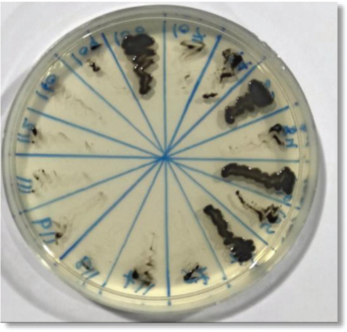
 Fig. 2:
 Case 1: material collected during surgery.
The second case involved a neck abscess. A 28-year-old man was admitted due to an abscess of the left half of the neck and facial skeleton with a large edema of this area. The patient complained of 2 days of pain in the neck and jaw, ranking 5/10 in the Numeric Rating Scale (NRS), fever up to 38°C, difficulty breathing and sore throat. Lockjaw precluded an examination of the oral cavity. In the laboratory tests, elevated inflammatory markers were observed (WBCs 16.61 103/μl, CRP 390 mg/l). Contrast-enhanced head and neck CT (Fig. 3) revealed signs of severe inflammatory infiltration with significant soft tissue edema in the left half of the neck and viscerocranium and with numerous, poorly separated dense-fluid compartments with peripheral enhancement. The abscess extended from the inferior pole of the left thyroid lobe to the head of mandible in the left temporomandibular joint. It was located in front of the left carotid vessels, in the left parapharyngeal space, deforming the pharyngeal air column and displacing the laryngeal part of the pharynx. On the left side, there were numerous enlarged lymph nodes of levels II and III. The patient was qualified for surgery. Simultaneously, an empirical intravenous antibiotic therapy with metronidazole (500 mg t.i.d.) and amoxicillin/clavulanic acid (1000 mg + 200 mg t.i.d.) was administered. Anesthesia consultation before the operation revealed carious tooth loss and purulent fistulas near the teeth roots. The patient was given ceftriaxone 1 g i.v. as a perioperative antimicrobial prophylaxis. During the intubation, a purulent collection emptied into the oral cavity and throat. During the surgery, after the preparation of the sternocleidomastoid muscle, the lateral wall of the pharynx and the larynx, followed by the elevation of the submandibular gland, another purulent collection in the parapharyngeal space including prestyloid compartment was revealed and drained. A second part of the neck abscess was found at the level of the hyoid bone, and a third-spreading along the wall of larynx and trachea into the subclavicular area. Purulent discharge was sent for bacteriological analysis. The peri- and postoperative course was uneventful. Microbiological analysis revealed Streptococcus group C sensitive to aminopenicillins, cephalosporins, carbapenems, clindamycin, levofloxacin and erythromycin. Antibiotic therapy was modified to metronidazole (500 mg t.i.d.) and ciprofloxacin (400 mg t.i.d.) lasting for 2 weeks of hospitalization. During hospitalization, inflammatory parameters normalized (WBCs 6.16 103/μl, CRP 3.52 mg/l). The patient was discharged in good general condition with a recommendation to continue the antibiotic therapy (cefuroxime p.o. 500 mg b.i.d.) for the next 7 days. At a first follow-up visit 1 week after the hospitalization, the patient remained in good condition, with the lockjaw effectively resolving, without fever. The surgical wounds were healing properly. Antibiotic therapy was continued with clindamycin p.o. (600 mg b.i.d.) for 7 days. After 3 more weeks, a physical examination showed no signs of inflammation. Oral cavity sanitation was planned.
 Fig. 3:
 Case 2: computed tomography of the head and neck – frontal and sagittal section.
DiscussionThe classic odontogenic infection is a mixed aerobic-anaerobic infection, with anaerobes predominating [1,4,5]. The main aerobic bacteria isolated are α-hemolytic Streptococci, microaerophilic Streptococci, and Staphylococcus aureus. The predominant anaerobic bacteria are Peptostreptococcus, Fusobacterium, and Gram-negative bacilli (Bacteroides, Prevotella, Porphyromonas, Propionibacterium). All these isolates are members of the natural oropharyngeal flora.
The etiological factors of odontogenic sinusitis differ from the typical microorganisms causing infectious sinusitis (Streptococcus pneumoniae, Haemophilus influenzae) [5]. This is caused by the impaired mucus drainage through the natural ostium into the middle meatus and increased intranasal pressure which result in a reduction of the oxygen tension in the sinus [1]. To achieve therapeutic success and prevent relapse, it is necessary to eliminate the source of infection [1,4,5].
The choice of antimicrobial treatment should be guided by the result of an antibiogram. Antibiotic therapy is based on penicillin (phenoxymethylpenicillin, aminopenicillin with beta-lactamase inhibitor). However, microbiological reports indicate that in 50% of cases of acute sinusitis and 75% of chronic sinusitis, beta-lactamase-producing bacteria were isolated [4,5]. Metronidazole, linkozamides, cephalosporins and macrolides are alternative antibiotics (following from the drug-resistance test), but metronidazole should be administered with an agent effective against the aerobic or facultative streptococci [1]. If conservative treatment fails, surgical intervention is indicated. The Caldwell-Luc operation, although largely replaced by less radical functional endoscopic sinus surgery (FESS) [6], remains the only procedure that ensures wide access to the sinus in the case of large odontogenic changes in the maxillary sinus [5].
Odontogenic infections can spread to adjacent tissues and lead to a deep neck space infection. The most common symptoms of deep neck infection are neck pain, fever, malaise, swelling, odynophagia, dysphagia, trismus, dysphonia, otalgia [2,7]. Deep neck abscess is potentially fatal and requires aggressive diagnostics and therapy to avoid life-threatening complications (including airway obstruction, jugular vein thrombosis, descending mediastinitis, pericarditis, pleural empyema, cavernous sinus thrombosis, sepsis, pleuropulmonary suppuration and hematogenous dissemination to distant organs) which have been reported to occur at a rate of 10–20%, even in the recent literature [2,3,8–10].
A contrast-enhanced head and neck CT scan is the examination of choice in such cases because it determines the type, localization and the extent of inflammation [11].
Deep neck space infections are generally polymicrobial, particularly Gram-positive. The most common findings are Streptococcus viridans, Streptococcus milleri, Prevotella spp., Peptosstreptococcus spp. and Klebsiella pneumoniae whereas Streptococcus pyogenes/S. pneumoniae is the most frequent polymicrobial culture. Management of deep neck space infection is traditionally based on prompt surgical drainage of the abscess followed by antibiotics IV, management of complications and supportive treatment [2,7]. Antibiotic therapy is usually started empirically, then subsequently updated based on a culture and sensitivity report [2]. Empiric antibiotics must cover Gram-positive and Gram-negative aerobic and anaerobic pathogens. For optimal coverage, a penicillin with a beta-lactamase inhibitor combination (such as amoxicillin/clavulanic acid), or a beta-lactam antibiotic resistant to beta-lactamase (such as cefuroxime, meropenem or imipenem) in combination with a drug highly effective against most anaerobe bacteria (such as metronidazole or clindamycin) is advised [7,12,13]. Usually second- or third-generation cephalosporins along with metronidazole are administered. Bakir et al.[8] and Wang et al.[14], however, preferred penicillin and metronidazole for initial antibiotic therapy, and clindamycin for severe deep-neck infection. In their studies, second- or third-generation cephalosporins were used instead if poor clinical response was noted or if complications had developed. Surgical management of deep neck space infections usually involves drainage of purulent abscesses via an external incision [15].
ConclusionsThe clinical manifestations of odontogenic infections vary depending on the anatomical spaces involved. The aggressive course of such infections requires comprehensive diagnostic and therapeutic management. Invasive abscess in the maxillary sinus may mimic a neoplastic process. A deep neck space abscess involving soft tissues surrounding the floor of the mouth or larynx might lead to upper airway obstruction. Therapy must include early administration of antibiotics and surgical intervention, if warranted.
AcknowledgementsThe authors thank the patients for allowing the publication of this case report.
Contributions of authors: All authors made substantial contributions to the present study. A.S. and M.R.C. conceptualized this case report. M.W. and J.S. performed the surgeries. A.S. and J.C. participated in the clinical management of the patients. J.S. supervised the clinical care of the patients. A.S. and M.R.C. analyzed and interpreted the patient data. A.S., M.R.C. and M.L. were responsible for the literature review and wrote the paper. P.S. and J.Sz. prepared histopathological images and description of histopathological analyses. M.W., M.L. and J.C. guided the manuscript draft before submission. All authors read and approved the final manuscript.
Funding: None.
Conflicts of interestThe authors declare that they have no competing interests in relation to the present work. This paper deals with patients’ treatment at the University Hospital in Krakow (Poland). The patients were treated by the authors.
References 1. Itzhak B. Sinusitis of odontogenic origin. Otolaryngol Neck Surg 2006; 135:349–355. 2. Brito TP, Hazboun IM, Fernandes FL, Bento LR, Zappelini CEM, Chone CT, et al. Deep neck abscesses: study of 101 cases. Braz J Otorhinolaryngol 2017; 83:341–348. 3. Bali RK, Sharma P, Gaba S, Kaur A, Ghanghas P. A review of complications of odontogenic infections. Natl J Maxillofac Surg 2015; 6:136–143. 4. Brook I. Microbiology of acute and chronic maxillary sinusitis associated with an odontogenic origin. Laryngoscope 2005; 115:823–825. 5. Mehra P, Jeong D. Maxillary sinusitis of odontogenic origin. Curr Allergy Asthma Rep 2009; 9:238–243. 6. Barzilai G, Greenberg E, Uri N. Indications for the Caldwell-Luc approach in the endoscopic era. Otolaryngol Head Neck Surg 2005; 132:219–220. 7. Han X, An J, Zhang Y, Gong X, He Y. Risk factors for life-threatening complications of maxillofacial space infection. J Craniofac Surg 2016; 27:385–390. 8. Bakir S, Tanriverdi MH, Gün R, Yorgancilar AE, Yildirim M, Tekbaş G, et al. Deep neck space infections: a retrospective review of 173 cases. Am J Otolaryngol 2012; 33:56–63. 9. Mathew GC, Kumar Ranganathan L, Gandhi S, Jacob E, Singh I, Solanki M, et al. Odontogenic maxillofacial space infections at a tertiary care center in North India: a five-year retrospective study. Int J Infect Dis 2012; 16:e296–e302. 10. Zhang C, Tang Y, Zheng M, Yang J, Zhu G, Zhou H, et al. Maxillofacial space infection experience in West China: a retrospective study of 212 cases. Int J Infect Dis 2010; 14:e414–e417. 11. Wabik A, Hendrich BK, Nienartowicz J, Guziński M, Sąsiadek MJ. Odontogenic inflammatory processes of head and neck in computed tomography examinations. Polish J Radiol 2014; 79:431–438. 12. Parhiscar A, Har-El G. Deep neck abscess: a retrospective review of 210 cases. Ann Otol Rhinol Laryngol 2001; 110:1051–1054. 13. Vieira F, Allen SM, Stocks RMS, Thompson JW. Deep neck infection. Otolaryngol Clin North Am 2008; 41:459–483. 14. Wang LF, Kuo WR, Tsai SM, Huang KJ. Characterizations of life-threatening deep cervical space infections: a review of one hundred ninety-six cases Am. J Otolaryngol 2003; 24:111–117. 15. Eftekharian A, Roozbahany NA, Vaezeafshar R, Narimani N. Deep neck infections: a retrospective review of 112 cases. Eur Arch Otorhinolaryngol 2009; 266:273–277.
留言 (0)