Objectives:
To report on baseline refractive and keratometric values and their correlation with tomographic characteristics of eyes with keratoconus (KC).
Methods:
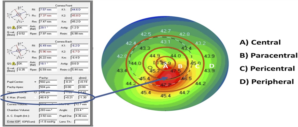
Retrospective chart review of patients treated in a single-center cornea and refractive surgery practice. Baseline topographic measurements were reviewed for 1,012 keratoconic eyes of 586 patients between 2008 and 2018. The manifest refraction, thinnest pachymetry (Pthin), corneal astigmatism (Kastig), and the maximum (Kmax), steep (Ksteep), flat (Kflat), and mean (Kmean) keratometry were analyzed. The location of Kmax (x, y) was used to determine central (<1 mm), paracentral (1–3 mm), pericentral (3–5 mm), or peripheral (>5 mm) cone locations.
Results:
In the entire cohort, the mean manifest sphere was −2.2±4.4 diopters (D) and the cylinder was −3.2±2.3 D. In total, 48.6% of patients had against the rule (ATR) manifest astigmatism (Mastig). The average Kastig was 3.8±2.7 D, and unlike the manifest axis, 50.2% of patients had with the rule (WTR) Kastig. Patients with a Kmax less than 50 D had an Mastig of −1.9±1.6 D, 45.9% of which was ATR Mastig. With respect to baseline tomography measurements, Kmax, Ksteep, Kflat, and Kmean were 58.0±9.4, 50.6±6.5, 46.8±5.9, and 48.6±6.1 D, respectively. There was a weak correlation between Kmax and simulated keratometry (Ksteep, Kflat, and Kmean) for patients with a Kmax less than 60 D.
Conclusions:
Simulated keratometry is poorly correlated with KC severity until the disease is more severe. Mastig ≥2 D and ATR Mastig were correlated with KC at all levels of severity. Mastig ≥2 D and ATR Mastig may serve as a simple, inexpensive, and widely available indicator for topographic analysis to identify possible KC and suggest further workup; however, further prospective studies are needed to confirm its utility.


留言 (0)