Ophthalmic involvement has been observed in up to 40% of VEXAS syndrome patients, with episcleritis, scleritis, and uveitis being the most common manifestations, and orbital and periorbital inflammation occurring less frequently.
A literature review for English language publications with the keyword “VEXAS” produced 85 results, of which 21 articles described ocular features in VEXAS syndrome (summarized in Table 2). Periorbital edema is the most common ocular adnexal finding, with other manifestations including orbital inflammation or cellulitis, orbital myositis, dacryoadenitis, optic perineuritis, or oculomotor nerve paresis.
Table 2 Summary of ocular features reported in VEXAS syndrome in the current literature
Systemic corticosteroids are the mainstay of management for VEXAS syndrome, [2] including its ophthalmic manifestations, although orbital involvement may be self-limiting, as observed in our case. Various steroid-sparing medications, such as mycophenolate and methotrexate, have been shown to be beneficial in the long-term management of VEXAS syndrome. Tocilizumab (a monoclonal IL-6 receptor inhibitor) and ruxolitinimib (a JAK inhibitor) have also been found to be effective in controlling the disease [25]. Azacitidine (a hypomethylating agent) has been reported to be effective in selected patients with VEXAS and associated myelodysplastic syndrome [26]. Furthermore, there have been promising results following allogeneic hematopoeitic stem cell transplant, with some patients achieving complete disease remission [2].
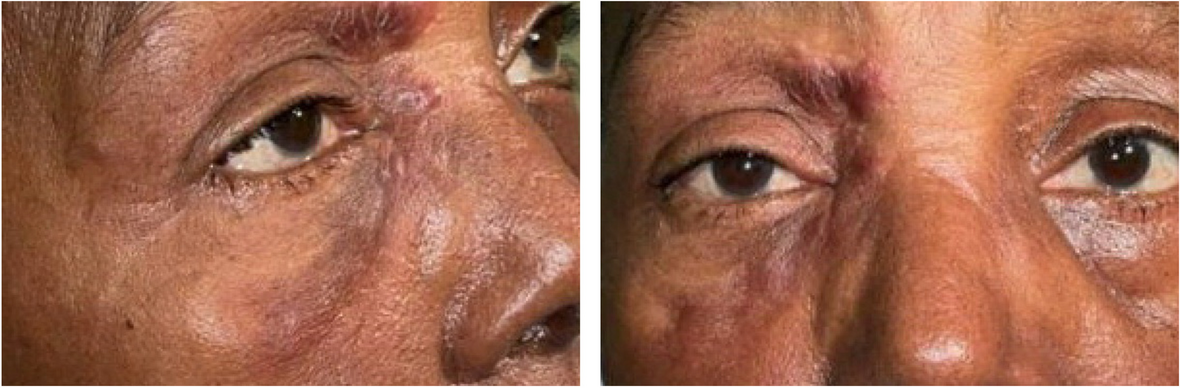
The current case represents a rare ophthalmic manifestation of VEXAS syndrome, with recurrent orbital and anterior cavernous sinus/superior orbital fissure syndrome, orbital myositis, and dacryoadenitis; the ophthalmoplegia being probably caused by a combination of myositic and paralytic factors. Despite the self-limiting inflammatory syndrome and long-term immunosuppressive therapy, the patient had mild persistent ophthalmoplegia and blepharoptosis.
When orbital inflammatory syndrome secondary to systemic disease is suspected, a detailed history, systemic review, and laboratory evaluations are paramount. This approach includes a full blood count, metabolic panel, thyroid function tests, inflammatory markers, and an autoimmune profile encompassing anti-nuclear antibodies (ANA), antineutrophil cytoplasmic antibodies (ANCA), serum angiotensin-converting enzyme (ACE) and immunoglobulin G4 (IgG4) levels. CT and MRI scans may reveal a spectrum of findings such as enhancing orbital mass, uveoscleral thickening, enlarged extraocular muscles or lacrimal glands, diffuse inflammatory changes, apical lesions, cavernous sinus or paranasal sinuses involvement. Positron emission tomography (PET) scans may also be useful in detecting multi-system inflammatory disease or malignancy. If clinical and imaging results remain ambiguous, orbital biopsy is advised to identify the cause of the orbital inflammation. In the current case, blood tests and orbital imaging indicated an inflammatory process, within the context of multi-system disease and constitutional symptoms such as fatigue, muscle wasting and weight loss. The diagnosis of VEXAS in the current case was ultimately confirmed by bone marrow biopsy and genetic testing.
In summary, VEXAS syndrome should be considered in the differential diagnosis of orbital inflammatory syndromes, particularly in older patients who have multi-organ inflammation and or haematological disorders. With a broad spectrum of clinical manifestations, patients may be under the care of multiple specialists for seemingly unrelated inflammatory conditions, until the unifying diagnosis of VEXAS is confirmed by genetic testing for the UBA1 mutation.


Comments (0)