Deep Learning–Based Image Noise Quantification Framework for Computed Tomography
Objective
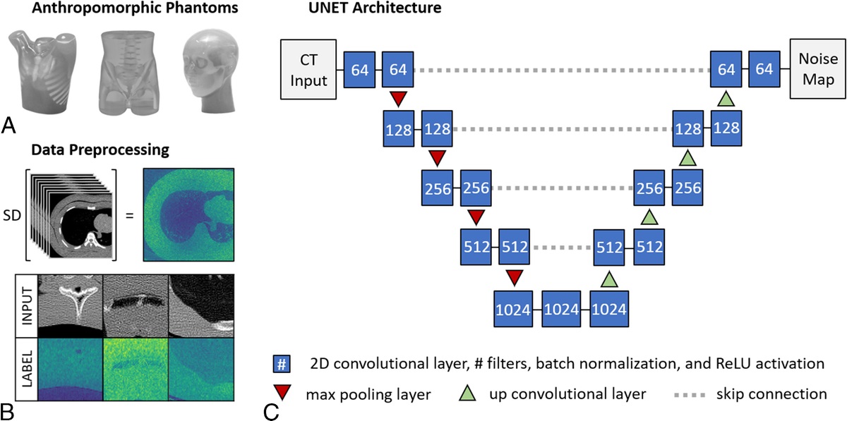
Noise quantification is fundamental to computed tomography (CT) image quality assessment and protocol optimization. This study proposes a deep learning–based framework, Single-scan Image Local Variance EstimatoR (SILVER), for estimating the local noise level within each region of a CT image. The local noise level will be referred to as a pixel-wise noise map.
Methods
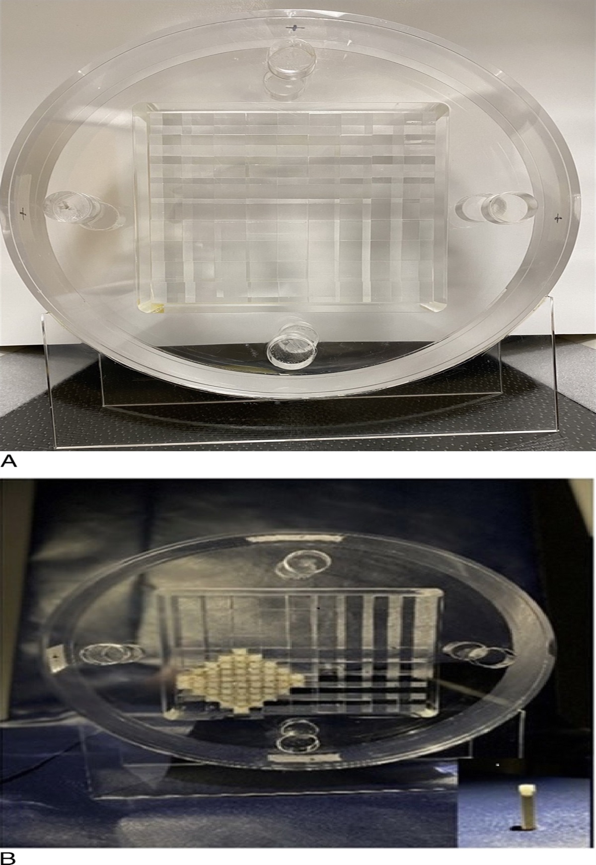
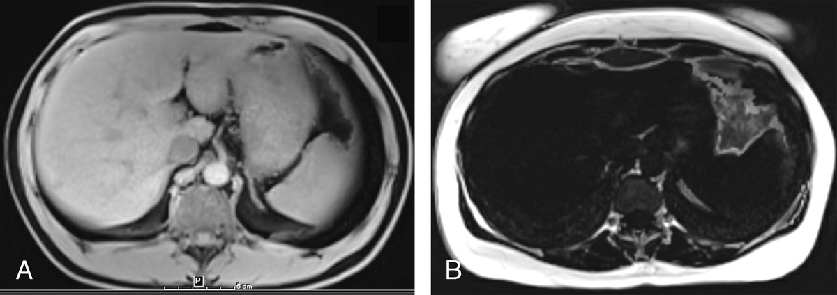
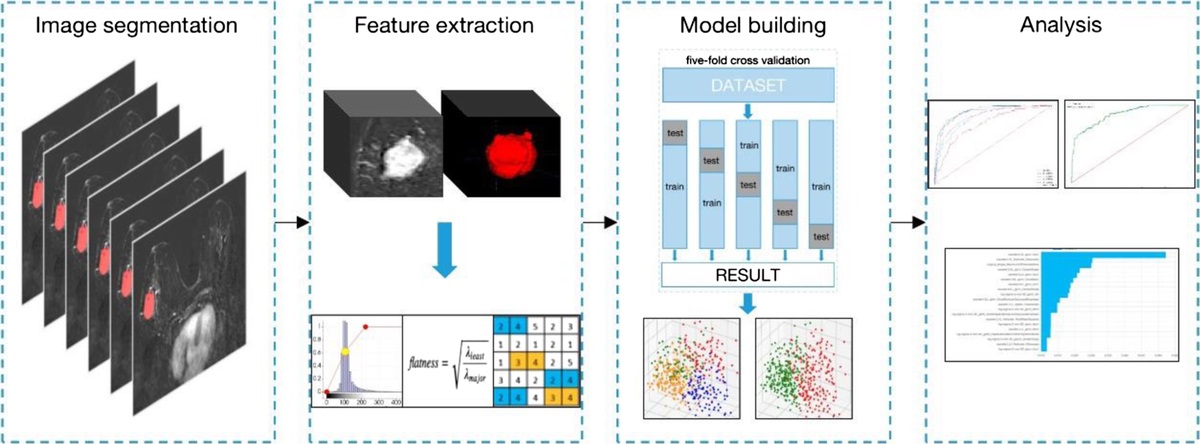
The SILVER architecture resembled a U-Net convolutional neural network with mean-square-error loss. To generate training data, 100 replicate scans were acquired of 3 anthropomorphic phantoms (chest, head, and pelvis) using a sequential scan mode; 120,000 phantom images were allocated into training, validation, and testing data sets. Pixel-wise noise maps were calculated for the phantom data by taking the per-pixel SD from the 100 replicate scans. For training, the convolutional neural network inputs consisted of phantom CT image patches, and the training targets consisted of the corresponding calculated pixel-wise noise maps. Following training, SILVER noise maps were evaluated using phantom and patient images. For evaluation on patient images, SILVER noise maps were compared with manual noise measurements at the heart, aorta, liver, spleen, and fat.
Results
When tested on phantom images, the SILVER noise map prediction closely matched the calculated noise map target (root mean square error <8 Hounsfield units). Within 10 patient examinations, SILVER noise map had an average percent error of 5% relative to manual region-of-interest measurements.
Conclusion
The SILVER framework enabled accurate pixel-wise noise level estimation directly from patient images. This method is widely accessible because it operates in the image domain and requires only phantom data for training.


留言 (0)