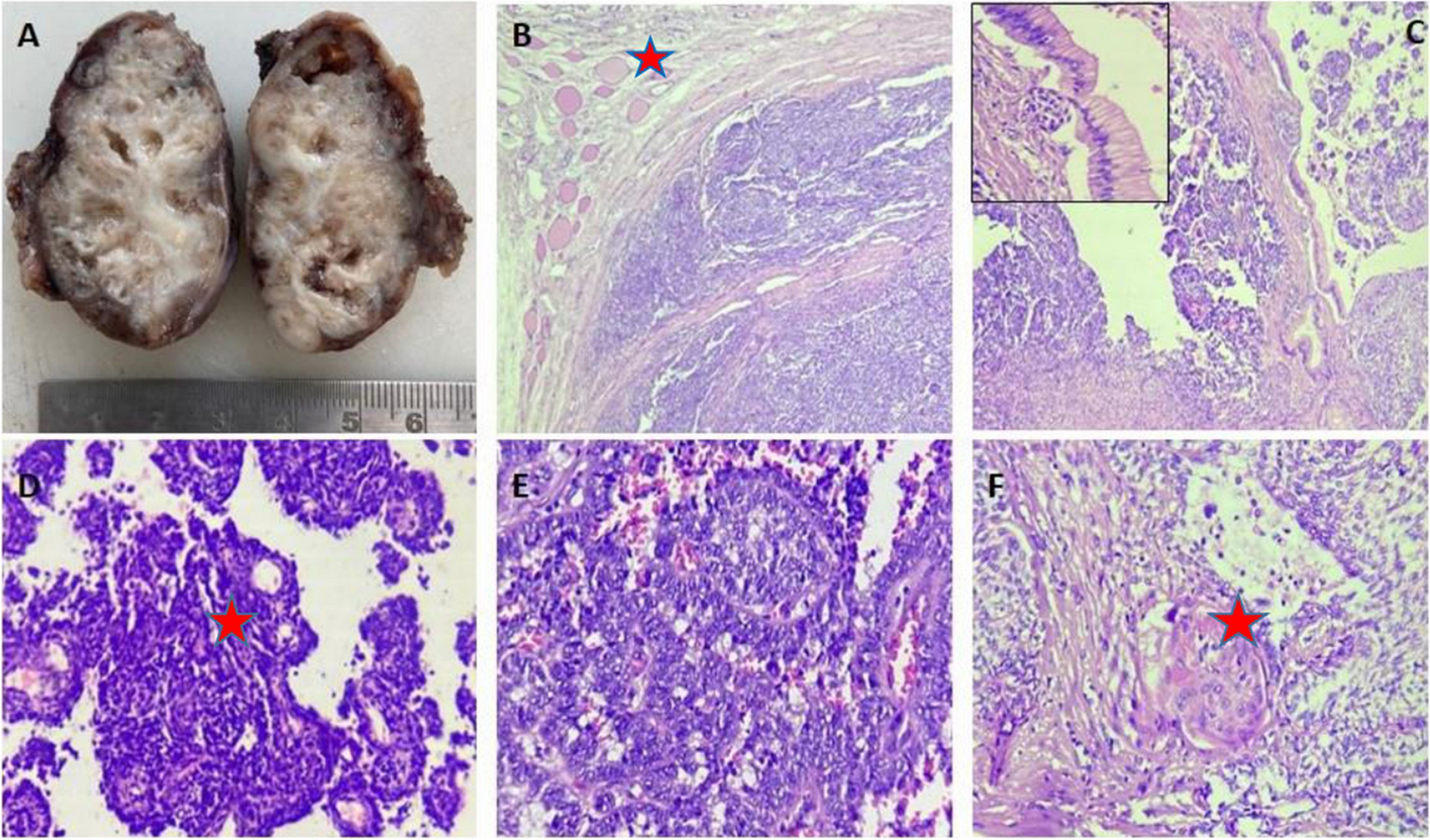
The thyroid is a unique endocrine organ where both benign and malignant lesions can co-exist. PTC is the most common thyroid malignancy and is often present along with BTLs like HT or NG more often than, without any pre-existing thyroid pathology [15,16,17]. Although the histopathological features of PTC have been very well defined, many BTLs do show some nuclear features mimicking PTC. Nuclear grooving is a longitudinal nuclear membrane invagination seen along the long axis of the nucleus of thyroid follicular cells with an oval rather than round appearance. These features are always given significant importance in thyroid cyto-histopathology in differentiating non-neoplastic from neoplastic lesions particularly, in the PTC. But, when nuclear grooves with oval shape, chromatin clearing, and nuclear overlapping, are seen in BTLs, they do cause a diagnostic dilemma, resulting in the diagnosis of “atypical cytology/indeterminate significance” on fine needle aspiration (FNA) samples [3, 18, 19]. C D Scopa et al. studied 80 non-papillary thyroid lesions both neoplastic and non-neoplastic and showed that nuclear grooving was present in a variety of thyroid lesions with 76% of the BTLs having grooves of ≤ 6/hpf [3]. The frequency of nuclear grooving among BTLs in our study was 36.8% but studies have reported the frequency of grooving to range between 3.6% to 52.4% based on the history of irradiation [6, 19]. Various studies have tried to ascertain the significance of nuclear grooving, which are functional channels connecting the nuclear envelope to the chromatin and have been proposed to have a possible role in Ca + + signaling ortransport from the cytoplasm to the nucleus [12, 20]. Some studies have put forth a semi-quantitative approach of counting the number of cells with nuclear grooves and categorizing the lesions as either PTC, indeterminate cytology or benign lesions [21]. Benign thyroid hyperplastic nodules are typically characterized by thyroid follicular cells with small, round, dark nuclei and a honeycombing pattern of arrangement. However, focal nuclear atypia, including grooves, oval shape, chromatin clearing, and overlapping, have been reported in hyperplastic nodules, which leads to diagnostic difficulties and can be mistaken for PTC [18, 19]. Studies have also shown that some of the BTL like FA, HT, and adenomatous goiter show positivity for RET/PTC gene translocation by RT-PTC [7]. Among the BTLs, HT is commonly associated with PTC and micro-papillary tumors [22, 23]. Small clusters of cells do show PTC-like nuclear features, particularly grooving in HT [21, 24, 25].
However, there is no definitive demarcation of cells with such PTC-like nuclear features to make a definitive diagnosis of PTC. It could be hypothesized that chronic inflammation with auto-antibody production and T-cell mediated cytotoxic effect in HT, might result in DNA damage and genetic alterations [24]. We observed that 6 out of 12 cases of HT, had grooving, with 3 out of 6 cases being positive for RET/PTC gene translocation. There was a statistically significant association of nuclear grooving in HT with genetic alteration like RET/PTC translocation. Apart from grooving, nuclear enlargement, irregular nuclear membrane, and clearing due to peripheral chromatin condensation were also commonly seen in our study suggesting the possibility of genetic or epigenetic alterations. The study by Dae-Young Kang et al. on normal thyrocytes, oxyphil cells, and PTC cells dissected by Laser capture to study the RET/PTC-RAS-BRAF cascade in these cells showed an increased nuclear expression of RET, RAS, and ERK proteins in oxyphil cells, PTC cells and concluded a molecular link between Hurthle cell metaplasia and PTC progression [26].
Our study also favors that genetic alterations like RET/PTC gene translocation results in PTC-like nuclear morphology in HT and that, there could be further additional mutations or alterations in downstream signaling pathways or unknown epigenetic alterations responsible for the overt development of PTC in HT. As studies have shown that PTC associated with HT are usually multifocal and aggressive [22], it is worthwhile looking for the PTC-like nuclear features in HT on cytology, particularly nuclear grooving, as regular follow-up with ultrasound and FNA can be warranted for any increase in the size of the lesion and early detection of PTC. As induction of RET/PTC alterations has shown to induce nuclear irregularities in some studies, genetic testing for RET/PTC alteration in an FNA sample of HT showing increased nuclear grooving with oval and elongated nucleus might aid in the early detection of PTC [27]. Apart from nuclear grooving, many studies have shown intra-nuclear pseudo-inclusions in HT [24]. The intra-nuclear pseudo-inclusions of PTC are distinct and appear as “punched out” areas in the nucleus, whereas in BTLs they appear as vague nuclear clearings.
Follicular neoplasms are another benign lesion showing nuclear grooving. In our study, all 8 cases of FA, showed grooving of varying degrees with RET/PTC gene translocation seen in 2 cases. The follicular lesions range from FA to the newer entities of WHO Classification, the borderline tumors like “Uncertain malignant potential (UMP)” and “noninvasive follicular thyroid neoplasm with papillary-like nuclear features” (NIFTP) [28]. The distinction between FA and the borderline entities is based on the scoring of PTC-like nuclear features, further pressing the significance of nuclear grooves. Although our study did not show any statistical significance between grooving and RET/PTC translocation in FA, closer observation for PTC-like nuclear features will aid in a definitive diagnosis, warranting regular follow-up of the patient for any rapid increase in the size of the lesion and planning further management.
Many non-thyroid lesions and tumors also show nuclear groovings like granulosa cell tumor of the ovary, mesothelioma, and Langerhans cell histiocytosis, to name a few. These lesions have different underlying genetic alterations, pointing to the fact that nuclear irregularities can have multiple causative factors like BRAF mutations in Langerhans cell histiocytosis and FOXL1 gene mutation in ovarian granulosa cell tumor [29].


留言 (0)