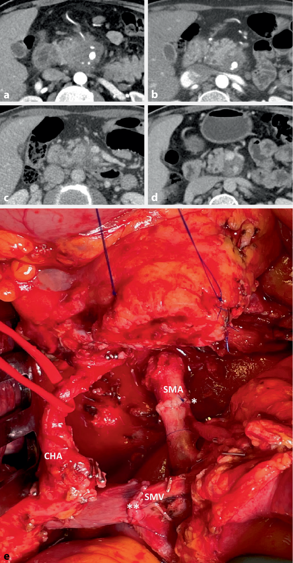
Simple liver cysts are often asymptomatic and have no malignant potential. However, symptoms from large cysts can arise and surgical therapy is warranted in these patients. Although there is no clear cut-off for defining a giant liver cyst, it has been reported that cysts > 10 cm in diameter are more likely to cause compression-related symptoms [6]. Cyst contents are usually serous, but proteinaceous material from previous intra-cystic hemorrhage may be present. Massive hemorrhage and/or spontaneous rupture of SLC has also been described and may require an emergency surgical procedure [3].
Treatment modalities for SLC range from percutaneous needle aspiration and sclerotherapy with ethanol injection to open or laparoscopic fenestration. Liver resection is usually performed in the setting of polycystic liver disease or when biliary cystoadenoma or carcinoma is suspected [1].
Laparoscopic deroofing, first described by Z’Graggen et al. [7] in 1991, has gradually become the surgical therapy of choice, especially for cysts located in the ventral and left lateral locations. Kisiel et al. [8] reported the long-term outcomes including symptom relief and quality of life in 48 patients operated in a single hepatobiliary unit between 2000 and 2012. There was one (2%) bile leak and no mortality. These patients were followed for a median of 62 (22–173) months and 60.4% were followed up for over 5 years. Of 46 patients who had initial symptom relief, 37 (80%) reported long-term symptomatic benefit and 2 underwent redo surgery with open fenestration and omentopexy. In a large meta-analysis including 62 studies with a total of 1314 patients, Bernts et al. [2] reported symptomatic relief in 90.2% and symptomatic recurrence in 9.6% of the patients, with a reintervention rate of 7.1%. The postoperative complication rate was 10.8% and the major complications rate 3.3%, with a procedure-related mortality of 1%.
A search of English-language case reports or case series published in PubMed and Scopus identified 35 publications, with a total of 148 patients treated by laparoscopic deroofing of large hepatic cysts. We restricted our search to January 2000 to December 2022. We included studies reporting on both conventional and single-port laparoscopy (Table 1).
Table 1 Summary of the selected publicationsStudies reporting procedures performed via open laparotomy or robotic surgery and/or associated with liver resections for polycystic liver disease (PLD) were excluded. Pediatric patients were also excluded. According to Murad et al. [44], we assigned a high, medium, or low quality score to each study according to clinical relevance, and reported surgical details and length of follow-up.
Based on the above criteria, one third of the studies (28.5%) were considered to be of high quality. Single-port laparoscopy was performed in 9 studies, for a total of 28 patients (18.9%). Simple liver cysts were more common in women, with a male to female ratio of 1:3.2. Patient age ranged between 47 and 88 years, with a mean age at diagnosis of 72 years. Hepatic cysts ranged in size from 5 to 28 cm and were slightly more frequent in the right (n = 42) than in the left liver segments (n = 37). The majority (78%) of patients had a single liver cyst. Seven studies reported a total of 12 patients with PLD [10, 12, 20, 26, 31, 38, 41]. Overall, 7 (4.7%) patients presented with acute abdomen and underwent emergency surgery for spontaneous cyst rupture, intra-cystic bleeding, or infection [11, 29, 30, 32, 41, 42]. Conversion to laparotomy was reported in only 1 patient (1/149, 0.6%) and was due to a challenging hepatic mobilization during excision of a single hepatic cyst located in segment 8 [10].
Surprisingly, only 22 publications included details of the surgical procedure and devices used to complete the fenestration. In a minority of patients (10.8%), laparoscopic deroofing was combined with omentopexy (n = 8), argon plasma coagulation (n = 4), or was preceded by ethanol sclerotherapy (n = 4). To date, there is no clear consensus in the literature regarding the real efficacy of these ancillary techniques in terms of the recurrence rate, and the evidence to support their use is limited. In addition, they are not free from complications such as cauterization of a major vascular structure or iatrogenic cholangitis due to the sclerosing agent [2].
Regarding the surgical device, the harmonic scalpel (UltracisionTM) was used in 13 centers and a vessel-sealing system (LigaSureTM) in 8. Both devices are considered effective and safe in liver surgery [45, 46].
On the other hand, we found only 3 publications reporting use of a stapling device during laparoscopic deroofing [13, 27, 35]. Stapling devices have gained popularity in laparoscopic liver surgery and are generally employed for major vessel division and less frequently for parenchymal transection [47, 48]. Some authors have used an endoscopic linear stapler to minimize any possibility of blood loss or bile leaks during excision of a thick cyst wall [27]. Interestingly, Umemura et al. [35] used a linear stapler to prevent bile leakage after injecting ICG through an endoscopic nasobiliary drainage. We used the harmonic scalpel to initially excise the cyst wall but then preferred to complete the deroofing with an endoGIA stapler due to the thickness of the cyst wall near the liver parenchyma.
For years, it has been known that intravenously administered ICG is excreted into the bile [49]. In this review, use of ICG was reported in only four studies, all published after 2016. In the case reported by Tanaka et al. [31], intravenous ICG allowed identification of an unexpected bile leakage, which could not be identified in the initial laparoscopic view. However, there is a lack of agreement on the timing of ICG administration. During laparoscopic cholecystectomy, ICG is administered between 0.5 h and 24 h before the procedure. In 3 patients undergoing laparoscopic deroofing [31, 36, 40], ICG was injected intravenously 1 h before surgery, after induction of anesthesia, and 24 h before surgery (followed by an intraoperative boost). We decided to administer ICG 4 h before induction of anesthesia, and there was no need for an intraoperative boost. Indocyanine green fluorescence helps to recognize the correct boundaries to liver parenchyma and to assess the real thickness of the cyst wall edges. Recent consensus guidelines on the use of ICG during open and laparoscopic hepatobiliary surgery state that intraoperative fluorescence imaging improves anatomical visualization, has the potential to guide surgical dissection, and is capable of augmenting safety, efficiency, and outcomes [49].
Finally, from our literature search, only 5 patients presented with a symptomatic recurrence during a follow-up ranging from 2 to 72 months from the index operation, and all required laparoscopic revision [10, 14, 25]. Limitations of this review are that some studies do not describe the extent of deroofing, a short follow-up may have underestimated recurrence rates, and postoperative imaging may have been undertaken only in patients with recurrent symptoms.


留言 (0)