Imaging of the biliary tract
Purpose of review
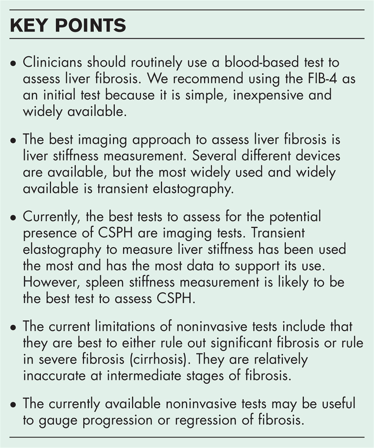
Magnetic resonance cholangiopancreatography (MRCP) has become the reference examination for the exploration of the biliary tract and has replaced endoscopic cholangiography for the analysis of the biliary tract because of its equivalent performance and its noninvasive character.
Recent findings
Based on the International Primary Sclerosing Cholangitis (PSC) Study Group recommendations for MR imaging in PSC, two protocols can be distinguished for the imaging of biliary tract: a basic protocol and a more complete protocol. It is essential to know the main pitfalls in order not to wrongly describe biliary anomalies. In addition to the excellent performance of MR imaging with MRCP in analyzing the anatomy and the anomalies of the biliary tree, complementary techniques have recently been developed. Several MR prognostic factors have been described. New hepato-specific contrast agents are now available for assessment of the general and segmental liver function. MR Elastography and Diffusion-weighted MR sequences are accurate to evaluate the degree of hepatic fibrosis. Finally, images obtained in MRCP can be postprocessed by a software that will analyze and model the biliary tree in order to quantitatively evaluate the biliary system.
Summary
Magnetic resonance imaging with its recent developments becomes by now an essential tool for the evaluation of biliary diseases


留言 (0)