
記住我
Binaural beats can serve as an effective modality for the non-pharmacological non-invasive reduction of anxiety in pediatric patients during dental treatment.
The reduction in anxiety was significantly greater as compared to white noise due to the brainwave entrainment effect of binaural beats
An exposure time of 10 min is sufficient to achieve the desired effect; which is also suitable for a routine dental clinical setting.
INTRODUCTIONManagement of apprehensive or anxious children in dental clinics is relatively difficult when compared with adults and can be quite time-consuming, costly, and demanding for the clinician.[1,2] Over time, a plethora of techniques have been developed for the management of the behavior of anxious pediatric patients during dental treatment. Although newer techniques such as general anesthesia and sedatives are quite promising with surety of results, non-invasive psychotherapeutic management techniques are always preferred as they help in avoiding adverse effects associated with the use of drugs and also the discomfort associated with invasive modalities.[3]
Among various non-pharmacological behavior management techniques, distraction with audio and videos is a simple and effective technique that focuses children’s attention away from noxious stimuli.[4] Based on this principle, audio analgesia has been previously used to diminish the fear and anxiety of the patients at the time of local anesthesia administration and dental extraction.[5] Audio analgesia, first introduced by Gardner and Licklider in 1959, is the relief of pain using audio means without any pharmacological agents while doing painful medical procedures such as dental treatments.[6]
Audio analgesia in the form of live music, familiar songs, calm sounds, and white noise has been previously utilized with satisfactory results in a reduction of anxiety and subsequently increased pain threshold in dental patients.[7,8] Although simple and effective, the conventional audio analgesia modalities relied mostly on the principle of distraction and thus, posed with certain limitations. Binaural beats, first described by Dove in 1939, occur when two sounds with steady intensities but different frequencies are presented separately, one to each ear.[9] The resulting perception in the brain equals the difference between them requiring a collective action of both ears.[10] For example, when a frequency of 152 Hz tone is presented to the left ear and a 150 Hz tone to the right, a beat of 2 Hz is perceived in the brain.[11] Depending on the difference in frequency, various types of brainwaves (alpha, beta, gamma, delta, epsilon, and theta) may be generated which, in turn, arouse corresponding cognitive and behavioral responses[12] and the technique is known as “brainwave entrainment.”
Overall, binaural beats provide additive benefits of conventional distraction audio analgesia along with brainwave entrainment which forms its primary scientific principle. Brainwave entrainment waves are widely used for relaxation in conjugation with meditation or yoga, and their use in dental patients has also been attempted.[13] The present study was conducted with the objective of further understanding and validating the clinical effectiveness of binaural beats in the reduction of anxiety in pediatric patients during dental treatment procedures ranging from minor procedures such as fluoride application to major invasive ones like extraction of teeth.
MATERIAL AND METHODS Preliminary preparationsThe study conducted was a randomized, clinical, and controlled trial. The study protocol was approved by the institutional ethical review board and has been registered in the Clinical trial registry of India (CTRI) database [Ref: CTRI/2021/05/033436]. The study sample comprised 120 anxious pediatric patients (American Society of Anesthesiologists [ASA] Physical Status 1) of age ranging from 3 to 10 years undergoing dental treatment that ranged from fluoride application or scaling to invasive procedures such as extraction or root canal treatment.[14] The patients were then assigned to either the study or control (n = 60 each respectively) group using a randomly generated code. Pure binaural beats of frequencies 344 Hz (left) and 340 Hz (right) that would result in a theta wave of 4 Hz were produced by “Brain Waves - Binaural Beats” (MynioTech Apps, Chapeco, Santa Catarina, Brazil) software compatible with smartphones and used in patients belonging to the experimental group. Similarly, a pure white noise of 432 Hz was used in the control group.[15] The patient’s parents/ guardians were explained about the complete procedure and allowed to listen to the type of audio that was going to be presented to the patient following which written consent was obtained. A brief medical, as well as dental history of the child, was obtained and patients with a history of underlying medical conditions such as epilepsy, psychiatric, or hearing disorders were excluded from the study.
Presentation of soundFive minutes before the commencement of the treatment procedure, the anxiety level of the patient was assessed using Venham’s picture test (VPT) which comprises eight panels, each comprising of an “anxious” and a “non-anxious” picture.[16] The child was asked to point out the most relatable picture in each panel that was shown in a fixed numbered order. The selection of the anxious pictures was scored as 1 and the non-anxious one was scored as 0 for each panel. The total score of all eight panels was recorded as V1 which provided the initial level of anxiety subjectively perceived by the patient. At this point, an additional assessment of the patient’s pulse rate was also carried out using a Finger Pulse Oximeter (PO) (NecLife [NL]-50D, NecLife, India) and the value was recorded as PO1. Alteration in pulse rate is often correlated with changes in the stress levels of an individual and thus, an objective evaluation of the change in anxiety levels could be made across the duration of the treatment procedure.[17]
Followed by the first set of assessments, the patients in the experimental group were presented with pure binaural beats, and those in the control group were presented with white noise using wireless headphones (boAt Rockerz 370, Delhi, India) or earphones (boAt Rockerz 255 Pro+, Delhi, India) [Figure 1]. Although a recommended volume was set uniformly for all the patients to minimize variation in the study settings, they were allowed to control the volume in case of discomfort. To minimize external interference, the clinic area was maintained quiet during the procedure. Patients requiring treatment procedures generating external sound from an airotor such as cavity preparation or access opening were also excluded from the study for the same reason.

Figure 1:: Armamentarium used in the present study.
Export to PPT
In addition, the use of other behavior management techniques would imply that the reduction in anxiety level could not be entirely attributable to the sound being presented. Therefore, the surgeon did not speak to the patients except for treatment-related instructions. Non-cooperative patients that required additional behavior management techniques during the treatment were excluded from the study.
To avoid bias in scoring and assessment, the same investigator performed treatment for all the cases and was blinded concerning the sound being provided. The patient’s level of anxiety was reassessed using VPT and PO after 5 min of presentation of the sound and the values were recorded as V2 and PO2, respectively [Figure 2]. After recording the second set of values for anxiety assessment, the treatment procedure was commenced while the sound continued to be presented to the patient.

Figure 2:: (a) Presentation of binaural beats to the patient before beginning the treatment procedure, (b) Subjective assessment of anxiety level by Venham’s picture test, (c) Assessment of pulse rate by pulse oximeter.
Export to PPT
Data from patients that perceived the sound as uncomfortable during the treatment and demanded that the headphones be removed were not included in the final results. A final assessment of anxiety level was carried out 10 min from the starting point by VPT and PO and the values were recorded as V3 and PO3, respectively. The headphones were then removed and cleaned using alcohol-based sanitizer wipes and the patient’s parent/guardian was provided post-operative instructions about the treatment once it was completed.
Statistical proceduresThe recorded data were compiled on a Microsoft Office Excel Sheet (v 2019, Microsoft Redmond Campus, Redmond, Washington, United States) and proofed for entry errors. Statistical analysis was carried out using the Statistical Package for the Social Sciences (SPSS) (SPSS v 26.0, International Business Machines Corporation). For demographic data, an inter-group comparison was done using a t-test. Comparison of frequencies of categories of variables with groups was done using the Chi-square test. The normality of numerical data was checked using the Shapiro–Wilk test and was found that the data did not follow a normal curve; hence, non-parametric tests were used for comparisons. Mann–Whitney U test was performed to obtain a comparison of parameters between the two groups. The intra-group comparison was done using Friedman’s (for >2 observations) followed by pairwise comparison using Wilcoxon Signed rank test. For all the statistical tests, P < 0.05 was considered to be statistically significant, keeping α error at 5% and β error at 20%, thus giving power to the study as 80%.
RESULTSThe study sample (n = 120) comprised of 56 females and 64 males with a mean age of 5.867 years (standard deviation [SD] = 1.864). The experimental group (n = 60) comprised 33 females and 27 males having a mean age of 5.975 years (SD = 2.0366) while the control group consisted of 23 females and 37 males with a mean age of 5.758 years (SD = 1.6862). When considering the experimental group, the p values of the Wilcoxon Signed Ranks Test as well as the Friedman Test (P < 0.01) between all three-time points indicated that there was a statistically highly significant difference for the values of PO (PO1-PO2, PO2-PO3, and PO1-PO3) and VS (VS1-VS2, VS2-VS3, and VS1-VS3) wherein the values were highest at first timepoint (PO1 and VS1) and lowest at final evaluation (PO3 and VS3). These findings suggested that there was a definite reduction in anxiety between all the intervals both by subjective as well as objective assessment. In contrast, the PO3 and VS3 values were found to be significantly higher (P < 0.01) in the control group as compared to the study group. The end-point PO3 values being higher than the initial PO1 suggested that the anxiety level of patients in the control group increased from the time of initiation of the treatment procedure until its completion [Figure 3].
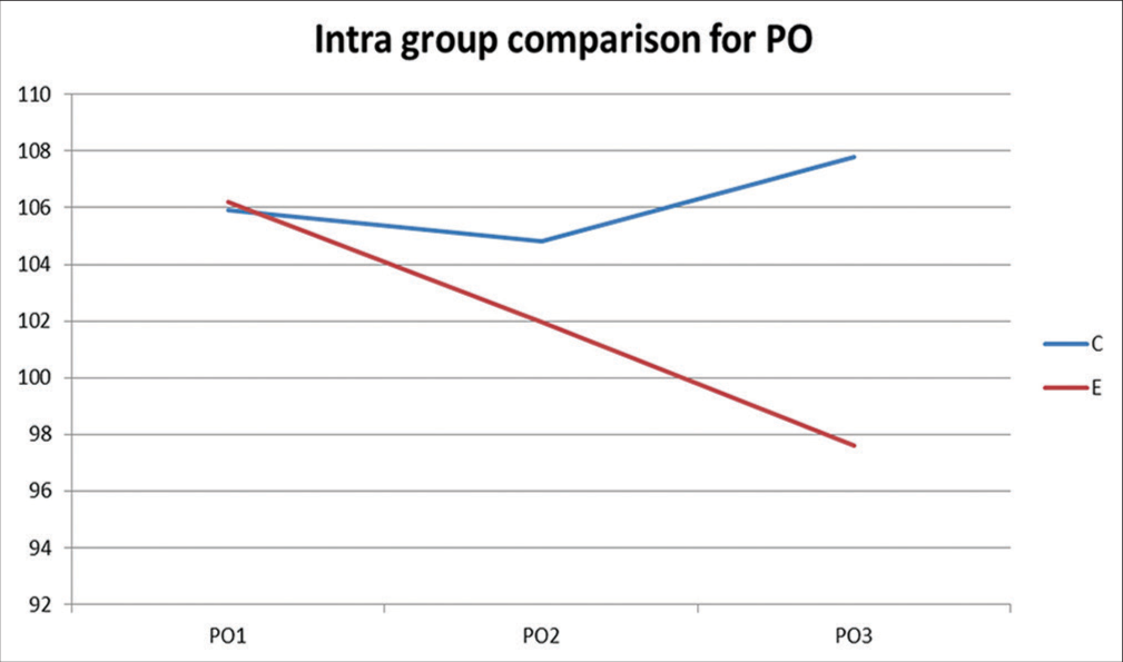
Figure 3:: Comparison of change in trend in pulse rate from first timepoint (PO1) to final timepoint (PO3) in experimental (blue) and control (red) groups.
Export to PPT
On comparison of both the groups using the Mann–Whitney U test, there was a statistically highly significant difference seen for the values between the groups (P < 0.01) for PO2, PO3, and VS3 with higher values in control while there was a statistically non-significant difference seen for the values between the groups (P > 0.05) for PO1, VS1, and VS2 [Table 1]. These findings indicated that the reduction in anxiety levels was statistically significant in the experimental group as compared to the control group. The non-significant differences for values of PO1 and VS1 were indicative of similar baseline characteristics of both groups at the initial time point. Thus, both the groups began with the same overall anxiety levels at the first time point and the reduction was found to be more significant in the experimental group.
Table 1:: Statistical analysis of pulse rate (PO) and anxiety level (VS) at different time points in control and experimental groups.
Group Mean Standard deviation Median Mann-Whitney U value Z value P-value of Mann-Whitney U test PO1 C 105.90 7.433 110 1755.500 −0.237 0.812# E 106.20 8.157 109 PO2 C 104.83 7.477 110 1341.000 −2.436 0.015* E 101.97 7.461 102 PO3 C 107.77 8.390 112 559.500 −6.544 0.000** E 97.63 6.894 98 VS1 C 3.47 1.432 4 1662.500 −0.747 0.455# E 3.72 1.585 4 VS2 C 3.53 1.384 4 1600.500 −1.100 0.271# E 3.23 1.619 4 VS3 C 3.65 1.716 4 1066.500 −3.925 0.000** E 2.43 1.577 2 DISCUSSIONThe concept of binaural beats is only valid when two sinusoidal waves of sounds with nearly-identical frequencies are presented to either of the ears separately. The exchange of sound is ensured by the olivary body, the auditory center that processes sound in association with the cochlear and colliculus inferior nuclei.[18] Once the two signals connect in the brain, the difference in their frequency manifests itself as a unified, third signal which is different from the original waves altogether, called “binaural integration.”[19] In this regard, binaural beats can produce signals in the brain through the generation of frequencies much below the human hearing threshold, which is 30 Hz.[20] A difference in frequencies beyond 30 Hz would, therefore, result in capturing the third sound independently with failure to generate any low-frequency signals.[21]
The characteristics of low-frequency auditory waves generated through the presentation of binaural beats are similar to inherent brainwave activity and thus, the reticular system processes the information as actual brain activity. Depending on the frequency generated, the waves are classified primarily as delta (0.5−3.9 Hz), theta (4.0−7.9 Hz), alpha (8.0−11.9 Hz), and beta (12.0−29.9 Hz).[22] The stimulus waves generated by binaural beats of these varying ranges can synchronize the listener’s brainwave activity and have corresponding psychophysiological effects on the individual. A recent meta-analysis of studies about binaural beats has concluded that waves in the alpha/beta range have been implicated in the improvement of vigilance, alertness, and creativity while those in the delta/theta range tend to induce relaxation and hypnosis.[23]
Tones with frequencies ranging from 200 to 900 Hz have been suggested to be more effective in the invocation of binaural beats.[17,24] Since music has also been implicated in reducing anxiety in general, various online sources offer binaural beats conjugated with light music for added beneficial effects. However, since the present study aimed at evaluating the effectiveness of binaural beats themselves, pure binaural beats were used instead of those superimposed on music. The frequencies present in the music, no matter how rhythmic, are not always strictly regular or periodic and thus, may interfere with the “entrainment” of the brain by binaural beats.[22] Therefore, we utilized pure binaural beats with frequencies of 350–354 Hz, respectively, to obtain waves within the theta frequency range as end-products.
This effect of binaural beats has also been exploited in studies related to dental settings to alleviate pre-operative anxiety in patients undergoing extraction of third molars.[13,25] However, the studies were concerned with the reduction of anxiety in adults. However, the present study focused on the pediatric population which is more vulnerable to odontophobia and presents with more serious consequences such as non-cooperative behavior during treatment or avoidance of treatment altogether.[26] Furthermore, the majority of studies on audio analgesia have employed the visual analog scale (VAS) for determining anxiety levels. Although widely accepted, the VAS is a scale that records the intensity of pain perceived by the patient, rather than gauging the level of anxiety. Therefore, we incorporated VPT in the present study which is more suitable for assessing dental anxiety levels.[27]
For a binaural beat to work effectively, it is critical for each respective frequency to be heard strictly by one ear only which is possible only using headphones or earphones, as used in the present protocol.[21] In dental settings, these have certain additional requirements such as being comfortable to the patients while not interfering with resting the head on the dental chair during treatment and also being esthetically appealing when being used in pediatric patients for more chances of acceptance. Furthermore, their size must match the patient’s head and ears to ensure they are not dislodged during the treatment procedure. In our study, the size of headphones posed an occasional problem in patients with relatively smaller head sizes leading to disengagement of the headphones from the head and also, interference with the hands of the dentist during treatment. Thus, wireless earphones were preferred by these patients. Regardless, wired devices must be avoided in the use of audio analgesia during dental treatment as the patient is required to make various types of movements such as raising their head to rinse in the spittoon.
The selection of the appropriate duration of presentation of binaural beats is also a factor that needs to be duly considered. A recent meta-analysis has found that a positive correlation exists between exposure time and the effectiveness of binaural beats.[22] Based on their findings, the authors have recommended of use of binaural beats for an induction period before the event or task and continuing the exposure for a longer duration for maximal benefit. It has been hypothesized that habituation occurs to the effect of binaural beats after a certain amount of exposure rendering prolonged administration without any added benefits. Thus, the duration should be adequate for induction of the entrainment effect and at the same time, minimal to be compliant with the routine dental clinical scenario.
A short time frame of 10 min has been proven suitable to significantly reduce pre-operative anxiety in dental settings.[13] Based on these findings, the two points of measurement were selected for evaluation in our study. The first time point was after 4 min from the point of initial assessment marking the end of the time allotted for induction following which the treatment was to be initiated. While the second time point was at the end of recommended 10-min duration. Once all the cortical regions of the brain are stimulated by exposure to binaural beats, their effects do not immediately vanish after the removal of the source. Instead, they are retained for a sufficient period in which the treatment procedure can be completed.[28]
Although pure binaural beats are employed in most of the studies similar to the present trial, complex-frequency multilayered binaural beats are suggested to have an improved effect wherein the patient is given multiple sets of binaural beats of varying frequencies periodically. Their increased effectiveness is also valid for surgical procedures and thus, future researchers could consider the incorporation of multilayered binaural beats in their study design.[22] It would also be interesting to understand variations resulting from different frequency ranges used, time of exposure to binaural beats, and superimposition of binaural beats with music.
Certain other factors that may affect the response to binaural beats also need to be studied. Although statistically insignificant in our findings, gender-based perception and susceptibility to the effects of binaural beats could vary their effectiveness. Similarly, introverted individuals are generally more responsive to changes induced in dopaminergic activity by binaural beats as compared to extroverts that possess a more resilient and compensating neurohomeostatic ability.[29] Modulation of the frequency of binaural beats based on these factors could aid in achieving optimal results. A thorough understanding of various other factors affecting response to binaural beats warrants further research.
CONCLUSIONBinaural beats are definitely effective in the reduction of dental anxiety due to their brainwave entrainment capability. Our findings demonstrated their effectiveness in anxiety reduction at both the time points, end of the induction phase, and after the recommended 10-min period of exposure. The use of such non-invasive and non-pharmacological psychotherapeutic modalities to allay patient anxiety should always be encouraged in dental settings. The future personalized modulations in binaural beats protocols during dental treatment could further optimize their efficiency.
留言 (0)