Microbiology Research, Vol. 14, Pages 21-33: Serum PCR Diagnosis of Brucella melitensis Infection in Rev. 1 Vaccinated Sheep

In a publication by Bardenstein et al., (2002) we identified separated by a t-RNAser. Whereas omp2b includes a conserved PstI site an omp2a—PstI site SNP difference between Israeli B. melitensis field strains and Rev. 1 vaccine strain upon which presence or absence of this SNP distinguishes between the two strains [22]. As shown in Figure 1A, the omp2 region comprises two closely identical omp2 gene copies among the strains omp2a varies by its PstI SNP according to the studied strain (broken arrow), Primers P1 and P2 identified by their locations on Chromosome I, 1,354,796 (P1) and 1,355,899 (P2), have been designed to amplify a 1233 bp sequence which includes the intact omp2a gene and flanking sequence [23]. By delineating into the nested PCR amplicon, primers P3 and P4 within omp2a amplify a 282 bp sequence at the 5′ end of the amplicon which may or may not include a PstI site (broken arrow), in accordance with the specific Brucella species and strains. Note that some of the genomes of these strains have been annotated at a reverse complementary sequence to that of B. melitensis 16M and therefore they are marked by an accompanying star to indicate their opposite alignment. As shown in Figure 1B, a zoom in profile into the PstI site identifies a “C/T” SNP within these sequences which differentiates between B. melitensis strain 16M and its cognate Rev. 1 strain and the other Brucella species. A Brucella BLASTn, taxid:234 analysis against the P3–P4 amplified sequence revealed that B. melitensis strains from all around the Eastern Hemisphere of the world including countries from Africa, Western Europe, Eastern Europe, Mediterranean region, and the Gulf [24], as well as China and Mongolia (data not shown) included an intact PstI site in omp2a. This indicated that the DIVA method which was primarily validated for B. melitensis infections in Israeli strains could be practically employed in most of B. melitensis DIVA queries among vaccinated populations worldwide, Unfortunately, the same could not be indicated in cases of Latin American B. melitensis infections as whole genome sequencing (WGS) has not been performed extensively on these strains [24,25,26].

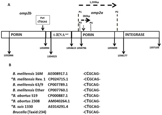
Figure 1. (A) Schematic presentation of the B. melitensis strain 16M omp2 region (arrows indicate locations in the chromosome of ORFs and primers). P stands for a primer. The size of the first amplicon (1233 bp) and nested PCR product (282 bp) as well as direction of gene transcription are indicated by broken arrows. Vertical broken short arrows stand for an apparent PstI site. (B) The specific omp2a sequence which aligns with the corresponding PstI site and accession numbers are given. Note that the PstI site is lacking exclusively from strains 16M and Rev. 1. Star (*) stands for a reverse complementary sequence in comparison to that of B. melitensis 16M. SNPs are depicted by letters with an increased font size.

Microbiolres 14 00002 g001 Figure 2A shows a schematic genetic map of the tRNA (uracil-5-)-methyltransferase gene and flanking sequences in chromosome I of B. melitensis strain 16M. Figure 2B focuses on two SNPs differences between Brucella species. Although this is not applied for B. microti which shares identical SNPs with B. suis, one can rule out involvement of the two species in most if not all brucellosis cases among small ruminants and cattle. Intriguingly, as can be seen in Figure 2A, these sequences are linked to a hypervariable Bruc04 sequence of the MLVA technique [27], possibly implying that this region is a hotspot of mutational events.

Figure 2. (A) Schematic presentation of the B. melitensis strain 16M tRNA (uracil-5) methyltransferase region (arrows indicate locations in the chromosome of ORFs and primers). The size of the first amplicon (785 bp) and nested PCR product (256 bp) as well as direction of gene transcription are indicated by broken arrows. Note, tRNA (uracil-5) methyltransferase is linked to an MLVA Bruc04 sequence. P stands for a primer. SNP locations on the nested PCR amplicon are indicated by short vertical broken arrows. (B) The specific tRNA (uracil-5) methyltransferase sequence which aligns with the corresponding SNPs and accession numbers are given. Star (*) stands for a reverse complementary sequence in comparison to that of B. melitensis 16M. SNPs are depicted by letters with an increased font size.

Microbiolres 14 00002 g002

留言 (0)

沒有登入
gif