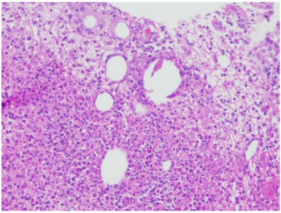
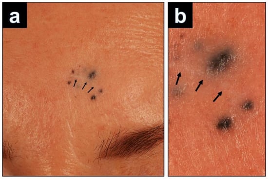
3. ResultsHistopathological examination of the relapsed cellular blue nevus showed solitary nodules in close vicinity to the scar (
Figure 2a). The nodules were constituted by a nodular dermal proliferation of ovoid melanocytes, with monomorphous nuclei and inconspicuous cytoplasm, intermingled with dendritic pigmented melanocytes. At immunohistochemistry, either the ovoid or the dendritic cells were positive with Melan-A (
Figure 2b). Nevus cells of the nodules were dispersed in the dermis and hyperpigmented, spindle-shaped melanocytes infiltrated among the collagen bundles. There were no features suggestive of malignancy, such as cytological atypia, atypical mitoses, or necrosis (
Figure 3a). Positivity in the melanocytes with the Melan-A immunostains (
Figure 3b), SOX 10 (
Figure 3b), HMB45, S100, and BAP1 (
Figure 3d) was seen.On histology of the primary lesion, the CBN was symmetric and revealed spindle-shaped and dendritic, strongly pigmented melanocytes, amid dense fibrous stroma and melanophages, predominantly occupying upper and mid dermis. (
Figure 4a–c). Mitoses were absent. A superficial subepidermal grenz zone without junctional involvement was present. Both the primary and the recurrent lesion showed identical infiltrates of a benign cellular blue nevus. Concentrations of the nevus cells were mainly observed in the primary lesion periappengeal and perivascular. Further, we could demonstrate melanocytes and melanophages in the wall of small peripheral nerves (
Figure 4d–f). 4. DiscussionWe performed a literature review using PubMed (search terms: blue nevus, satellitosis) to identify earlier reported cases of BN with satellitosis between 1999 and 2022. We only became aware of 10 BN cases with satellitosis, making our case the 11th report. The most important features of these cases are summarized in
Table 1.
Histopathologically, only three cases were CBN. The mean age of the patients was 47.8 years, with the youngest patient being 15 and the oldest 71 years. The predilection sites for BN with satellitosis are the scalp, forehead, and the upper extremities. Tumor sizes ranged from 6.0 mm to 18.0 mm (average 10.5 mm). In almost all reports, melanoma or a malignant BN was the initially favored clinical diagnosis. In all 11 cases, it was difficult to distinguish BN with satellitosis from malignant melanoma without histological examination as satellite lesions of pigmented tumors are clinically often considered as a sign of malignant transformation.
Max Tieche first described BN in 1906 [
13]. BN evolve as the result of an ectopic accumulation of melanocytes retained in the dermis during their migration from the neural crest to the epidermis. BN and related entities represent a heterogeneous group of congenital and acquired melanocytic tumors that includes dendritic (“common”) blue nevi (DBN), cellular blue nevi (CBN), and variants such as atypical cellular blue nevi (ACBN) and malignant BN/melanoma.Desmoplastic BN is considered as another important variant of BN, which must not be confused with desmoplastic melanoma [
14]. Epithelioid BN is a diagnostic challenging entity because of its rarity and histological overlap with CBN and malignant BN [
15].CBN differs from the classic DBN by its large size, cellularity, strong pigmentation, and growing pattern with subcutaneous infiltration. Additional atypical features associated with a variety of CBN histological patterns, but without clear-cut evidence of malignancy, have been referred to as ACBN. Clinically, ACBNs resemble CBNs, but histologically, they contain worrisome features, such as asymmetry, hypercellular foci, focal cytological atypia, and occasional mitoses (2) [
16,
17]. Malignant BN is a very rare form of melanoma, arising in association with or exhibiting some morphologic similarities to BN. Pigmented epithelioid melanoytoma (previously termed animal-type melanoma) is another rare variant of cutaneous melanoma that may also mimic melanoma associated with blue nevus [
18].The variant described here, a CBN with satellite lesions mimicking malignant melanoma with cutaneous metastases, is extremely rare. The term “satellite” is usually defined as a skin cancer that has spread from the primary tumor through the lymphatic system to a distance of no more than 2 cm [
19]. In benign tumors such as BN, the term “satellite” is used regardless of the dissemination route of the cells or the distance between the satellite and the primary tumor. Since satellite lesions usually occur in malignant neoplasms, the differential diagnosis of BN with satellitosis is locally advanced melanoma or malignant BN. Another differential diagnosis of BN with satellitosis is agminated blue nevus. Only a few cases of multiple, agminated, or plaque-type blue nevi have been reported [
20]. In contrast to cases of BN with satellitosis, the agminated subtype is formed when bluish-pigmented lesions cluster together in a circumscript area without a central main papule or nodule [
21].The etiology and pathogenesis of satellitosis in BN is still unclear. In several studies, a spread of the melanocytes along small hair vessels and skin appendages starting from the primary lesion was observed [
1,
4,
8]. The fact that the nevus cells were densely aggregated around the blood vessels in the main papule and in the satellite lesions implicated spreading of the nevus cells along the perivascular space to manifest clinically satellite lesions. It is therefore conceivable that, in our case, the scarred sections in the area of the pre-existing primary lesion could have served as a guide for the spread of melanocytes. Further, it is also known that nerve sheaths within BN contain dendritic melanocytes that could also migrate [
22]. The primary lesion of our case showed melanocytes in the wall of small peripheral nerve fibers. We speculate that this could be another possibility of nevus cell spread in cases of BN with satellitosis, especially when CBN are involved.Recurrent melanocytic nevus is a proliferation of melanocytes arising from a partially excised melanocytic nevus. Recognizing this phenomenon is important, because recurrent melanocytic nevi often display atypical borders and irregular pigmentation, which can lead to diagnostic confusion with melanoma. The pathologic features of recurrent nevus and melanoma are often very similar. Kelly et al. claimed that the most suspicious feature for melanoma in these cases is the growth of the lesion beyond the confines of the initial scar into the surrounding normal skin [
12]. To differentiate recurrent nevus from melanoma, other diagnostic tools such as immunohistochemistry, dermoscopy, and confocal microscopy should be used in addition to standard (immuno)histologic work-up.Recurrence of BN has rarely been reported. Harvell et al. performed a study to better define these entities [
11]. Clinical recurrence is often considered to be associated with malignant transformation in BN, but a study by Harvell et al. has shown that malignant tumor progression is not necessarily the case. In the absence of necrosis, marked cytological atypia, and frequent mitotic figures, atypical morphologic parameters in BN are probably reactive and “pseudomalignant”. In our case, the recurrence of the cell-rich BN is characterized by a significant extension of melanocytes beyond the scar, but no increased cytological atypia or mitotic activity could be detected. The follow-up of our case (currently 36 months) showed an inconspicuous course without evidence of recurrence or metastasis.
In conclusions, satellitosis is a very rarely reported phenomenon in BN (and even rarer in CBN) that may lead to clinical appreciation as melanoma. On histopathologic examination of our case, we found malignant changes neither within the primary CBN nor in the recurrent CBN-associated satellite lesions. It is important to realize that a recurrent pigmented lesion with satellitosis does not necessarily represent malignant melanoma: it may rather also be an unusual manifestation of a recurrent BN.


留言 (0)