
記住我


Angiomyolipoma (AML) is a benign mesenchymal neoplasm composed of variable proportions of abnormal thick-walled blood vessels, spindle and epithelioid smooth muscle cells, and adipocytes and is usually found in the kidney. One-third of patients with AML present with manifestations of tuberous sclerosis.1 Recently, immunohistological studies have revealed that AML belongs to the family of perivascular epithelioid cell tumors (PEComas).2 The occurrence of AML in the head and neck region is rare and it is very rare in the larynx. Although benign laryngeal tumors are not a primary cause of death, they may interfere with the three major functions of the larynx, namely, breathing, speech, and swallowing, so treatment is often necessary. In addition, laryngeal tumors are sometimes found incidentally during tracheal intubation or upper gastrointestinal endoscopy, and thus are a concern not only for otorhinolaryngologists but also for physicians in other specialties. In this report, we describe a rare case of AML of the larynx and review the literature on PEComas, including their genetics and histopathological findings.
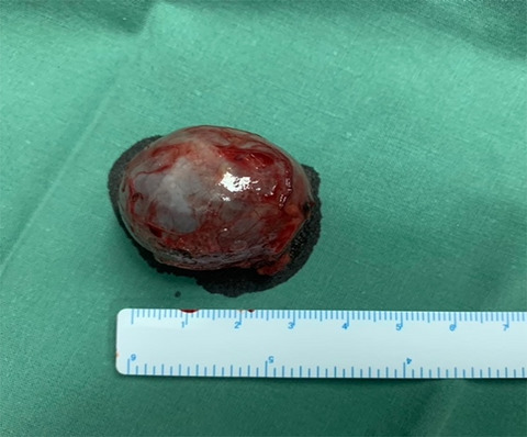
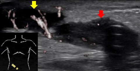
2 CASE REPORTA 69-year-old man was admitted to our department with a right-sided laryngeal tumor that had been discovered incidentally during upper gastrointestinal endoscopy. He did not report any history of laryngeal symptoms such as hoarseness or dysphonia. His medical history included hypertension and hyperlipidemia. Endoscopic examination using a flexible fiberscope revealed a mass approximately 10 mm in size that was loosely attached to the right aryepiglottic fold and covered by smooth mucosa (Figure 1). Since it was covered by mucous membrane by naked eye, we thought it would be difficult to diagnose by biopsy and performed surgical operation for diagnosis and treatment purpose. Following total mass excision by endolaryngeal microsurgery, pathologic analysis revealed a reddish-brown, submucosal, encapsulated tumor measuring 8 mm in diameter (Figure 2).

Laryngoscopic image of a smooth submucosal tumor located in the right aryepiglottic fold

Histopathologically, the resected lesion contained numerous smooth muscle cells (white arrowheads), mature adipose tissue (black arrows), and numerous thin and large irregular vessels (black arrowheads). Hematoxylin and eosin staining; magnification ×100
Histopathologically, the tumor was well demarcated and covered with a layer of squamous epithelium consistent with the laryngeal mucosal surface layer. The presence of adipocytes and large meandering arterial and venous blood vessels interspersed with spindle-type smooth muscle cells led to a diagnosis of AML (Figure 3). Immunohistochemically, the tumor cells were positive for smooth muscle actin (SMA) and desmin but negative for human melanoma black (HMB)-45 and Melan-A. The patient was followed for 14 months and showed no signs of local recurrence.

Immunohistochemically, the tumor was negative for (A) HMB-45 and (B) Melan-A but the smooth muscle component was positive for (C) smooth muscle actin and (D) desmin
3 DISCUSSIONThe larynx is a luminal structure extending from the laryngeal inlet, which is surrounded by the epiglottis, aryepiglottic folds, and interarytenoid notch, to the inferior margin of the cricoid cartilage. The laryngeal lumen contains the laryngeal folds and vocal cords. Histologically, most of the laryngeal mucosa is covered by multilineage epithelium, whereas the vocal cords and laryngeal surface of the epiglottis are covered by multilayered squamous epithelium. The most common malignant tumor of the larynx is squamous cell carcinoma, but benign tumors such as papilloma, lipoma, fibroma, and myxoma also arise. Although the occurrence of benign tumors other than papilloma is rare, when a tumor occurs in the larynx, it may cause airway narrowing, dysphonia, and dysphagia. Therefore, treatment is often necessary even for benign tumors.
Angiomyolipoma is a benign mesenchymal tumor composed of variable amounts of smooth muscle, adipose tissue, and thick-walled blood vessels, and is generally regarded as a type of PEComa.1 PEComas share overlapping histopathological features with epithelioid cells and have a perivascular distribution and characteristic immunohistochemical findings with co-expression of myoid markers (SMA and/or desmin) and melanocytic markers (HMB-45 and/or Melan-A). PEComas include AML, lymphangioleiomyomatosis (LAM), pulmonary and extrapulmonary clear cell “sugar cell” tumor (CCST), clear cell myomelanocytic tumor of the falciform ligament/ligamentum teres, and rare clear cell tumors of other sites.2 Six cases of AML of the larynx have been reported previously.3-8 In three of these cases, the tumors were negative for HMB-45. Many cases of AML occurring in the head and neck region are negative for HMB-45, and it has been argued that the entity in these cases may be different from renal AML. In recent years, however, the histopathology of PEComa has been investigated further, and it has been reported that AML with negative HMB-45 also belongs to the PEComa family because of its similarity to PEComa in terms of chromosomal imbalance.9 The HMB-45 negativity may be explained by the relative rarity of cells with the morphology of PECs. Not all PECs are epithelioid and some have spindled or adipocyte-like morphology. Spindled PECs strongly express myogenic markers, whereas epithelioid PECs strongly express HMB-45.10, 11 In addition, AML occurring in the head and neck region is usually relatively small, in contrast to AML arising in the kidney or liver, which is often large.12 The CT findings of AML occurring in the head and neck region that have been reported so far are characterized by well delimited and unenhanced.4, 5, 12 We did not perform imaging for the risk of airway narrowing and the urgency of the surgery.
Renal AML has been reported in 80% of patients with tuberous sclerosis, which is characterized by mutations in either the TSC1 gene (9q34) or the TSC2 gene (16p13.3).13 The TSC1/TSC2 proteins are essential nodes in a signaling pathway that regulates the activation state of mTOR kinase. This pathway is involved in the regulation of cell growth, proliferation, and survival, and the TSC1/TSC2 proteins exert inhibitory control this pathway. Therefore, the presence of TSC1 or TSC2 gene mutation activates the mTOR pathway, leading to tuberous sclerosis and the complication of renal AML. The mTOR pathway was also activated in renal AML and LAM without tuberous sclerosis and in PEComas in organs other than the kidney and liver. Taken together, these studies indicate very consistent involvement of this pathway in the pathogenesis of AML/PEComa, but also suggest that other genetic lesions besides the TSC genes may be important pathogenic factors.13 In all six of the previously reported cases of AML of the larynx tuberous sclerosis was absent and genetic testing was not performed. Further study of AML of the larynx is needed.
Although the larynx is one of the smaller organs in the body, it can be the site of various very rare tumors and plays key roles in important functions. Laryngeal tumors are encountered by not only otorhinolaryngologists but also by physicians in other specialties. For this reason, we think that all physicians should be aware that that AML and other types of PEComa can occur in the larynx.
4 CONCLUSIONWe reported a rare case of AML of the larynx. AML is a member of the PEComa family, and AML and other types of PEComa can occur in any organ. This case highlighted differences in immunohistochemical findings between renal AML and AML in the head and neck region and demonstrated that not only otorhinolaryngologists but also physicians in other specialties as well, should be aware of AML of the larynx.
CONFLICT OF INTERESTThe authors have no funding and conflict of interests directly relevant to the content of this article.
AUTHOR CONTRIBUTIONSHN involved in project development and manuscript writing. YT served as a surgeon. TO, YN, SM, and YF involved in manuscript editing.
CONSENTWritten informed consent was obtained from the patient for publication of this case report.
留言 (0)