
Remember me

The etiology of Alzheimer’s disease (AD) has traditionally been linked to the presence of amyloid-β 42 (Aβ42), a protein widely recognized as a key marker of the disease. However, a growing body of recent scientific evidence suggests that it may be the amyloid oligomers with smaller molecular weight, rather than the more conspicuous amyloid fibrils, that play a pivotal role in the development and progression of various protein misfolding diseases, including neurodegenerative disorders and type-II diabetes. Numerous studies have highlighted a disconnect between the accumulation of amyloid plaques observed in post-mortem examinations and the neurological deficits experienced by patients during their lives. To explain this lack of correlation, multiple hypotheses have been proposed. Among these, the “oligomer hypothesis” has recently emerged as a leading explanation, positing that the toxic effects of these small oligomers may be more critical to the pathology of AD than the larger aggregated plaques. This shift in focus highlights the need for a deeper understanding of how these oligomers contribute to the disease process . The proposition regarding amyloid oligomers has garnered significant attention over time, primarily because of three key observations related to drug candidates for AD therapy. These observations are (i) ineffectiveness of plaque-targeting therapies, that is, therapeutic agents that focus solely on the removal of amyloid plaques or amyloid fibrils have not demonstrated substantial improvements in patients’ cognitive behaviors; (ii) efficacy of oligomer-targeting drugs, that is, drug candidates that specifically target amyloid-β oligomers (AβOs) have shown greater clinical effectiveness in treating AD patients; and (iii) influence of the APOE4 allele, that is, positive clinical trial outcomes tend to have a higher concentration of AβOs in the brain of individuals carrying the E4 allele of apolipoprotein E (APOE4). The origins of AβOs in AD patients remain a subject of debate and require further extensive research for a definitive understanding. Genetic studies on AD patients indicate that mutations in the amyloid precursor protein (APP), such as the Osaka and Arctic mutations , lead to an overproduction of soluble AβOs. These mutations are associated with an earlier onset of AD, often occurring before the age of 50 , suggesting that certain genetic factors can significantly accelerate the development of the disease. In contrast, the Icelandic mutations appear to have a protective effect, reducing both the overall levels of AβOs and the concentrations of amyloid fibrils, which are another form of amyloid aggregation linked to AD . Additionally, the presence of the APOE4 genotype is notable, as it is found in approximately 65% of AD patients . This genotype is associated with an increased tendency for amyloid monomers to aggregate into AβOs, potentially contributing to the pathology of the disease . Comparisons between AD patients who carry the APOE4 allele and those who do not reveal that the former group has about three times the concentration of AβOs in the brain. This suggests that the APOE4 genotype plays a significant role in the progression of AD by facilitating the accumulation of these toxic oligomers . AβOs exhibit several distinct characteristics that set them apart from amyloid-β fibrils (AβFs). They are small, globular aggregates that display a metastable and transient nature, along with a higher content of β-sheet structures . These small protein aggregates can arise from specific interactions between n-mers (oligomers formed from a defined number of monomers), or from non-specific interactions, akin to micelles. The precise mechanisms underlying the formation of AβOs during the growth of AβFs remain elusive. However, researchers have identified three main pathways to explain this process, as illustrated in Figure 1. The first two pathways fall under the “on-pathway” model, which includes nucleated polymerization and nucleated conformational conversion. These models suggest that oligomers are transient species that form as intermediates during the transition from monomers to mature AβFs. In this context, AβOs are considered stepping stones on the pathway to fibril formation. In contrast, the third model, known as the “off-pathway” formation of AβOs, asserts that oligomers represent a separate class of aggregates that do not progress to form fibrils. This model highlights the possibility that AβOs may have distinct properties and biological implications that differ from those of amyloid fibrils, suggesting a more complex relationship in the pathology of amyloid-related diseases. Understanding these pathways is crucial for unraveling the role of AβOs in the development and progression of AD .
![[2190-4286-16-44-1]](https://www.beilstein-journals.org/bjnano/content/figures/2190-4286-16-44-1.png?scale=2.0&max-width=1024&background=FFFFFF)
Figure 1: Schematic representation of the hypothesized pathways leading to the formation of toxic amyloid-β oligomers and their detrimental effects on neuronal health. The diagram outlines the key steps in the aggregation of amyloid-β peptides into oligomers, highlighting the underlying model mechanisms that contribute to neurotoxicity. (Figure 1 was redrawn from using Microsoft PowerPoint and ChemDraw Professional (Version 20.1.1.125)).
Numerous studies have indicated a strong association between neuronal dysfunction and the presence of oligomeric species. Researchers are increasingly coming to a consensus that the neurotoxicity observed in neurodegenerative diseases (NDs) is not attributable to a single, isolated toxic conformer of amyloid oligomers. Instead, it appears to be the result of a diverse and heterogeneous population of oligomers. Understanding the factors that influence their toxic nature is crucial for developing tools for early detection and treatment. Despite years of research, targeting AβOs has yielded limited clinical success, primarily because of the tendency to treat patients at later stages, when extensive brain damage has already occurred. However, monoclonal antibodies targeting AβOs such as aducanumab, have demonstrated promising efficacy, leading to its FDA approval .
Early diagnosis of AD by targeting AβOs is crucial for improving outcomes. Current imaging methods, such as computed tomography (CT)/positron emission tomography (PET) with FDA-approved 18F-radiotracers (e.g., Amyvid™ and Tauvid™), detect plaques and tau tangles but not AβOs . There is an urgent need for agents targeting AβOs to enable earlier and more accurate diagnosis and treatment. Despite extensive research, most AD treatments only offer temporary symptom relief and fail to target the root causes of the disease. This limited effectiveness stems from AD’s complex and multifactorial nature, which complicates early detection and the identification of reliable biomarkers and therapeutic targets. Moreover, the blood–brain barrier (BBB) poses a significant obstacle to effective drug delivery, further hindering the development of successful treatments.
Nanomaterials (NMs) offer promising solutions for the early detection and treatment of AβOs in AD. Because of their nanoscale size, NMs can interact with biological systems in ways that traditional treatments cannot. Their unique properties such as high surface area, quantum effects, and specific physicochemical traits make them ideal for developing advanced biosensors for early diagnosis and improving the sensitivity of AβO detection. In imaging, nanoparticles (NPs) can help to visualize localized protein accumulation, complementing existing diagnostic methods. Materials such as carbon-based NMs (e.g., graphene oxide) and metal NPs (e.g., gold and silver) enhance imaging sensitivity because of their distinct electrical or photoluminescent properties. For treatment, NPs can serve as drug carriers, improving delivery across the BBB and reducing side effects. Their large surface area allows for controlled drug release and targeted therapy, enhancing treatment efficacy. Additionally, NMs can interact directly with tissues and cells, potentially halting disease progression by preventing protein misfolding and the formation of toxic oligomers, a hallmark of AD pathology. Overall, nanotechnology holds significant potential to advance both the diagnosis and treatment of AβO-driven AD , and we will discuss these topics in the following.
Mechanisms of neuronal cell toxicity induced by AβOsResearch has shown that AβOs possess a remarkable ability to penetrate cell membranes, largely due to their capacity to form porins within the lipid bilayer. This ability arises from the increased presence of β-sheet structures in AβOs, which can create distinct rafts in the membrane. These ring-shaped oligomers adhere to the cell membrane and inflict damage either by directly penetrating the membrane or by aggregating into fibrils that disrupt cellular integrity. Once internalized, AβOs activate N-methyl-ᴅ-aspartate-type glutamate receptors (NMDARs) located on neuronal membranes. This activation triggers endoplasmic reticulum (ER) stress through the stimulation of phospholipase C, leading to an influx of calcium ions (Ca2+) into the cytosol . Elevated Ca2+ levels result in the accumulation of reactive oxygen species (ROS) and reactive nitrogen species, contributing to oxidative stress within the cell . The increase in cytosolic Ca2+ also promotes the phosphorylation of ATP proteins, which, in turn, leads to the enhanced production of Aβ42 and AβOs, creating a vicious cycle. This cascade ultimately results in further spikes in intracellular Ca2+ concentrations sourced from the ER, which is linked to memory impairments commonly associated with AD. Additionally, elevated cytosolic Ca2+ activates the enzyme calcineurin, which is implicated in the activation of the Bcl-2-associated death promoter (BAD). This process, coupled with oxidative stress pathways originating from mitochondrial dysfunction, facilitates the release of cytochrome c from the mitochondria. This release is a key event that promotes caspase activation, initiating pro-apoptotic signaling that drives neuronal apoptosis . Furthermore, AβOs can disrupt the membranes of endosomes and lysosomes, exacerbating neuronal cell death .
In another dimension, AβOs exhibit a strong affinity for cellular prion protein (PrPC) receptors, binding to them irreversibly. The formation of the oligomer–PrPC complex, together with the co-activation of the mGluR5 receptor, leads to the activation of intracellular Fyn kinase. This activation causes dysregulation of calcium ion homeostasis, hyperphosphorylation of tau protein, and disruption of synaptic functions. Together, these processes, as depicted in Figure 2, contribute significantly to the neurodegenerative pathways associated with AD, highlighting the multifaceted role of AβOs in neuronal dysfunction and cell death .
![[2190-4286-16-44-2]](https://www.beilstein-journals.org/bjnano/content/figures/2190-4286-16-44-2.png?scale=2.0&max-width=1024&background=FFFFFF)
Figure 2: Illustration of calcium-mediated toxicity induced by oligomers. Oligomers initiate a calcium cascade that results in a series of harmful effects on cellular health. Elevated intracellular calcium levels activate phospholipase C (PLC), triggering endoplasmic reticulum (ER) stress and additional calcium release. This positive feedback loop exacerbates calcium dysregulation, leading to increased amyloid-β formation, oxidative stress, mitochondrial dysfunction, and ultimately apoptosis. (Figure 2 was created using Microsoft PowerPoint and Servier Medical Art (https://smart.servier.com)).
Conventional methods for addressing the presence and toxicity of AβOsAβOs are small aggregates formed from the misfolding and aggregation of amyloid-β (Aβ) peptides, primarily Aβ40 and Aβ42. These oligomers typically consist of a limited number of Aβ monomers, often ranging from trimers to tetramers, but they can form larger aggregates under certain conditions. Their small size and unique structural properties contribute to several challenges in therapeutic targeting. They are considerably smaller than fibrillar aggregates and plaques, making them difficult to target with conventional binding agents. AβOs exhibit significant heterogeneity in size and conformation. This variability means that a single therapeutic agent may not effectively recognize all oligomeric forms, complicating the development of broad-spectrum therapies. Unlike larger aggregates, which may present multiple binding sites, AβOs have fewer defined surface characteristics that can be targeted. AβOs can interconvert between different oligomeric states and may also exist in equilibrium with monomeric and fibrillar forms. This dynamic nature poses a challenge for therapies that rely on specific binding, as the target may change rapidly in response to environmental factors or therapeutic intervention. As a result, the predominant strategies for targeting AβOs have largely been confined to biologics, particularly monoclonal and polyclonal antibodies. The advantages of using antibodies stem from their remarkable capacity to recognize conformational epitopes that are unique to various oligomeric forms, thereby facilitating the selective targeting of pathogenic AβOs.
The most widely utilized monoclonal antibodies in AD research are 6E10 and 4G8 . These antibodies were generated by immunizing mice with specific peptide fragments of Aβ, allowing them to bind effectively to amyloid aggregates. Importantly, the development of “conformation-dependent” antibodies, such as A11 and OC, marked a significant advancement in the field, as they were among the first to differentiate between AβOs and AβFs. This distinction is crucial for understanding the pathophysiology of AD, as oligomers are believed to be more toxic than fibrils .
In addition, the polyclonal antibody M94 has demonstrated high selectivity towards pathogenic AβOs, while not recognizing physiological Aβ monomers. This selectivity is vital, as it helps to minimize potential off-target effects and enhances the therapeutic profile of the antibodies . Additionally, the monoclonal antibody mAb158 selectively targets soluble AβOs, including protofibrils, rather than monomeric Aβ or APP, highlighting its specific ability to focus on oligomers . Recent developments have introduced novel approaches to target AβOs, such as the work by Haynes et al., who reported the creation of a unique anti-soluble AβO (E3) nanobody derived from an alpaca immunized with soluble AβOs. This E3 nanobody, conjugated with carboxyfluorescein (FAM), demonstrated effective recognition of both soluble AβOs and Aβ plaques, highlighting the potential for nanobody technology to complement traditional antibody approaches . Conventional methods for targeting AβOs primarily rely on the use of antibodies because of their ability to recognize specific conformational epitopes associated with oligomers. The development of monoclonal and polyclonal antibodies has significantly advanced our understanding of AβOs in AD, allowing for targeted therapeutic and diagnostic strategies. While traditional antibody-based approaches remain valuable, ongoing innovations, such as the development of nanobodies and nanochaperones, promise to enhance the specificity and efficacy of oligomer targeting. Ongoing research into traditional therapeutic approaches, such as small molecules/compounds, immunotherapy, peptidomimetics, and chaperon proteins, as outlined in Table 1, continues to be a critical part of the effort to tackle AD. These conventional strategies, which target different aspects of AβO formation and accumulation, have shown promise in managing AD pathology. However, they face significant challenges, including limited efficacy and specificity in targeting the diverse forms of AβOs. By combining the strengths of both established and new technologies, researchers aim to develop more effective therapies for AD that can specifically target AβOs and ultimately slow or halt the progression of the disease.
Table 1: Classification of therapeutic strategies targeting AβOs and their mechanisms of action.
Class Compound Target Mechanism of action Reference immunotherapy aducanumab AβOs and AβFs aducanumab interacts with the area covering residues 3–7 in the N-terminal region of Aβ crenezumab Aβ aggregates (including AβOs) crenezumab, a fully humanized IgG4 monoclonal antibody, reduces the activation of Fc-gamma receptors (FcγRs) while preserving FcγR-mediated microglial phagocytosis and facilitating the clearance of AβOs gantenerumab AβOs, plaque, and AβFs mechanism of action involves binding with high affinity to both the N-terminal and central regions of Aβ peptides bapineuzumab soluble AβOs and AβFs changes in APOE4 carrier expression PMN310 AβOs humanized PMN310 inhibits AβO-induced memory impairment and diminished synaptic loss and inflammation small molecule/compounds curcumin AβOs, plaque, and AβFs curcumin directly interacts with small amyloid species to inhibit aggregation and fibril production both in vitro and in vivo epigallocatechin gallate (EGCG) AβOs and AβFs the interactions of EGCG are determined by hydrophobic π–π and hydrophilic interactions with the aromatic side chains and Aβ backbone, respectively melatonin AβOs melatonin treatment inhibits the Aβ1–42-induced decline in Notch1, NTMF, and NICD both in vivo and in vitro trodusquemine AβOs selectively binds to oligomeric species and reduces the toxicity methylene blue AβOs inhibits the oligomer formation by selectively inducing the fibril formation sulforaphane AβOs decreases oligomer production, tau phosphorylation, oxidative stress, and inflammation, while enhancing cognition in PS1V97L Tg mice transthyretin (TTR) AβOs TTR tetramers inhibit Aβ aggregation in vitro through an interaction between the thyroxine binding pocket of the TTR tetramer and Aβ residues 18–21 peptidomimetic RI-OR2 AβOs and AβFs attaches itself to its corresponding region (KLVFF, residues 16−20) in native Aβ and disrupt Aβ self-association LPFFD Aβ plaque and AβFs the absence of a proton on the secondary substituted nitrogen in the peptide bond of the proline residue may impede the development of intramolecular hydrogen bonds inside fibrils D3 AβOs D3 derivative peptides bind to Aβ in monomeric stage and stabilize these species within the diverse equilibria of Aβ assemblies, ultimately resulting in the eradication of AβOs APPI Aβ plaque and AβFs the binding of this 20-mer cyclic peptide to Aβ42 (in a 1:1 molar ratio) promotes the development of Aβ42 aggregates, thereby ameliorating Aβ42-mediated cellular toxicity chaperon proteins BRICHOS domain Aβ plaque, AβOs, and AβFs interferes with Aβ in the nucleation process and extends the lag phase HSP104 AβOs, protofibrils, and AβFs the inhibition of Aβ fibrillization by Hsp104 is evident at Hsp104/Aβ, indicating a selective involvement of Hsp104 with aggregation intermediates (such as oligomers and protofibrils), during amyloid formation αB-crystallin AβOs αB-crystallin interacts with the monomer and oligomeric state of the proteins via capping the β-sheet elongation surfaces; it restricts the nucleation phase, which in turn does not allow the oligomer to form fibrils Emerging therapeutic approaches in clinical trials for targeting AβOsRecent advancements have positioned immunotherapeutic approaches utilizing anti-Aβ antibodies as some of the most promising strategies for the treatment of AD. Notably, the first generation of anti-Aβ antibody therapies, including aducanumab, lecanemab, and donanemab, has demonstrated significant therapeutic potential in combating AD. Aducanumab and lecanemab have already received FDA approval, while donanemab is currently undergoing clinical evaluation . In a noteworthy study by Sandberg et al., an oligomer-specific antibody known as ALZ 201 was reported to effectively mitigate the toxic effects associated with extracts from AD brains. This research confirmed that ALZ 201 selectively recognizes AβOs and, interestingly, demonstrated efficacy in protecting neurons exposed to AD brain extract. ALZ 201 is presently in preclinical development, highlighting its potential as a therapeutic candidate . Lecanemab, another anti-Aβ antibody, has been found to exhibit a higher affinity for Aβ protofibrils characterized as “beaded” curvilinear fibrils and recognized as a specific form of AβOs than other known antibodies such as aducanumab or gantenerumab. Furthermore, clinical observations have indicated that treatment with lecanemab is associated with a reduction in cognitive decline, underscoring its promise as a viable therapeutic option in the fight against AD .
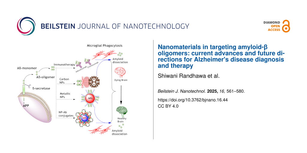
The landscape of AD treatment is evolving, with emerging therapeutic approaches in clinical trials targeting AβOs offering new hope for patients. Monoclonal antibodies and oligomer-specific antibodies are at the forefront of this research. As clinical trials progress, these therapies hold the potential to significantly improve cognitive outcomes and quality of life for individuals affected by AD, underscoring the importance of targeted interventions in combating this complex neurodegenerative disorder. Conventional approaches have shown promise in the detection and treatment of AβOs in AD. However, NP-based approaches present a complementary and potentially more versatile strategy for addressing AβOs in AD. In our review, we discuss a range of strategies aimed at targeting AβOs, and these approaches are illustrated in Figure 3. Figure 3 provides an overview of the different therapeutic strategies, highlighting both conventional and emerging methods for addressing the challenges posed by AβOs in AD pathology.
![[2190-4286-16-44-3]](https://www.beilstein-journals.org/bjnano/content/figures/2190-4286-16-44-3.png?scale=2.0&max-width=1024&background=FFFFFF)
Figure 3: This figure summarizes the key strategies discussed in our review for targeting AβOs in AD. Aβ monomers, generated from APP through β-secretase activity, aggregate to form oligomers and subsequently amyloid plaques. The diagram highlights various therapeutic approaches, such as immunotherapy and antibody-conjugated NPs designed to enhance microglial phagocytosis for Aβ clearance, as well as NP-based techniques aimed at disrupting Aβ aggregates and preventing their toxic effects. (Figure 3 was created using ChemDraw Professional (Version 20.1.1.125) and Servier Medical Art (https://smart.servier.com)).
Nanoparticle-based approaches for the diagnosis and dissociation/inhibition of AβOsAlthough conventional approaches for diagnosing and targeting AβOs have laid the foundation for AD treatment, they often face limitations. For example, monoclonal antibodies, while capable of binding AβOs, may also interact with other forms of Aβ, including fibrils or monomers, leading to off-target effects and reduced efficacy. Recent advancements in nanotechnology offer a promising alternative with NPs specifically designed for AD diagnosis and AβO inhibition. These NPs possess unique properties, including variable size and shape and readily modifiable surfaces. These features allow for targeted and effective therapeutic strategies. In this section, we discuss NPs specifically designed for the diagnosis and inhibition of AβOs.
One example of this innovative approach is demonstrated by Viola et al., who designed mono-dispersed nitro-dopamine (nDOPA)- and polyethylene glycol (PEG)-stabilized magnetic nanostructures (MNSs) in a size range of 12–16 nm. The surface of these MNSs was further modified with oligomer-specific antibodies, creating a stable nanobioconjugate for both in vitro and in vivo applications. The MNSs could detect toxic AβOs present on nerve cell surfaces in vitro, demonstrating their specificity and effectiveness. Additionally, upon intranasal delivery in a mouse model, these MNSs rapidly targeted AβOs under in vivo conditions, providing strong MRI contrast, thus supporting their potential for non-invasive diagnostic imaging of early-stage AD . In another study, a similar approach was utilized by Wang et al.; they developed a AβO-targeting gadolinium-based NIR/MR dual-modal theranostic nanoprobe. The developed nanoprobe was used as an efficient and sensitive MR/NIR contrasting agent for the detection of AβOs in different age groups of transgenic AD mice. The theranostic nanoprobe also showed strong inhibitory effect against Aβ fibrillation and improved associated neurotoxicity . Shifting from imaging to electrochemical approaches, researchers have developed biosensors comprising immobilized thiolated PrPC peptides on a graphene oxide/gold nanoparticle hydrogel electrode. This nanobiosensor displayed high specificity and sensitivity for detecting soluble AβOs in artificial cerebrospinal fluid (CSF) or blood plasma. It was capable of effectively distinguishing AβOs from Aβ monomers and fibrils, indicating its utility for accurate and selective detection of AβOs . Another group of researchers utilized a similar approach of electrochemical detection of AβOs via a nanobiosensor consisting of gold nanoparticles (AuNPs) embedded in a conductive polymeric matrix. For this, the surface of the AuNPs was further modified with PrPc, which acted as the biorecognition element for the specific detection of AβOs in ex vivo real samples, viz., CSF and blood tests . Researchers have also employed the surface plasmon resonance (SPR) of citric acid-coated AuNPs, to specifically detect and quantify Aβ40 oligomers, as the SPR absorption band of AuNPs was found to be sensitive to the presence of AβOs . While exploring the range of AβO detection methods, Liu et al. developed a fluorescence-based system using a FAM-labeled DNA aptamer fluorophore along with a nanoquencher attached to self-assembled polydopamine nanospheres. This nanosystem showed selective recognition of AβOs through a “fluorescence-signal on” mechanism, where the FAM-DNA aptamer interacted with AβOs, causing a hairpin-like conformational change that triggered a fluorescent signal. This approach combined sensitivity with specificity, providing a promising tool for the detection of AβOs . While the aforementioned NP-based systems excel in identifying and imaging AβOs, these technologies also pave the way for developing therapeutic strategies aimed at inhibiting the formation of toxic AβOs. Liu et al. synthesized multifunctional superparamagnetic iron oxide nanoparticles (SPIONs) conjugated with a specific scFv antibody (W20) targeting AβOs and a class-A scavenger receptor activator (XD4). These W20/XD4-SPIONs demonstrated promising results in mitigating the cytotoxicity induced by AβOs and enhancing microglial phagocytosis of these toxic aggregates . Previously, the same NPs were found to show promising early diagnostic potential for AD . Brambilla et al. employed a combination of experimental and computational approaches to investigate the interaction between PEGylated NPs and Aβ monomers. Their findings revealed that surface interactions between NPs and Aβ monomers effectively inhibited the formation of AβOs . Building upon their previous research, Parikh et al. developed a curcumin-loaded self-nanomicellizing solid dispersion system (Cur-SNSDS) to significantly enhance the in vivo bioavailability of curcumin. This novel NP system demonstrated superior safety and efficacy in mitigating Aβ42 oligomer-induced toxicity in SH-SY5Y695 APP human neuroblastoma cells compared to pure curcumin. Moreover, the Cur-SNSDS system effectively prevented cognitive decline in aged APPSwe/PS1deE9 mice, suggesting its potential as a therapeutic intervention for AD . Ultimately, these diverse examples illustrate the transformative, versatile, and effective approach of NP-based strategies for the detection and inhibition of AβOs (Table 2). Their ability to target and interact with AβOs presents a highly promising avenue for future therapeutic development. As research continues in this area, NPs show promising potential to revolutionize how we diagnose and manage NDs such as Alzheimer’s.
Table 2: NP-based strategies for diagnosing and inhibiting AβO formation, as well as mitigating the associated toxicity.
Nanoparticles Conjugation/modification Mechanism of action Reference poly(dopamine) nanospheres conjugated with carboxyfluorescein-labeled DNA aptamer detection of AβOs with high sensitivity using carboxyfluorescein-labeled DNA aptamer–polydopamine nanospheres, capable of identifying concentrations as low as 20 nM gold nanoparticles casein-coated gold nanoparticles chaperones together with gold nanoparticles effectively neutralized Aβ aggregates, reducing their toxic effects iron oxide nanoparticles conjugated with Aβ oligomer-specific scFv antibody W20 and class-A scavenger receptor activator XD4 mitigation of AβO-induced cell toxicity and stimulate microglial phagocytosis of Aβ single-wall carbon nanotubes (SWCNTs) hydroxylated SWCNTs disruption of the beta-sheet conformation of Aβ16–22 oligomers, leading to the formation of less structured, disordered aggregates PLGA NPs — interaction with AβOs during the elongation phase via π–π and hydrophobic interactions, destabilizing their harmful structure gold nanoparticles conjugated with oligomer specific antibody detection of Aβ1–40 with exceptional sensitivity, capable of identifying concentrations as low as 1 fg/mL PLGA NPs conjugated with 83-14 mAb and encapsulated with rosmarinic acid (RA) and curcumin (CUR) enhanced cellular uptake of RA and CUR when delivered using these nanocarriers, indicating that the antibody is crucial for improving nanoparticle delivery to the brain exosomes (EXOs) M2 microglia-derived exosomes M2-EXOs were found to decrease Aβ plaque formation and Aβ oligomer expression in AD cell models, suggesting a protective role in AD pathogenesis through the enhancement of PINK1/Parkin-mediated mitophagy AuNPs conjugated with chiral ʟ- and ᴅ-glutathione ᴅ-enantiomer showed a stronger binding affinity to Aβ42 and demonstrated improved reversal of behavioral deficits in mice modeling Alzheimer's disease AuNPs conjugated with glucosamine the abundance of carbohydrate groups on the nanoparticle surface formed robust hydrogen bonds with protein oligomers, preventing their aggregation PLGA NPs conjugated with PEG and encapsulated with indirubin-3′-monoxime (I3M) nanoparticles continuously released I3M, improving the ability to inhibit Aβ aggregation; additionally, PLGA-PEG nanoparticles enhanced the uptake of I3M by PC12 cells, demonstrating their potential to protect neurons from AβOsIn this review, we systematically categorize NPs used for the diagnosis and inhibition of AβOs based on their composition and functionalization. This bifurcation allows for a clearer understanding of the diverse mechanisms and applications of NPs in addressing AD. We have organized the NPs into four primary categories, namely, carbon based nanomaterials (CNMs), metal based NMs, biomimetic NMs and antibody-functionalized NMs.
Carbon-based nanomaterials for the detection and inhibition of AβORecent advances in nanomedicine have spotlighted CNMs because of their remarkable physicochemical properties, diverse structural forms, and potential applications in combating NDs. The unique characteristics of CNMs, including their hydrophobic surfaces and variable dimensions, enable them to interact effectively with biomolecules, making them valuable tools in biomedical research and therapeutic applications. CNMs can be categorized into three primary forms, namely, zero-dimensional fullerenes (e.g., C60), one-dimensional carbon nanotubes (CNTs), and two-dimensional graphene. Each of these NMs possesses distinct attributes that facilitate their engagement with proteins and peptides, particularly those associated with NDs like AD. Research has demonstrated the ability of fullerenes to prevent the aggregation of Aβ peptides. For instance, molecular dynamics simulations have shown that fullerenes inhibit the fibrillation of the hydrophobic KLVFFAE peptide by disrupting the formation of β-sheet oligomers. This property is particularly significant as β-sheet formation is a critical step in the aggregation pathway leading to neurotoxic amyloid fibrils . Further investigations revealed that fullerene C60 interacts strongly with non-polar aliphatic groups in polar residues of the GNNQQNY peptide, effectively redirecting the formation of potentially toxic oligomers towards disordered coil structures. This mechanism not only hinders fibril formation but also shifts the balance toward less harmful aggregates . Single-walled carbon nanotubes (SWCNTs) have emerged as another promising CNM for the detection and inhibition of AβOs. Studies indicate that hydroxylated SWCNTs significantly inhibit the β-sheet formation of Aβ peptides . By facilitating the formation of disordered aggregates, these nanomaterials diminish the aggregation propensity of Aβ peptides, thereby mitigating their neurotoxic effects . In addition to their inhibitory capabilities, SWCNTs can serve as effective sensors for AβOs. Their ability to interfere with β-sheet formation, a hallmark of Aβ aggregation, has been confirmed through comprehensive molecular dynamics simulations. These studies reveal that SWCNTs interact with the hydrophobic residues of Aβ peptides, particularly through π-stacking interactions with aromatic amino acids such as phenylalanine. This interaction destabilizes the prefibrillar β-sheet structures, preventing the formation of toxic oligomers and promoting the aggregation of less harmful conformations . Moreover, the development of positively charged carbon quantum dots has shown promise in preventing the aggregation of amyloid proteins, specifically by inhibiting the formation of hetero-oligomers between islet amyloid polypeptide (IAPP) and Aβ42. Such findings highlight the versatility of CNMs in addressing different aspects of amyloid aggregation . In summary, CNMs present a multifaceted approach to the detection and inhibition of AβOs. Their unique structural properties and interactions with amyloid peptides hold significant potential for the development of innovative therapeutic strategies aimed at combating NDs. As research in this field continues to advance, the integration of CNMs into clinical applications may offer new avenues for early detection and intervention in AD and related disorders.
Metal nanomaterials for detection and inhibition of AβOsMetal NPs have emerged as pivotal tools in the detection and inhibition of Aβ1–42 oligomers. Their unique optical and electrical properties, particularly those of gold and silver NPs, enhance sensitivity and specificity in identifying the early stages of Aβ aggregation. By binding to AβOs, these NPs facilitate label-free detection methods such as SPR, colorimetric changes, and fluorescence amplification, enabling straightforward real-time monitoring of oligomer formation. This innovative approach not only deepens our understanding of amyloid pathology but also contributes to the development of diagnostic strategies for NDs. Zhou and colleagues introduced an advanced electrochemical aptasensor that utilizes metal-organic frameworks (MOFs) as signal probes for detecting AβOs. They engineered an electrode modified with gold nanoflowers to capture targets, employing aptamer-tagged gold nanoparticle/Cu-MOFs conjugates to produce sensitive signals. This resulted in a highly effective sandwich sensor capable of detecting AβOs in a linear range from 1 nM to 2 μM, demonstrating a correlation coefficient of 0.996 and a low detection limit of 0.45 nM . Phan and team developed a robust and straightforward method for creating multichamber paper devices using wax printing techniques, which they applied to detect AβOs. This approach leverages copper-enhanced gold nanoprobe colorimetric immunoblotting, achieving detection limits as low as 23.7 pg/mL, visible through a smartphone camera, and up to 320 pg/mL with the naked eye . Zhao and collaborators utilized AuNPs embedded in various matrices to construct three-dimensional layers for detecting AβOs. Among their innovations, PrPC/AuNPs embedded in a Ppy-3-COOH matrix (AuNPs-E-Ppy-3-COOH) exhibited superior sensitivity, with a detection range spanning from 10−9 to 103 nM . An electrochemical, label-free aptassay developed by Gallo-Orive et al. incorporates graphene oxide–gold nanoparticles/nickel/platinum nanoparticles for the rapid and accurate detection of AβOs in complex clinical samples, such as brain tissue and CSF from Alzheimer’s patients. This method showcased exceptional sensitivity with a limit of detection of 0.10 pg/mL, demonstrating reproducibility and rapidity .
Metallic NPs have gained considerable attention as potential therapeutic agents regarding AβO formation. Their unique surface characteristics enable specific interactions with amyloid fibrils, effectively inhibiting oligomerization and reducing neuronal cell death. Moreover, these NPs can be functionalized with targeted ligands, enhancing their selectivity and efficacy in therapeutic applications aimed at Aβ-induced neurotoxicity. Recent research has focused on cyclometallated palladium complexes (Pd-1, Pd-2, and Pd-3), which incorporate anthracene and pyrene within a tridentate ligand framework. These complexes specifically target the oligomerization of soluble Aβ1–42 peptides. Among them, Pd-3 has shown significant promise, exhibiting the greatest reduction in Aβ1–42 peptide-induced cytotoxicity in Neuro-2a cell lines. Structural studies indicate that these palladium complexes interact with both the fibrillar (PDB: 2BEG) and monomeric (PDB: 1IYT) forms of the Aβ1–42 peptide. This interaction occurs through a variety of binding modalities, including hydrophobic and hydrogen bonding, leading to substantial inhibition of peptide aggregation . In another innovative approach, Javed et al. evaluated the inhibitory potential of casein-coated AuNPs against oligomers through molecular dynamics simulations. Their findings demonstrated that these NPs effectively bind to oligomeric species, preventing the formation of fibrillar structures . The influence of varying diameters and lengths of cetyltrimethylammonium bromide-stabilized gold nanorods (AuNRs) on Aβ oligomerization and fibrillation has also been thoroughly investigated. Fluorescence studies revealed that the presence of the AuNRs significantly inhibits the development of larger oligomers and fibrils, with inhibition efficacy diminishing as the diameter of the NPs decreases . In a different approach, Randhawa et al. designed glucosamine-conjugated gold nanoparticles (Gln@CA-AuNP), which demonstrated strong inhibition of hen egg-white lysozyme oligomers (HEWLO) in comparison to fibrils (HEWLF) (Figure 4). The high density of carbohydrate moieties on the NP surface facilitated strong hydrogen bonding with protein oligomers, preventing their aggregation. Additionally, Gln@CA-AuNP was found to enhance the production of sulfated glycosaminoglycans, bolster extracellular matrix generation, and confer neuroprotection against oligomeric protein aggregates . These findings collectively underscore the potential of metal-based NPs in not only inhibiting Aβ oligomerization but also in paving the way for effective therapeutic strategies against NDs.
Comments (0)