
記住我
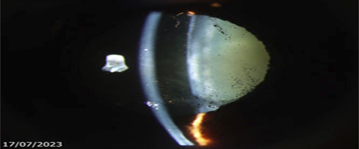

A 56-year-old man with a history of myopic LASIK presented with left eye gradual blurring of vision over 3 months. There was no history of trauma. His uncorrected visual acuities were 20/25 in the right eye and 20/400 in the left eye. The right eye was normal except for an early cataract. The left eye had a moderate cataract that was phacodonetic. Vitreous was present in the shallow anterior chamber (AC) (Figure 1 Figure 1.:
Slitlamp photograph of the left eye showing a shallow anterior chamber with vitreous present, and a moderate cataract.
). The fundus was normal. The intraocular pressures (IOPs) were 14 mm Hg in the right eye and 20 mm Hg in the left eye.Ultrasound biomicroscopy of the anterior segment in the left eye revealed near total zonular loss with few intact zonular strands at the 6 and 10 o'clock regions (Figure 2 Figure 2.:
Ultrasound biomicroscopy radial scan of the left eye at the 3 o'clock position showing the absence of zonular fibers and vitreous herniation into the anterior chamber.
). Vitreous was observed in the AC, herniating mostly from the 3 o'clock region. The endothelial cell density and optical coherence tomography (OCT) of the macular and disc in both eyes were normal. Central corneal thickness was 527 µm in the right eye and 520 µm in the left eye.Describe how you would manage this case surgically. Optical biometry had been obtained, and the axial length in both eyes was similar. Discuss how you would select the monofocal intraocular lens (IOL) diopter (D) targeted for −1.50 D if the AC depth in the left eye was 2.48 mm and in the right eye was 3.25 mm.
留言 (0)