Infective endocarditis (IE), a systemic disease with diverse and multiorgan clinical manifestations, was first described by Osler in 1885.1 In the 20th century, the discovery and utilization of penicillin for treatment and prevention reduced mortality to approximately 30%. In the 1960s, removal of the damaged infected valve and its replacement by a valve prosthesis constituted an approach toward cure of IE and marked the beginning of the association between valve prosthesis and IE. The routine use of surgical indication in patients with IE complicated by severe congestive heart failure reduced mortality from 90% to 10%.2 Over the past 4 decades, changing epidemiology and microbiology has led to the mortality rate reports between 14% and 37%.3–7 Chronic hemodialysis is the most common risk factor, followed by intravenous drug use. In addition, a worldwide increase in infective endocarditis caused by Staphylococcus aureus and Enterococci has been documented to contribute to the current day morbidity and mortality despite advances in diagnostic and therapeutic modalities.8 Most of the studies have been reported from large tertiary care centers including ours and patients have been selected for referral to such institutions because of complications and needed surgical interventions.
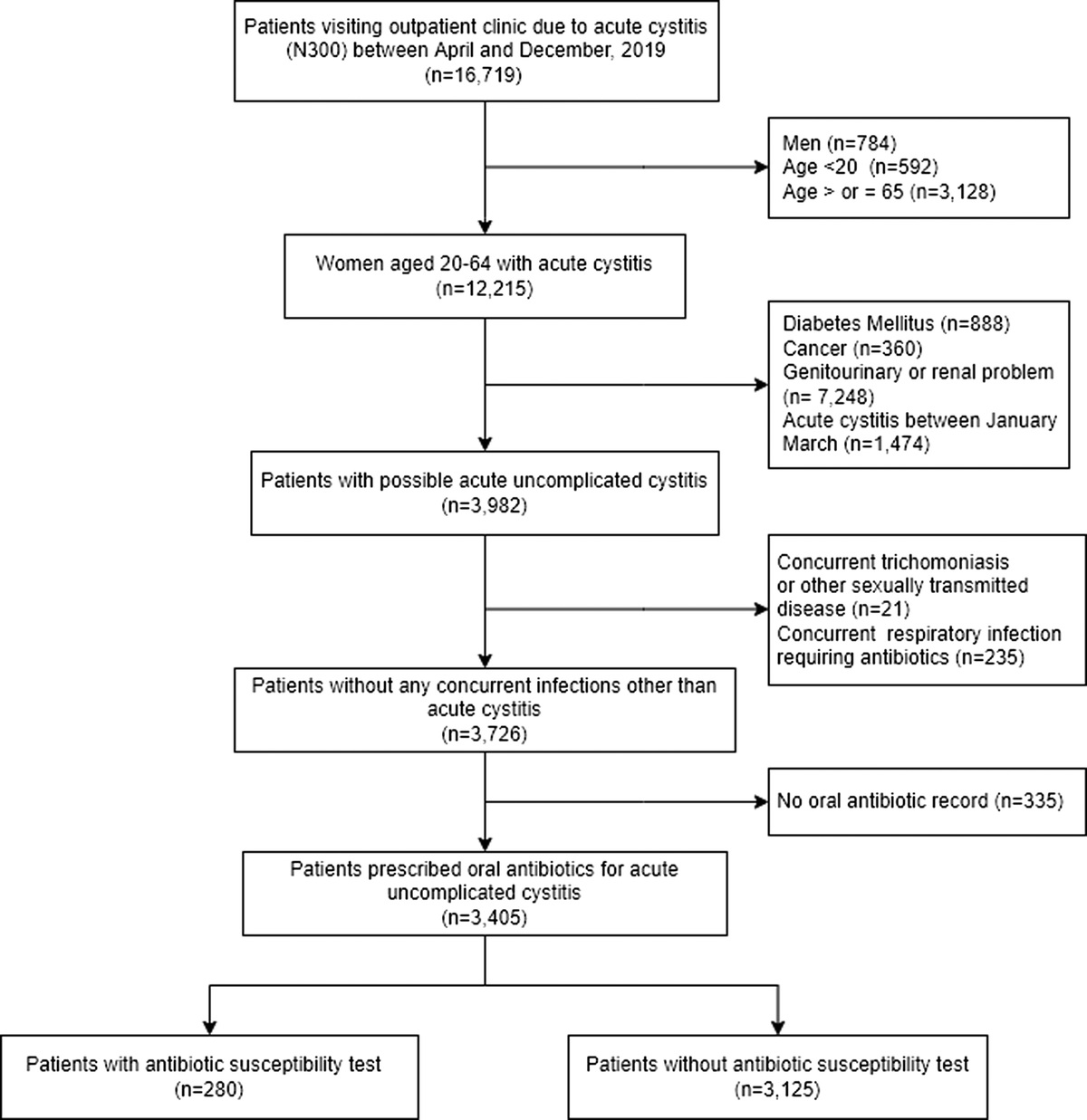
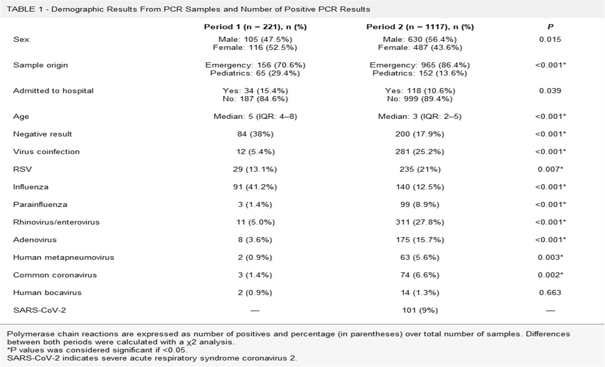
In this issue of the Infectious Diseases in Clinical Practice, the authors present data describing the clinical characteristics, microbiology, and outcomes of infective endocarditis from a small community teaching hospital with no on-site cardiothoracic surgery service.9 There is a clear rationale behind such studies. They give an overall picture of the disease in diverse patient populations and define the impact of decision making at the initial point of care. This retrospective observational study is based on patients with bacterial growth in blood cultures and a potential of IE over a 5-year period. Given the type of study, this would be unavoidable. However, it is important to point out that blood cultures without growth remain a major challenge in the diagnosis of infective endocarditis. The authors calculated Charlson's Comorbidity Index using clinical presentation, comorbidities, echocardiogram findings, surgical indications, microbiology results, antibiotic management, and discharge or death. The factors recorded under the noncardiac predisposing conditions included any invasive procedures, urinary or vascular catheter placement, any surgical interventions within 6 months of admission, history of intravenous drug use, any immunocompromising conditions, any hospital admissions, or antibiotic use within a month of admission. Although a reasonable criterion to include only patients with positive blood cultures there are instances with no growth in blood cultures, the diagnosis of infective endocarditis is clear based on detection of multiple embolic phenomena, clinical course, complications, and response to antimicrobial therapy (personal observation). In such patients' heart, valve vegetations may also not be present, a phenomenon I have tried to explain away as “spent vegetation” on rounds. Prior infective endocarditis, mitral valve prolapses or regurgitation, bicuspid aortic valve, aortic stenosis or regurgitation, and presence of prosthetic valve or intracardiac device were included as cardiac predisposing factors. To determine surgical indication, refractory heart failure, aortic regurgitation or mitral regurgitation with shock, multidrug resistant pathogen, persistent bacteremia, and paravalvular abscess or valvular vegetation >10 mm in size were the criteria used in this study. Definitive antibiotic choice based on the susceptibility results of the organism grown in blood cultures or presumptive antibiotic choice before transfer or death were recorded. The outcomes studied included transfer to a tertiary care facility and in-hospital, 30-day, and 1-year mortality. The final number of patients included in the study is 99, a small sample size compared with previous such studies, pointed out as a limitation by the authors. They also add as a study limitation, a selection bias against transfer of patients with surgical indications to tertiary care based on patient declination. The results of the study are elaborately described in 8 tables in the publication and compared with a study of infective endocarditis from a community hospital published 4 decades ago. The notable findings in this study are (1) a bimodal age distribution with a higher peak in the decade of 80 and a lower peak at decade 30 (median age, 71 years) and (2) preexisting conditions were present in more than half the patients. Expectedly, older patients had more comorbidities, more concomitant cardiac conditions, and a higher Charlson's Comorbidity Index. The younger cohort had more intravenous drug users (IVDUs) and higher surgical indication rate. (3) Staphylococcus was the most common (60%) organism grown in blood cultures followed by streptococci, enterococci, and coagulase-negative staphylococci. (4) The in-hospital mortality was strikingly different between the 2 groups 0% in younger than 75-year age group and 17% in older than 75-year age group. The 30-day mortality was 5% versus 35% and 1-year mortality was 18% versus 53% in the younger versus older age group. The overall 1-year mortality rate remained at 33% similar to the studies published in the past 4 decades. (5) Charlson's comorbidity score of 6.5 and infection with methicillin-resistant S. aureus were predictive of in-hospital, 30-day, and 1-year mortality. (6) Compared with a similar study published 4 decades ago, the current study demonstrates the increasing role of advanced age, prosthetic devices, comorbidities, S. aureus (methicillin-susceptible S. aureus and methicillin-resistant S. aureus), and of IVDUs in younger age group.10
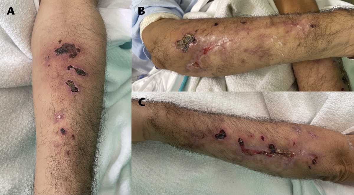
Infective endocarditis is a worldwide disease and is defined as infection of a native or prosthetic heart valve, the endocardial surface, or an indwelling cardiac device. The incidence of IE has been reported to be 1.7 to 11.2 per 100.000 population per year in the western world.11 The American Heart Association estimates the annual number of new cases in the United States to be 100,000 to 200,000. Compared with the preantibiotic era, the percentage of acute IE cases has increases significantly from 20% to 75%.12 The paradigm shift to more acute presentation and lack of classical signs (described previously for the subacute form of the disease) of IE is associated with significant in-hospital and 1-year mortality. The second change is the advancing age, increasing medical comorbidities, and exposure to health care environments leading to change in the microbial etiology of IE. Staphylococcus aureus associated with higher mortality rate is now the leading cause of IE. In the United States, the increase in the incidence of Staphylococci in IE is reported to be associated with chronic hemodialysis access, diabetes mellitus, and more frequent use of intravascular devices. It is apt to say that all these changes are a price for medical progress. The pathogenesis of device related infections involves the adherence and biofilm formation by staphylococci and infections caused by small colony variant S. aureus.13 Although the tricuspid valve is more commonly involved in IVDU's (73%), left-sided IE with polymicrobial infection and systemic embolization is on the rise in this patient population. The HACEK group (non–Haemophilus aphrophilus, Actinobacillus actinomycetemcomitans, Cardiobacterium hominis, Eikenella corodens, and Kingella) of gram-negative organisms has been reported to be responsible for less than 2% cases of IE and therefore not a significant reason for culture negative IE. However, rare organisms, for example, Erysipelothrix rhusiopathiae (nonsusceptible to vancomycin) should be considered based on work and recreational history since presumptive therapy of IE with vancomycin is a widespread practice.14 Keeping in view the published studies including the one in this issue of the Infectious Diseases in Clinical Practice from a smaller community teaching hospital, IE today causes significant morbidity and mortality despite much more advanced diagnostic, medical, and surgical interventions. It seems like more than an intuition that Osler in 1885 called this disease “malignant endocarditis.” Besides accepting the obvious impact of advancing age and comorbidities, any improvement over the status quo will be worthwhile. (1) At the diagnostic level, there is room for better microbiology laboratory (bench) to clinician (bedside) communication, coordination, and discussion especially in institutions with limited infectious diseases presence. This includes review of locally generated antibiograms to monitor antimicrobial resistance in and around the place of patient care. (2) In starting presumptive and then selecting definitive antimicrobial therapy based on available guidelines, there still is significant variation leading to adverse events and use of less effective agents that contribute to morbidity and mortality.15 Trained pharmacists with interest in infectious diseases are remarkably familiar with differences in pharmacokinetic and pharmacodynamics between and within classes of antimicrobial agents. (3) Infective endocarditis patients treated with 6 weeks of intravenous antibiotics had no recurrence compared with those treated for less than 6 weeks. Within that framework, Iversen et al16 reported that changing intravenous antibiotic therapy after at least 10 days to oral formulations was noninferior to continued intravenous treatment in stable patients with left-sided endocarditis. At a minimum, this approach would bring down the complications related to continued intravenous access. (4) It remains without saying that there is a need for more prudent use of indwelling vascular and cardiac devices especially in the elderly patients. (5) A recently published study is the first systemic review and meta-analysis to assess the impact of dedicated multidisciplinary teams (MDTs) on the management of IE.17 Of the 2343 studies screened, 60 full-text reviews were analyzed. Of the 18 studies summarized narratively, 16 reported earlier surgical intervention and 13 reported increased rates of surgery when MDT approach was used. There was an overall significant association between MDTs and short-term survival. Whether this approach is feasible and/or cost effective in smaller hospitals with limited resources remains to be seen. To end on a positive note, well-structured MDTs in large referral hospitals could establish a formal but virtual relationship with the smaller community hospitals that refer to them. In the event a transfer is subsequently needed, the receiving team would be familiar with the clinical scenario.
1. Osler W. The Galtonian lectures, on malignant endocarditis. Br Med J. 1885;1(264):577–579.
2. Grinberg M, Solimene MC. Historical aspects of infective endocarditis. Rev Assoc Med Bras (1992). 2011;57(2):228–233.
3. Cresti A, Chiavarelli M, Scalese M, et al. Epidemiological and mortality trends in infective endocarditis, a 17-year population-based prospective study. Cardiovasc Diagn Ther. 2017;7(1):27–35.
4. Moreillon P, Que YA. Infective endocarditis. Lancet. 2004;363(9403):139–149.
5. DeSimone DC, Tleyjeh IM, Correa de Sa DD, et al. Temporal trends in infective endocarditis epidemiology from 2007 to 2013 in Olmsted County, MN. Am Heart J. 2015;170(4):830–836.
6. Cabell CH, Jollis JG, Peterson GE, et al. Changing patient characteristics and the effect on mortality in endocarditis. Arch Intern Med. 2002;162(1):90–94.
7. Correa de Sa DD, Tleyjeh IM, Anavekar NS, et al. Epidemiological trends of infective endocarditis: a population-based study in Olmsted County, Minnesota. Mayo Clin Proc. 2010;85(5):422–426.
8. Annie Lo HY, Khardori N. A retrospective epidemiological study to define risk factors, microbiology, and clinical outcomes of infective endocarditis in a large tertiary-care teaching hospital. Sage Open Med. 2017;5:1–9.
9. Yang JH, Tavares L, Moon SJ, et al. Endocarditis in a community teaching hospital: the Framingham experience. Infect Dis N Clin Pract. (in press).
10. Venezio FR, Westenfelder GO, Cook FV, et al. Infective endocarditis in a community hospital. Arch Intern Med. 1982;142(4):789–792.
11. Htwe TH, Khardori NM. Cardiac emergencies: infective endocarditis, pericarditis, and myocarditis. Med Clin North Am. 2012;96(6):1149–1169.
12. Murdoch DR, Corey GR, Hoen B, et al. Clinical presentation, etiology, and outcome of infective endocarditis in the 21st century: the International Collaboration on Endocarditis-prospective cohort study. Arch Intern Med. 2009;169(5):463–473.
13. Gandhi T, Crawford T, Riddell J 4th. Cardiovascular implantable electronic device associated infections. Infect Dis Clin North Am. 2012;26(1):57–76.
14. Nandish N, Khardori N. Valvular and myocardial abscess due to Erysipelothrix rhusiopathiae. Clin Inf Dis. 1999;29:1351–1352.
15. Mostaghim A, Annie Lo HY, Khardori N. A retrospective of antibiotic management of infective endocarditis in a large tertiary-care hospital. Am J Infect Dis and Micro. 2017;5(3):120–125.
16. Iversen K, Ihlemann N, Gill SU, et al. Partial oral versus intravenous antibiotic treatment of endocarditis. N Engl J Med. 2019;380(5):415–424.
17. Roy AS, Hagh-Doust H, Abdul Azim A, et al. Multidisciplinary teams for the management of infective endocarditis: a systematic review and meta-analysis. Open Forum Infect Dis. 2023;10(9):ofad444. PMID: 37674631.


Comments (0)