
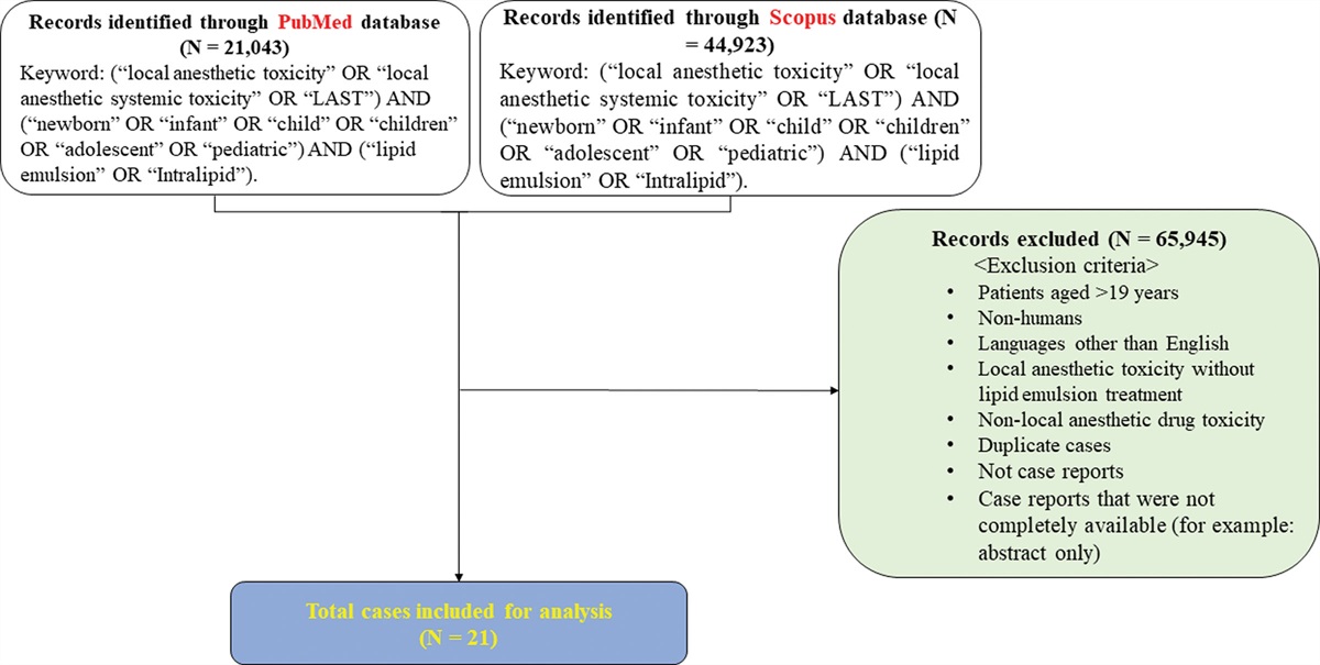
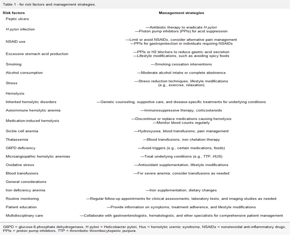
記住我

Skeleton is the third common site of malignant tumor metastasis after lung and liver. Meanwhile, 50% of bone metastases occur in the spine, among them 60% to 80% metastasized to the thoracic vertebra, 15% to 30% to the lumbar vertebra and 10% metastasized to the cervical vertebra.[1] In general, spinal metastasis occurs in 70% of patients with cancer, and 10% to 20% of these patients suffered from clinical symptoms caused by spinal cord compression.[2,3] There were 787,000 cases of lung cancer were newly diagnosed in 2015 China, current estimates maintain that as many as 25%, namely 200,000, of lung cancer patients will develop metastasis to the spine. This condition will inevitably bring huge economic and mental burdens to patients’ familied and the society.
Spinal metastasis is still a clinically challenging condition, with a morbidity rate of approximately 30% among cancer patients. However, With the widespread adoption of targeted therapy, immunotherapy and stereotactic body radiotherapy, the prognosis of patients with malignant tumor have improved prominently, and makes more patients with spinal metastasis have the opportunity to receive surgical treatment. The main manifestations following spinal metastasis are vertebral compression and symptomatic neurothlipsis, which contributes to server pain and significantly compromised the quality of life for these patients.
The incidence of complications following posterior fixation surgery in spinal metastases patients reported by previous literature was as high as 39%. Different from other surgical treatment patients, patients with spinal metastasis have poor physical conditions, and at the same time, radiotherapy and chemotherapy further damage the body immunity function, therefore, the incidence of postoperative wound complications in these patients is higher than that in other surgical disciplines. It is of great significance to understand which perioperative variables can predict the occurrence of surgical site infection (SSI), and provide clinical data support for the subsequent development of a new prognosis evaluation system for spinal metastases.
To our knowledge, there were few articles reviewed prognostic factors of SSI in patients with spinal metastases and underwent surgery. The purpose of the present study was to systematically: (1) investigate the incidence rates of SSI following spinal metastases surgery; (2) identify the factors which were independently associated with postoperative wound infection.
2. Patients and method 2.1. Study designAfter approved by the Ethics Committee of our hospitals this retrospective study was conducted, consecutive patients with spinal metastasis and underwent surgeries from January 2011 to February 2022 were recruited. Data of these patients were extracted and collected from the electronic medical records and picture archiving and communication system by well-trained investigators.
The inclusion criteria were: (1) patients with spinal metastases from solid malignant tumors confirmed by preoperative pathological biopsy and/or postoperative pathological examination; (2) multidisciplinary comprehensive evaluation showed that the patient’s expected survival time is more than 6 months; (3) the patient’s liver, kidney, and bone marrow function were in the roughly normal range; (4) patients underwent posterior thoracolumbar surgery, anterior cervical surgery, posterior cervical surgery, total en bloc spondylectomy, percutaneous vertebroplasty, and other surgical treatment. (5) Patients aged 18 year or older.
2.2. Definition of surgical site infectionDefinition of SSI was based on the criteria of the United States Center for Disease Control and Prevention.[4] Superficial infection: (1) infection occurred no more than 30 days postoperatively; (2) redness, swelling, pain of the incision, purulent discharge, spontaneous wound dehiscence or positive results of bacterial culture around the surgical site skin or subcutaneous were observed or extracted. Deep infection: infection occurs within 90 days postoperatively which involves the fascial and muscular layer and requiring surgical debridement and implant exchange or removal.
Regular observation of the wound was carried out by ward staff while patients were resident in the hospital. Patients were discharged from hospital were followed up for any evidence of SSI occurrence via telephone assessment or promptly clinical interview.
2.3. Surgical techniqueThe patients included in the study generally underwent 4 types of surgical treatment: (1) patients with multi segment spinal metastases or those with multiple comorbidities and poor physical condition who cannot tolerate open surgery should undergo minimally invasive procedures such as percutaneous vertebroplasty or percutaneous kyphoplasty. This type of surgery can effectively alleviate local pain in tumors, restore compressed vertebral height, increase spinal stability, and prevent further vertebral collapse. (2) Anterior vertebral tumor resection was applied to Tomita I, II, III and some IV and V patients with metastatic lesions located in the intervertebral space. During the surgery, the tumor lesion is removed based on the involvement of the spinal cord, and titanium mesh or artificial vertebral body implantation is used accompanied by anterior plate or anterior screw rod system. (3) Tomita VI, VII, and some IV or V patients with metastases located outside the intervertebral space or accompanied by jumping lesions were underwent posterior laminectomy combined with vertebroplasty and radiofrequency ablation was also performed if necessary. Posterior laminectomy was performed firstly during surgery, and pedicle screw rod internal fixation system was used to reconstruct spinal stability. Spinal metastases accompanied by large soft tissue masses and/or metastases with abundant blood supply require radiofrequency ablation or arterial embolization treatment. (4) Anterior and posterior spinal tumor resection: For some patients with Tomita IV and V spinal metastases, and metastases located outside the intervertebral space and obvious nerve root symptoms was observed. Firstly, the pedicle and adnexal lesions were excised through the posterior approach, and the spinal stability was reconstructed using the pedicle screw rod internal fixation system; and anterior resection of vertebral lesions and implantation of titanium mesh or artificial vertebral body were performed subsequently.
2.4. Postoperative protocolAnesthesiologist would evaluate the patient’s basic signs such as breathing, heart rate, blood pressure, and consciousness after the surgery for patients underwent simple single segment decompression and internal fixation surgery, as well as minimally invasive surgeries, then patients would return to the inpatient ward from operating room. Patients with multiple segments surgery and accompanied by significant intraoperative bleeding would enter into intensive care unit under the support of assisted mechanical ventilation, and then transferred back to the general ward after 1–2 days of treatment.
After surgery, vital signs of the patients were stably maintained, conventional dose of antibiotics was used to prevent infection, hormones and nutritional neurotrophic drugs were given to patients when spinal cord injury occurred. Drainage tube would remove when the drainage volume is <50 mL/d for thoracic spinal metastatic tumor. A brace can be used in assisting patients’ restorative exercise 2 to 3 days after surgery, patients with mobility were encouraged to engage in early partial or complete weight-bearing walking on the ground. Corresponding radiotherapy, chemotherapy, and immunotherapy treatment was also introduced sequentially based on postoperative histopathological results.
2.5. Data collectionData were collected from patients’ electronic medical records. The demographic data: age, gender, height; weight; body mass index (BMI, kg/m2), site of primary tumor, tobacco and alcohol consumption; surgical related variables: primary operative spinal region (cervical, lumbar or thoracic; categorized according to the location of the majority of the vertebrae operated on), operation time (min), intraoperative blood loss (mL), surgical incision length (cm), interoperative body temperature (°), drainage usage, American Society of Anesthesiologists (I–IV) classification, preoperative albumin (ALB) (references 40.0–55.0 g/L), protein, lymphocyte and c creative protein level, and other laboratory indexes such as white blood cell (references 3.5–9.5 109/L), red blood cell (1012/L), hemoglobin (references 3.5–9.5 115–150 g/L), blood platelet (109/L), globulin (references 20–40 g/L), blood glucose (references 3.90–6.10 mmol/L) were collected. Meanwhile, Karnofsky Performance Status score (KPS, 0–100) was introduced to evaluate the general preoperative status of patients. This variable mainly evaluates the general functional status of patients before surgery, the higher KPS score, the better health condition of spinal metastases patients, therefore, it is possible for patients to receive thorough treatment. If the score is below 60, many effective antitumor treatments cannot be implemented.
2.6. Statistical analysisStatistical procedures were performed by SPSS 20.0 software package (SPSS Inc., Chicago, IL). Continuous variables were expressed as the mean ± SD, Whitney U-test was used for non-normally distributed continuous variables, t-test for normally distributed variables and the Chi square test for categorical data. Multivariate logistic regression analysis was performed for the factors which with statistical significance in univariate analysis, and the OR value and 95% confidence interval were calculated respectively. Values of P < .05 were considered to indicate a significant difference.
3. Result 3.1. Demographic dataIn total, 167 patients with a mean age of 61.3 ± 12.7 years were identified for inclusion, there were 70 male and 97 females in this study. Table 1 shows the site of primary tumors in those patients. Among the 167 cases, lung was the most common primary site and accounted for 41.9% (n = 70), Esophagus was the least common primary site (1.2%, n = 2), no original tumor site was found in 30 patients (18.0%) with spinal metastases. The most frequent spinal metastatic site was thoracic spine (39.5%) followed by lumbar vertebra (38.3%), sacral vertebra (10.2%), cervical vertebra (12.0%). KPS score were 20–49 in 9 patients, 50–79 in 28 patients and 80–100 in 130 patients, and 88.6% (148/167) of the spinal metastasis patients had KPS score higher than 60.
Table 1 - Demographic data of patients included in this study. Age (year) 61.3 ± 12.7 Gender43
124
70 (41.9%)
15 (9.0%)
2 (1.2%)
10 (6.0%)
11 (6.6%)
7 (4.2%)
6 (3.6%)
13 (7.8%)
3 (1.8%)
30 (18.0%)
17 (10.2%)
Thoracic vertebraeSeventeen patients suffered from wound related complications, with an infection rate of 10.2%; prolonged hospitalization was needed in 9 patients to receive intravenous antibiotics treatment and wound disinfection to promote wound healing; debridement and continuous negative pressure suction was applied in 5 patients and additional revision surgery in 3 patients. For the infected patients, pathogenic microorganism was collected in 12 cases, Staphylococcus aureus being the most common one (7, 58.3%).
3.3. Risk factors of SSIUnivariate regression analysis showed that age (P = .028), preoperative ALB level (P = .024), operation time (P = .041), intraoperative blood loss (P = .030), KPS (P = .000), BMI (P = .013), American Society of Anesthesiologists > 2 (P = .010), Tobacco consumption (P = .035) and number of spinal levels involved in surgical procedure (P = .007) were associated with wound infection (Table 2). All those variables mentioned above were carried forward into the multivariate logistic regression analysis and a stepwise backward modeling strategy was introduced, the result demonstrated that BMI (P = .043; OR = 1.038), preoperative ALB level (P = .018; OR = 1.124) and number of spinal levels (P = .003; OR = 1.753) were found to be significantly associated with SSI occurrence (Table 3).
Table 2 - Statistical significance, odds ratios and confidence interval of univariate regression analysis. Variables P value OR 95%CI AgeASA = American Society of Anesthesiologists, BMI = body mass index, GLU = blood glucose, HGB = haemoglobin, KPS = Karnofsky Performance Status score.
BMI = body mass index.
The incidence of spinal metastases has shown an increasing trend in recent years due to improvements in the treatment of cancer and patients’ prolongation of survival.[5] Advantages of surgical intervention along with the prominent elevation of patients’ postoperative function making more and more patients with spinal tumors can receive effective surgical treatment,[6] meanwhile, indications of operation have been extended to symptomatic alleviation which including neurological dysfunction caused by epidural tumor compression. At the same time, incidence of perioperative complications has also increased.
SSI was one of the most common postoperative complications in spinal metastases patients who underwent surgery, it is reported that wound infection rate ranges from 3.5% to 20.0%.[7,8] In this present study, SSI rate after spinal metastases surgery was 10.2%, this incidence was confirmed by previously reported data.[9] Although, some studies have shown that wound complication does not compromise the long-term functional outcomes,[10] length of hospital stay is prolonged and some patients would have to undergo additional surgery to control infection of soft tissue, more importantly, postoperative life expectancy will inevitably affected.[11]
Results of the present study suggest that patients undergoing spinal metastases with the characteristic of higher BMI and lower preoperative ALB level were at great risk of developing wound infection, meanwhile, operation on multiple vertebral levels was independent risk factors of SSI. Relationship between BMI or obesity and wound complications has been demonstrated by authors in different surgical disciplines.[12–15] In our study, BMI was independent risk factor of SSI after spinal metastases and incidence rate would goes up to 1.496 and 1.038 times in univariate and multivariate logistic regression analysis models respectively. Obesity increases incidence of SSI may through the following mechanisms: (1) poor vascularization of adipose tissue resulting in decreased blood circulation to the surgical site tissue; (2) collagen fiber production was reduced and the growth of granulation tissue around the wound was correspondingly restricted; (3) ability of wound healing was decreased owing to the impaired inflammatory response at the surgical site. What’s more, tissue necrosis caused by the decreased retraction ability of adipose tissue may also contribute to the occurrence of wound infection. Therefore, perioperative weight management have positive role on accelerating wound healing and decreasing SSI rate, dietary consultation and exercise intervention were recommended for obese patients undergoing elective spinal metastases surgery and lumbar internal fixation should be considered after proper weight control.
Assessment of nutritional status and nutritional interventions is always one of the focuses in orthopedic surgery. Especially, malignant tumors belong to chronic consumptive diseases, spinal metastases patients were often accompanied by malnutrition, which indicates that body immunity and resistance of those patients were depressed. Blood loss in spinal surgery was considerably, therefore postoperative hypoproteinemia was more likely occurred in spinal metastases patients. The colloid osmotic pressure in patients with hypoalbuminemia is reduced, which can easily lead to exudation of tissue fluid and cause local edema of the surgical incision. At the same time, due to the increased exudate from the incision, bacteria parasitic around the wound can easily invade the surgical site, thereby increasing the probability of SSI incidence. Results of logistic regression analysis showed a lower ALB level was associated with 1.124 folds rate of SSI, tumor patients are in most cases nutritionally depleted, may be under radio/chemotherapy, which all represent risk factors for wound healing problems.[5,16] Oe S et al[17] conducted a retrospective study which enrolled 258 consecutive patients who have undergone adult spinal deformity surgery, they found that prognostic nutritional index was significant risk factor for postoperative medical complications, and the risk would go up to 2.9 times for poor nutritional status patients. Similarly, ALB was confirmed to be associated with postoperative complications in patients received one- or two-level posterior lumbar fusion (simple group, SG) and patients who had undergone fusion over 3 or more levels, or combined anterior and posterior surgery (complex group, CG) which consisted 584 patients and conducted by Kang et al,[18] moreover, the adverse events incidence would increase to staggering 11-fold and 9-fold for SG and CG respectively. The complexity of the surgery, as well as the preoperative nutritional status of patients, should be considered when determining if it is safe to proceed with spinal surgery, and nutritional support is of great clinical significance in patients with spinal metastases after surgery.
For the current study, risk of SSI of patients received operation on multiple vertebral levels was 1.753 times higher than the single level ones. Demura et al[19] reported that there were 1.01 (SD 1.0) perioperative complications per operation in the single vertebral resection group and 1.56 (SD 1.2) in the group with more than two vertebral resections in the spinal tumors patients who underwent total en bloc spondylectomy surgery. Atkinson and his colleagues[20] have undertaken a retrospective case note review on 152 adult patients, they found overall SSI rate was 9.7 per 100 procedures and an increased risk of SSI was observed when surgery involved a greater number of vertebral levels, when controlling for primary spinal region the odds of wound infection increase by approximately 26% for each additional spinal level involved in the surgical procedure. Number of vertebral levels was a comprehensive variable, which representing a larger surgical incisions, longer operation time, more intraoperative blood loss and poorer postoperative physical condition and so on, all these conditions may provide greater opportunity for the invasion of pathogenic bacterium. However, length of incision, operation time and blood loss were not associated with the occurrence of SSI. According to the findings, scholars and physicians should pay attention to the significance of minimally invasive spine surgery and palliative surgery in the treatment of metastatic tumors. When compared with traditional open surgery, minimally invasive spine surgery accompanied by the characteristics of less trauma and shorter recovery time, and were conducive to early rehabilitation and subsequent therapy.
There were some limitations of this study: first of all, the sample size of this research was relatively small and the role of some demographic data and surgery-related indexes in the occurrence of SSI after spinal metastases have not been confirmed, and due to the absence of auxiliary decision-making software such as APACHE II, which has positive clinical guidance significance, in our center, we could not analyze the correlation between this score and SSI; in addition, the retrospective nature of this investigation makes the present study inevitably possess some selection bias; finally, the pathological and clinical classification of tumors, preoperative radiotherapy and chemotherapy strategy of enrolled patients were not extracted and analyzed, the clinical significance of variables mentioned above in the occurrence and prevention of wound infection can also not demonstrate. Despite these shortcomings, the present study reported the incidence and risk factors of wound infection after spinal metastases, confirming the role of some intervening perioperative variables which associated with the occurrence of SSI.
5. ConclusionIncidence of SSI after surgery of spinal metastases was 10.2%, surgery on multiple vertebral levels for spinal metastases significantly increases the risk of SSI and weight management, nutritional support and palliative surgery have the positive significance in reducing wound complications. Orthopedist should focus on identifying such high-risk patients and decrease the incidence of wound infection by formulating comprehensive and multi-disciplinary care scheme.
Author contributionsData curation: Chen Song, Wanxi Zhang.
Formal analysis: Chen Song, Cheng Luo.
Investigation: Chen Song.
Methodology: Chen Song.
Software: Wanxi Zhang, Xiaoyong Zhao.
Writing – original draft: Chen Song, Wanxi Zhang.
Writing – review & editing: Chen Song.
References [1]. Bartels RH, van der Linden YM, van der Graaf WT. Spinal extradural metastasis: review of current treatment options. CA Cancer J Clin. 2008;58:245–59. [2]. Wong DA, Fornasier VL, MacNab I. Spinal metastases: the obvious, the occult, and the impostors. Spine. 1990;15:1–4. [3]. Choi D, Crockard A, Bunger C, et al. Review of metastatic spine tumour classification and indications for surgery: the consensus statement of the Global Spine Tumour Study Group. Eur Spine J. 2010;19:215–22. [4]. Horan TC, Gaynes RP, Martone WJ, et al. CDC definitions of nosocomial surgical site infections, 1992: a modification of CDC definitions of surgical wound infections. Infect Control Hosp Epidemiol. 1992;13:606–8. [5]. Igoumenou VG, Mavrogenis AF, Angelini A, et al. Complications of spine surgery for metastasis. Eur J Orthop Surg Traumatol. 2020;30:37–56. [6]. Yoshihara H, Yoneoka D. Trends in the surgical treatment for spinal metastasis and the in-hospital patient outcomes in the United States from 2000 to 2009. Spine J. 2014;14:1844–9. [7]. Sponseller PD, LaPorte DM, Hungerford MW, et al. Deep wound infections after neuromuscular scoliosis surgery: a multicenter study of risk factors and treatment outcomes. Spine. 2000;25:2461–6. [8]. Clarke MJ, Vrionis FD. Spinal tumor surgery: management and the avoidance of complications. Cancer Control. 2014;21:124–32. [9]. Luksanapruksa P, Buchowski JM, Zebala LP, et al. Perioperative complications of spinal metastases surgery. Clin Spine Surg. 2017;30:4–13. [10]. Quraishi N, Ahmed M, Arealis G, et al. Does surgical site infection influence neurological outcome and survival in patients undergoing surgery for metastatic spinal cord compression? Eur Spine J. 2019;28:792–7. [11]. Omeis IA, Dhir M, Sciubba DM, et al. Postoperative surgical site infections in patients undergoing spinal tumor surgery: incidence and risk factors. Spine. 2011;36:1410–9. [12]. Winfield RD, Reese S, Bochicchio K, et al. Obesity and the risk for surgical site infection in abdominal surgery. Am Surg. 2016;82:331–6. [13]. Gurunathan U, Ramsay S, Mitrić G, et al. Association between obesity and wound infection following colorectal surgery: systematic review and meta-analysis. J Gastrointest Surg. 2017;21:1700–12. [14]. Park H, de Virgilio C, Kim D, et al. Effects of smoking and different BMI cutoff points on surgical site infection after elective open ventral hernia repair. Hernia. 2021;25:337–43. [15]. Wilson CJ, Georgiou KR, Oburu E, et al. Surgical site infection in overweight and obese total knee arthroplasty patients. J Orthop. 2018;15:328–32. [16]. Ter Gunne AFP, Mohamed AS, Skolasky RL, et al. The presentation, incidence, etiology, and treatment of surgical site infections after spinal surgery. Spine. 2010;35:1323–8. [17]. Oe S, Yamato Y, Hasegawa T, et al. Association between a prognostic nutritional index less than 50 and the risk of medical complications after adult spinal deformity surgery. J Neurosurg. 2020;33:219–24. [18]. Kang T, Park SY, Lee JS, et al. Predicting postoperative complications in patients undergoing lumbar spinal fusion by using the modified five-item frailty index and nutritional status. Bone Jt J. 2020;102-B:1717–22. [19]. Demura S, Kato S, Shinmura K, et al. Perioperative complications of total en bloc spondylectomy for spinal tumours. Bone Jt J. 2021;103-B:976–83. [20]. Atkinson RA, Stephenson J, Jones A, et al. An assessment of key risk factors for surgical site infection in patients undergoing surgery for spinal metastases. J Wound Care. 2016;25(Suppl 9):S30–4.
留言 (0)