
記住我


Key Points Sleep-EEG components provide neurophysiological basis for the association of sleep and pain. Sleep-EEG components may help clarify mechanisms by which insomnia increases risk for chronic pain.
1. IntroductionDisturbed sleep is a common feature of chronic pain, with a point prevalence between 67% and 88%.31,71,94 In short, sleep disturbances and chronic pain share bidirectional relationships in experimental and clinical contexts. A specific sleep disturbance common in chronic pain is insomnia, a chronic subjective dissatisfaction with sleep duration and quality that is associated with daytime dysfunction. Insomnia is common; approximately 30% of the general population complain of transient insomnia, and approximately 10% experience chronic insomnia that disrupts daytime function.71 Patients with chronic insomnia experience less work productivity, more absenteeism, more accidents, and more hospitalizations than those without insomnia, leading to direct and indirect treatment costs of $150Bn annually.83 Importantly, over 50%99 of patients with chronic pain meet diagnostic criteria for chronic insomnia. Given the substantial functional impairments commonly engendered by chronic pain, the high prevalence of comorbid insomnia reinforces the clear public health significance of understanding how and why sleep and pain are so strongly related and how to leverage that knowledge to improve treatment outcomes for this population.
Evidence from experimental and longitudinal studies suggests that an increased magnitude of sleep disturbance leads to increased pain sensitivity and that recovery of sleep continuity and quality leads to reduced pain sensitivity.1,4,31,35,86 These findings support the premise that sleep disturbance—insomnia specifically—is a modifiable factor that can be improved through cognitive, behavioral, or pharmacological means to improve pain outcomes.
Because sleep interacts with pain through both direct (eg, physiology47) and indirect (eg, improved quality of life and emotional well-being26) pathways, the effects of improved sleep may synergize with pain interventions by both reducing pain sensitivity66 and clinical severity,93 increasing intervention engagement,52 and improving pain coping strategies.77,100 Yet, despite this strong theoretical rationale, the reduction in insomnia symptoms engendered by the gold standard treatment for insomnia—cognitive–behavioral therapy for insomnia (CBT-I)—is not consistently associated with improvements in chronic pain outcomes in samples with comorbid chronic pain and insomnia.30 Consequently, the assessment of sleep in patients with chronic pain and the identification or relevant biomeasures to both trace and predict therapeutic trajectories is a rising interest. In this brief review, we outline current advancements in sleep-EEG assessments for pain and provide research recommendations to progress the field towards a deeper understanding of their utility and potential future applications in clinical practice, specifically with respect to adults with chronic pain. Although sleep disturbances in adolescent and pediatric pain disorders match those of adult populations for prevalence and interference, differences in sleep physiology between these age groups, as well as the issues of parental influences in pediatric insomnia,54 necessitate careful consideration regarding transferability. As such, a detailed discussion of pediatric and adolescent populations is beyond the scope of this brief review.
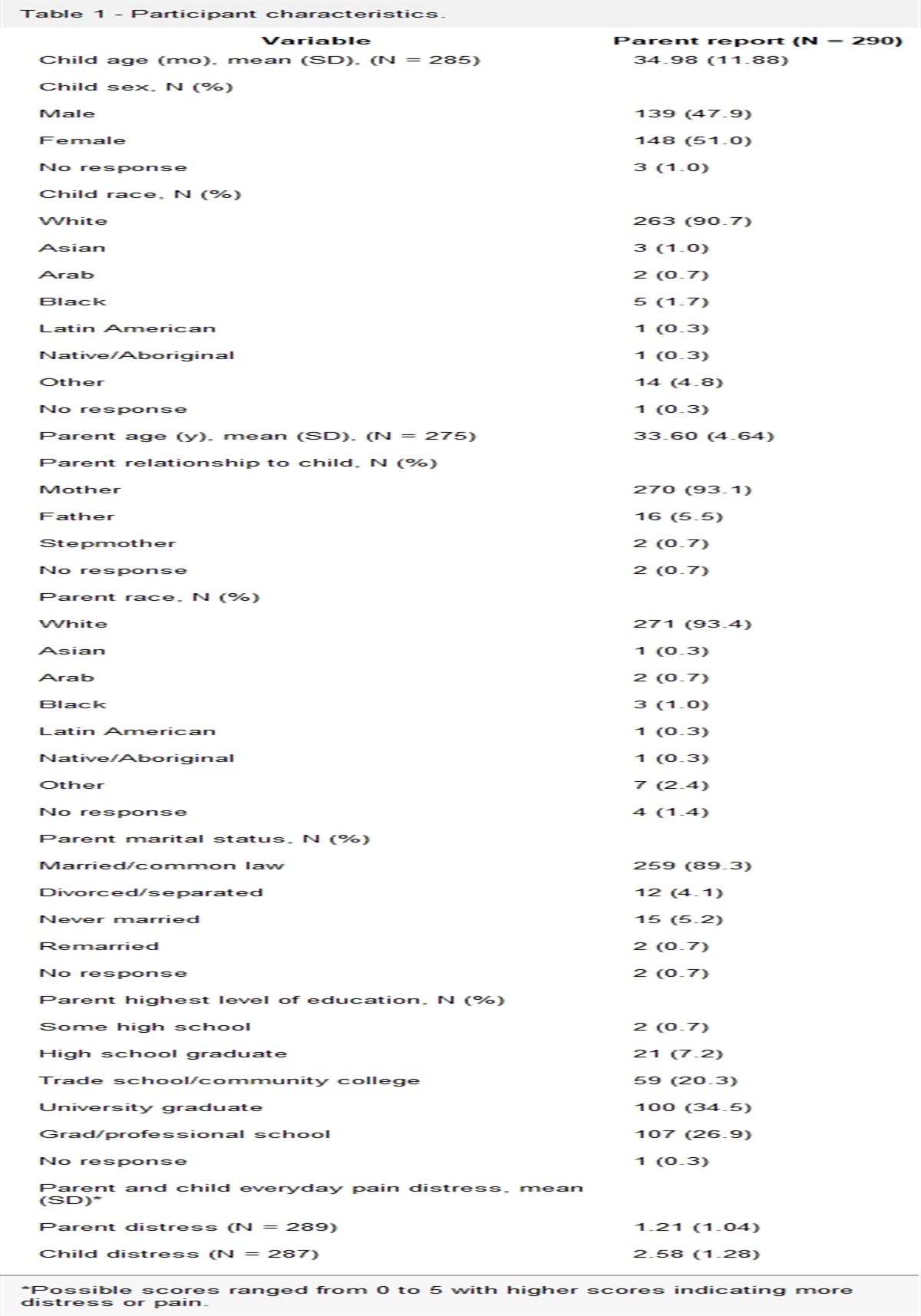
2. Objective assessments of sleepPolysomnography (PSG) is the gold standard in clinical assessment of sleep. Polysomnography is a multimodal assessment which includes EEG to measure brain activity, electrooculography (EOG) to measure eye movements, electromyography (EMG) to measure muscle tone, and respiratory measurements. In clinical use, PSG provides information necessary for sleep scoring by trained sleep technologists to identify sleep stages [W, non-rapid eye movement (NREM) (N1, N2, N3), and rapid eye movement] necessary to describe sleep architecture and identify primary or secondary sleep disorders. Although PSG provides the “full picture” of sleep necessary to diagnose a variety of sleep disorders and to provide markers for sleep staging, the quantitative EEG obtained during sleep may provide sufficient information relevant for the characterization of disordered sleep in chronic pain. Figure 1 presents an overview of the types of data that can be recorded and analyzed with PSG and other sleep EEG devices.
 Figure 1.:
 Summary of sleep-EEG techniques. (A) Visualization of the most common sleep-EEG recording methodologies. Traditional sleep-EEG or polysomnography (PSG) uses wired electrodes placed in standardized locations according to the 10 to 20 EEG system. Wireless devices (both wet and dry) usually deploy headband link devices and leave the user free to move easily and are minimally intrusive. (B) Visual scoring: EEG traces recorded by either wireless sleep-EEG or traditional sleep-EEG are displayed in standardized 30-second epochs and scored according to a set of standardized scoring rules (typically the American Academy of Sleep Medicine scoring criteria). Each epoch is assigned a stage through visual identification of waveforms associated with canonical sleep stages. These epoch scores are then compiled to create a hypnogram (Bi, Bii) and produce sleep continuity statistics. (Bi) Hypnogram recorded in a female with temporomandibular disorder (TMD) demonstrating the insomnia with normal sleep duration (INSD) phenotype. (Bii) Hypnogram from a female with TMD demonstrating the insomnia with short sleep (ISSD) phenotype, characterized by PSG determined total sleep time <6 hours. (C) Spectral Analyses: a quantitative approach used to deconstruct the raw EEG signal into functionally independent EEG power bands. Data are analyzed in short (typically 2–4 s) windows with overlap and passed through Fourier transforms or other spectral analysis methods to derive total power/activity in the delta (0.5–4 Hz), theta (4–8 Hz), alpha (8–12 Hz), sigma (13–18 Hz), beta (18–32 Hz), and gamma (>32 Hz) power bands (D). (E) Two whole night spectrograms from one female with TMD, with one night following a high pain day (bottom panel) and another night following low pain (top panel) nights. (F) Whole night average spectrum of healthy (30 yo/male) [light blue] individual vs patient (32 yo/female) with TMD [red]. REM, rapid eye movement; TST, total sleep time.
In addition to assessing EEG, some sleep monitoring devices are focused on the diagnosis of sleep apnea, using respiratory effort and flow, oximetry, and electrocardiography to provide a multidimensional assessment of sleep disordered breathing. Such devices and associated biomarkers are beyond the scope of this review. Actigraphy—the basis for many commercial exercise monitors—uses wrist accelerometry sensors to identify rest–activity which, in turn, can be used as a surrogate measurements for sleep–wake states. However, actigraphy has known limitations in the assessment of insomnia.49,56 For example, a recent meta-analysis indicates that actigraphy overestimates total sleep time (TST) and sleep efficiency (SE) and underestimates wake after sleep onset (WASO) in patients with chronic pain.16 Accurate scoring of actigraphy also relies on the use of sleep diaries and bedtime markers, compliance with which is typically poor in insomnia.109 Therefore, despite actigraphy's possible advantages, sleep-EEG, by virtue of directly measuring cerebral activity, may provide more precise sleep parameters for the purpose of sleep physiology assessment.
For the purposes of this review, we define spectral and architectural EEG components obtained from analysis of EEG obtained during sleep as “sleep-EEG.” So far, there is no consensus on what defines sleep-EEG for scalp EEG application techniques, scalp EEG coverage or selection, artifact reduction or exclusion, signal quality assessment, and algorithmic details.
Despite these limitations, reviewed studies share general principles. One important quantitative technique is spectral analysis; computations decompose the raw EEG signal into component frequencies of delta (0.5–4.5 Hz), theta (4.5–8 Hz), alpha (8–12 Hz), sigma (12–16 Hz), beta (16–32 Hz), and gamma (32–100 Hz) (Fig. 1). Other studies use some components of PSG (eg, elimination of non-EEG inputs) or quantitative EEG to provide the estimates of TST or other facets of sleep architecture traditionally derived from PSG that do not require traditional sleep scoring. Some advantages of limiting acquisition to EEG include the ability to acquire data outside of a laboratory, thereby avoiding the “first night effect” of sleeping in a new environment. Sleep-EEG implies a quantitative approach that avoids the need for skilled scoring inherent in PSG. Although these deviations from PSG have limitations (eg, the difficulty in identifying rapid eye movement as distinct from other low-amplitude stages or the inability to identify causes of arousals), lower costs, ease of use, and quantitative techniques make sleep-EEG desirable in studies of pain and sleep. For the purposes of our discussion, we will focus on sleep-EEG applications relevant to ambulatory pain medicine.
2.1. Wireless sleep-EEG devicesImprovements in technology, advances in analysis, and an explosion of capital investment have brought a variety of ambulatory sleep devices into public and clinical consciousness. Some devices avoid EEG altogether (eg, peripheral arterial pressure), but we concentrate on sleep-EEG devices to maintain consistency with the mechanistic focus of this review. Most implement frontal electrodes which collect EEG data from bilateral forehead electrode sites (typically AF7 and AF8) and deploy validated scoring algorithms to provide automatic outputs concerning sleep continuity, duration, and architecture. Some devices may incorporate other scalp electrode sites or types of sensors, such as pulse oximetry, photoplethysmography (PPG), and/or actigraphy, to provide additional data on sleep continuity and proxies of heart rate, heart rate variability, and respiratory function. The largest distinction among the most popular devices currently on the market is the use of “dry” vs “wet” EEG sensing approaches. Traditional sleep-EEG relies on conductive electrode paste to optimize signal conduction from the scalp to sensor. This “wet” approach is echoed in a number of devices (eg, Sleep Profiler,34 Hypnodyne ZMax73) through the use of proprietary and disposable electrodes which maximize user friendliness. The alternative “dry” approach (eg, Dreem,3 Muse46) relies on nanocarbon or silicon-coated sensors which are positioned to maintain contact with the skin or scalp. This approach has substantial benefits from a patient comfort and cost effectiveness perspective because the incurred cost of replacing disposable electrodes (∼$10–$20/night) can quickly surpass the initial cost of the device with large studies or regular clinical use. Nevertheless, “dry” EEG signals suffer from the limitation of higher signal impedances and greater liability to movement artifact. Furthermore, many of the signal processing methodologies used to address artifacts across all devices have been developed using “wet-EEG” approaches, and as such, there are limited validation data on their application to “dry” technology.
The wireless and automated nature of these devices has rapidly progressed the feasibility of implementing these technologies into clinical practice. However, a great deal of work must first be done to increase our understanding of the validity and clinical utility for patients with chronic pain and clinical use more generally. Therefore, we recommend clinicians to proceed cautiously with their use. These devices have been reviewed comprehensively in dedicated reviews,18,42,48 to which we direct any reader interested in further details. In addition to autoscoring algorithms, many such devices also implement validated (yet often proprietary) algorithms which can automatically calculate spectral values. Incremental progression in the sensitivity of these algorithms may drive the movement towards automated vs manual scoring in clinical settings. However, for the purposes of reproducibility, open source algorithms are better suited for sleep-EEG research purposes for those with programming skills. A wealth of validated toolboxes are available in MATLAB (eg, EEGLAB,20 SSAVE97), Python (eg, YASA104), C+ (eg, LunaC21), and R (eg, LunaR21) languages.
3. Nocturnal spectral EEG components: associations with sleep and pain 3.1. Beta and gamma powerParticular frequency bands of sleep-EEG offer utility in the studies of insomnia and chronic pain. Scalp-derived fast frequencies (beta 16–32 Hz, gamma 32–100 Hz) are commonly a feature of waking states; beta-power has been associated with active cognitive processing,75,76 whilst gamma power seems as an analog of memory, learning, and sensory processing and attention.43,76 Elevated fast frequencies during wake, however, seem to be a neurophysiological correlate of pain in the somatosensory40 and prefrontal cortices,65 both in healthy participants and those with chronic pain disorders,65 and seem to be associated with increased attention towards painful stimuli.102
During sleep, fast frequencies have been reliably observed during NREM in patients with chronic insomnia disorder.75,76,96 With respect to both gamma and beta frequencies, one study45 in patients with pain associated with traumatic brain injury (TBI) demonstrated greater beta and gamma power during sleep (across all sleep stages) relative to both healthy controls and patients with TBI without pain. A separate experimental study of healthy controls showed that experimental sleep restriction led to increased gamma power during the application of noxious stimuli.62 Another experimental study in healthy controls demonstrated an increase in fast frequency activity (particularly beta power) during the application of painful muscle, joint, and cutaneous stimuli during stage N3 sleep, which is typically dominated by slow waves.22 Synthesizing these findings with the role of gamma power in attention towards painful stimuli,102 it is reasonable to draw the hypothesis that gamma power during sleep could serve as a neural signature of nighttime pain sensations in patients with chronic pain.
Concerning the role of beta power as an EEG feature which is potentially useful in a clinical context, previous work demonstrates that both single component (sleep restriction therapy63) and multicomponent15 cognitive behavioral therapy for insomnia (CBTi) results in the reduction of beta power. Tracking clinical and fast frequency powers before and after treatments may quantify treatment response and reduced hyperarousal in patients undergoing sleep interventions, thereby supplementing traditional subjective pain or attention scales. To better understand the potential clinical applicability of these hypotheses to patients with chronic pain, research should focus on understanding the relationship between gamma and beta power during sleep and clinical pain severity in patients with chronic pain.
3.2. Delta power and alpha powerLow-frequency activity (delta 0–4 Hz), the main identifier of NREM slow wave sleep, may also provide insights into the ongoing status of a patient's response to interventions. Delta activity is believed to be a marker of sleep depth, quality, and neuroplasticity.88 Accordingly, delta power is diminished in both patients with insomnia5 and chronic pain,13 linked to increased fatigue and less restorative sleep.60 Conversely, alpha power reflects increased arousal and wake state and is used to identify periods of wakefulness (whether brief or sustained) during sleep or sleep onset. Early work by Moldofsky et al.69 identified the delta:alpha power ratio during sleep as a promising biomarker for pain.87 This was originally observed in investigations of fibromyalgia, which identified alpha intrusions during NREM sleep as a biomarker of disturbed sleep2,19 that, in turn, was correlated with increased pain severity.69 However, subsequent findings demonstrated the presence of alpha‐delta intrusions in healthy sleepers,89 and that frontal alpha has a sleep promoting role, contrary to occipital alpha which represents the degree of wake-state,78 indicating that not all alpha intrusions during delta signal arousal-related events. Nevertheless, alpha power (particularly peak alpha frequency) has emerged as a crucial area in the field of wake-EEG in relation to pain. Research has shown that the peak frequency of alpha power, which refers to the frequency within the alpha range with the highest absolute power, was inversely associated with the intensity of pain experienced during induction of thermal hyperalgesia.36,37 Moreover, the degree to which peak alpha frequency shifted from high to low, between pain-free and pain states, was independently correlated with the intensity of pain, potentially reflecting neurophysiological alterations indicative of pain chronification. Although this hypothesis remains untested in sleep-EEG, future investigations may tease out the nuances of the complex relationship between alpha power during sleep and chronic pain.
Other studies suggest that diminished delta power, irrespective of the presence or absence of alpha frequency intrusions, may bear relevance to pain. Findings from a small sample of patients with fibromyalgia (n = 8) identified an association between lower delta power and higher next day pain severity.69 One further study has replicated these findings in a larger sample of patients with temporomandibular disorder (TMD; n = 110), demonstrating that activity in delta power is directly linked to nocturnal, next morning, and next day pain severity reported in a pain diary.81,82 However, given the limited nature of the current research, further studies are required to substantiate these findings.
Studies of sleep-derived delta power (or delta:alpha ratios) could focus on replicating these findings in other chronic pain conditions to determine whether these phenomena are associated with the characteristics of “nociplastic pain” that are common among patients with fibromyalgia and TMD or generalize to other pain etiologies and sequelae. Delta power analyses could drive future innovation by providing a framework to use objective microlongitudinal measures of sleep-related treatment response to inform pain treatment decisions in real time, according to markers of response. For example, technology already exists to support the implementation of real-time ambulatory monitoring designs to facilitate the delivery of just-in-time digital interventions for pain, although this is still a nascent area of research.85
4. Insomnia and development of phenotypic biomeasures 4.1. Physiological insomnia phenotypesSleep-EEG, beyond frequency analysis, can supply information on sleep architecture or the basic distributions of TST, SE, WASO, or sleep onset latency. These measurements may be helpful providing quantifiable markers that differentiate the insomnias. However, quantitative approaches have some limitations. For example, it is intuitive to assume that insomnia is principally characterized by insufficient sleep duration. However, large-scale meta-analyses have failed to identify substantial differences in sleep duration between insomnia patients and controls using PSG.5 Total sleep time, for example, fails to capture the subjective dissatisfaction with sleep that is the hallmark of chronic insomnia. Consequently, survey measures, such as the Insomnia Severity Index (ISI70), are preferred as outcome measures in clinical trials. Overreliance on physiological markers could lead to unrecognized and untreated sleep difficulties that do not necessarily track with sleep duration. In fact, practice parameters of the American Academy of Sleep Medicine recommend against the use of PSG for the diagnosis of insomnia unless other sleep disorders are suspected.57
Although it is important to acknowledge the above limitations, sleep-EEG features may serve as a means of insomnia classification. Accordingly, 2 key insomnia phenotypes have been differentiated by their objective sleep duration (TST): (1) insomnia with objective normal sleep duration (INSD) and (2) insomnia with objective short sleep duration (ISSD) (Fig. 1B). Studies demonstrate that patients with insomnia classified as short sleepers using one night of PSG maintained their short sleeper status after 2 nights of subsequent sleep-EEG assessment.28 In another study, cluster analyses also identified that one night of PSG can be used to derive 2 phenotypes of individuals with insomnia (n = 110): those with (n = 43) and without (n = 53) objective short sleep duration.68 The above suggests, therefore, that a single night sample of PSG (or sleep-EEG) may be sufficient for insomnia classification.
4.2. Insomnia with objective short sleep durationInsomnia with objective short sleep duration (ISSD) is a “subtype characterized by insomnia complaints, along with an objectively documented average sleep time less than 6 h per night.”101 Physiological measurements, such as sleep-EEG derived TST, are important in the identification of ISSD since self-report alone is unreliable.9
Those with the ISSD subtype seem to share a common pathophysiology akin to physiological stress after acute sleep deprivation, such as heightened stress/inflammatory responses (such as cortisol, interleukin-6,51 and C-reactive protein28). Accordingly, chronic stress increases the risks of related comorbid diseases, such as cardiovascular disease,12 hypertension,106 diabetes,107 cognitive decline,44 and chronic musculoskeletal pain disorders.38
Relationships between TST and pain seem to be bilateral; meta-analyses suggest that patients with chronic pain have significantly lower TST than healthy controls.61 Daily variability in TST in patients with chronic pain is also associated with worse next day pain severity and interference.17,53,80 Finally, in healthy volunteers, sleep deprivation23 and truncation of TST results in decreased pain inhibition and increases in spontaneous pain.92
Although several studies have examined continuous associations between TST and pain,61 we are aware of only one that has specifically examined the ISSD phenotype in relation to pain. In a sample of women with TMD and comorbid insomnia, patients characterized by the ISSD phenotype (as objectively defined using PSG) demonstrated greater self-reported pain severity, pain sensitivity, and sensitization assessed through quantitative sensory testing, inflammatory markers (eg, interleukin-6), and functional severity of the jaw than those who failed to meet the ISSD criteria.51
Given the dearth of research specifically focusing on the ISSD in comorbid pain and insomnia, further investigations with proper controls are warranted. Such work should build on preliminary findings in TMD by expanding to further chronic pain populations and tracking the trajectories of pain-related symptoms after initial identification of ISSD.
4.3. Insomnia with objective normal sleep durationIn contrast to ISSD, patients with INSD have no objective decrement of TST and accordingly may have differing pathophysiological profiles. Vgontzas105 characterized rumination, depressed mood, poor coping skills, and sleep state misperception as the dominant phenomenological traits of the INSD phenotype. Such a symptom profile fits into the hyperarousal model of insomnia,84 which posits that obsessive preoccupation with sleep leads to cognitive distortions and subjective perceptions of sleep which are misaligned with actual obtained sleep. According to this model, acute cognitive arousal states transform into chronic physiological hyperarousal,84 which further contributes to disorder pathology. Although TST is normal in these patients, other sleep-EEG components (such as spectral analyses) may reflect the pathophysiology of the INSD phenotype. For example, as discussed above, elevated fast frequencies correlate with chronic pain, specifically in those with INSD-type insomnia.96 In fact, elevated beta activities during sleep or particular scenarios during wake seem to be a general biomarker for dysregulation of cortical arousal and inhibition,7,29 which in turn may indicate a “final common pathway” for those with INSD. Indeed, elevated beta power is commonly observed in patients with insomnia disorder96 and normal sleepers undergoing experimental models of insomnia10,14,59 and has been used by EEG studies as an index of cognitive hyperarousal during sleep.63,64 It has been proposed105 that the difference in arousal profiles between INSD (presenting with cortical hyperarousal) and ISSD (presenting with physiological hyperarousal and inflammation) may represent a means through which to differentiate these phenotypes. However, more work is needed to evaluate the specificity of these findings to INSD vs general insomnia profiles.
A related phenomenon reflected by the elevation of beta power is sleep misperception,42 the subjective feeling of short sleep despite physiological evidence to the contrary. Theoretical models propose that the elevated fast frequency activity, a marker of nighttime cognitive hyperarousal, renders those experiencing insomnia a mixed state of consciousness, simultaneously perceiving themselves as both awake and asleep. In a related hypothesis, hyperarousal and state misperception may also affect sensory gating mechanisms, facilitating pain throughout the sleep. For instance, patients with TBI and acute pain demonstrate elevated beta frequency activity during NREM sleep relative to patients with TBI and no pain complaints.45 This suggests that pain concurrent with sleep may increase hyperarousal. Combined with the theoretical concepts of the INSD phenotype, pain-related hyperarousal may drive sleep state misperception in patients with chronic pain and that this effect may increase the risk for patients with chronic pain to develop the INSD phenotype. Indeed, limited data in patients with chronic pain show that sleep state misperception mediates the relationship between presleep cognitive arousal and next day pain symptoms.11
Future work should seek to consolidate these findings by investigating the relationship between beta power and sleep state misperception in patients with chronic pain comorbid with insomnia as well as how this psychophysiological phenomenon maps onto pain symptoms.
5. Sleep discontinuity and microarousalsIn traditional PSG, information on sleep discontinuity is provided by calculating the proportions of 30-second epochs that contain wake vs sleep or observing the number of awakenings or arousals; for example, those with sleep maintenance insomnia experience an increased number of awakenings during the night.3,24,98 Sleep-EEG can also provide the measures of sleep fragmentation.
Fragmented sleep may be more potent than continuous sleep deprivation in worsening pain perception. For example, low SE is the sleep parameter most severely affected in patients with chronic pain disorders.61 In healthy sleepers, experimental sleep disruption through forced nocturnal awakenings has a more pronounced decremental effect on pain inhibition, spontaneous pain, pain sensitivity,41,92 and positive mood32,33 than continuous sleep restriction of equivalent duration.32,33,91 In a daily diary study of patients with chronic pain deriving from sickle cell disease, higher WASO on a given night was associated with greater next day pain72 and greater next day prescription opioid requirements.74 The latter effect was mediated by increased pain severity and pain catastrophizing—a cognitive/affective pain coping style characterized by rumination, magnification, and/or helplessness about pain—suggesting that the effects of sleep discontinuity on pain-related processes may have downstream implications for opioid use behaviors.
Sleep fragmentation may occur in time increments shorter than the traditional 30-second epochs of sleep scoring. For example, cortical microarousals—brief events (3–10 seconds) marked by increases in heart rate, shift to higher frequencies on EEG, and increase in muscle tone not accompanied by clinical change in sleep state—are elevated in somatosensory brain regions during NREM sleep in rodents with neuropathic pain.11 These decrements may also extend to drug sensitivity, as sleep discontinuity has been linked to blunted opioid analgesia, and increased “drug liking” using analogous paradigms.95
These findings build a strong case for the assessments and monitoring of sleep discontinuity in both research and practice. Sleep maintenance insomnia, characterized by frequent nocturnal awakenings and/or extended periods of WASO (>30 minutes), is a common insomnia variant observed in patients with chronic pain. Indeed, sleep consolidation is a primary goal of the stimulus control and sleep restriction components of CBTi, which is generally effective at reducing WASO in patients with chronic pain and comorbid insomnia.93 Although reductions in WASO have been associated with reductions in knee osteoarthritis pain in prior clinical trials,33 many patients emerge from CBTi as “nonresponders,” leaving open the question of how to optimize CBTi for maximum impact on pain outcomes in a greater proportion of patients. EEG may offer opportunities to more precisely identify who is most likely to achieve pain-related benefits from sleep treatment. For example, power spectral analyses have shown that the frequency of alpha oscillations slows during periods of WASO relative to daytime wake periods and that WASO-related alpha slowing was more pronounced in patients with insomnia relatively to healthy individuals.90 Furthermore, lower resting state peak alpha frequency reliably predicts increased pain sensitivity in healthy individuals and patients with chronic pain.36 Future studies may, therefore, consider the potential for variation in WASO-related alpha frequency to predict pain-related treatment response in trials of CBTi among patients with chronic pain.
Future lines of research should also consider using microlongitudinal designs incorporating objective sleep assessments over multiple nights to understand how night-to-night variability in sleep continuity relates to daily opioid use and craving. Studies should also seek to resolve whether sleep discontinuity measured using EEG may serve as an identifying feature or phenotype for increased opioid use, using prospective, observational designs.
6. Future innovations in sleep-EEG for clinical practice 6.1. Treatment selection and customization based on phenotypesSleep-EEG may help identify likely treatment responses among patients with comorbid chronic pain and insomnia. For example, although CBTi is the gold standard first-line intervention for those with chronic insomnia,79 some fail CBTi.6,67 Although a variety of factors may contribute to treatment response, patients with the ISSD phenotype seem to be at greater risk for worse responses and higher remission8 to CBTi.58,103 Conversely, some studies suggest that those with the ISSD phenotype may respond better to pharmacotherapy or combined therapy rather than CBTi alone. For instance, one small study by Vgontaz et al.108 found that trazodone increased TST in patients with the ISSD phenotype, whilst CBTi did not. A second study by Edinger et al.25 used an adaptive trial design in 211 patients with insomnia to better understand patient characteristics contributing to response to common treatment sequences involving varying orders of behavioral therapy, cognitive therapy, and pharmacotherapy. Interestingly those with ISSD, as well as those with a pain disorder, demonstrated higher remission rates in response to treatment sequences starting with behavioral therapy for insomnia (BTi) but demonstrated more a favorable response to a subsequent course of zolpidem. Further mechanistic findings also indicate that trazodone, but not CBTi, increased delta power and decreased beta and gamma power during NREM sleep in patients with ISSD.55
Although preliminary, these findings outline the potential utility of sleep-EEG in identifying the optimal course of treatment for patients with chronic pain and sleep disturbance. Further research with an a priori focus on comorbid pain and insomnia is required to understand how treatment response to both behavioral and pharmacological interventions differs as a function of sleep-EEG components. Such studies may wish to focus on the use of adaptive trial methodology with the aim of outlining treatment response options for those who respond poorly to an initial intervention (eg, CBTi). Future studies might also consider the directionality of these EEG-features' correlation with pain through either upregulation or downregulation (eg, boosting power in a specific band) using noninvasive stimulation techniques.27,39,50 Not only would this work provide an important test of their causal nature and role in disease pathophysiology but would also provide us preliminary data on their utility as putative therapeutic targets.
7. Summary and conclusionRecent advancements in sleep-EEG research have identified sleep-EEG features that may clarify the mechanisms linking sleep, sleep disturbance, clinical insomnia, and pain. Preliminary work suggests that power in beta, gamma, alpha, and delta bands of the EEG spectrum as well as features of sleep architecture (eg, objectively defined total sleep time) could be applied to identify neurophysiological signatures relevant to pain, facilitate insomnia classification, and perhaps guide treatment selection and effectiveness in the treatment of chronic pain. Future work should seek to expand on existing data by characterizing signatures across broad pain diagnoses and examining relationships with clinical outcomes in prospective studies. Future work should also attempt to address these questions among diverse populations throughout the lifespan to provide further clarity on the transferability of these findings to differing age groups.
DisclosuresM.J.R. received nonfinancial support from Oura Health Ltd and Whoop, Inc. and Big Health ltd. M.J.R also has a patent pending for an automated sleep decoding system. M.Q. is a consultant for Natus. P.H.F. is on the Scientific Advisory Board of Ninnion Therapeutics.
AcknowledgmentsThe authors wish to acknowledge funding from NIH/NIDA (R01DA048206; Smith/Finan) in support of this manuscript.
References [1]. Afolalu EF, Ramlee F, Tang NK. Effects of sleep changes on pain-related health outcomes in the general population: a systematic review of longitudinal studies with exploratory meta-analysis. Sleep Med Rev 2018;39:82–97. [2]. Anch AM, Lue FA, MacLean AW, Moldofsky H. Sleep physiology and psychological aspects of the fibrositis (fibromyalgia) syndrome. Can J Psychol 1991;45:179–84. [3]. Arnal PJ, Thorey V, Debellemaniere E, Ballard ME, Bou Hernandez A, Guillot A, Jourde H, Harris M, Guillard M, Van Beers P, Chennaoui M, Sauvet F. The Dreem Headband compared to polysomnography for electroencephalographic signal acquisition and sleep staging. Sleep 2020;43:zsaa097. [4]. Arnison T, Schrooten MGS, Bauducco S, Jansson-Fröjmark M, Persson J. Sleep phase and pre-sleep arousal predicted co-developmental trajectories of pain and insomnia within adolescence. Sci Rep 2022;12:4480. [5]. Baglioni C, Regen W, Teghen A, Spiegelhalder K, Feige B, Nissen C, Riemann D. Sleep changes in the disorder of insomnia: a meta-analysis of polysomnographic studies. Sleep Med Rev 2014;18:195–213. [6]. Baron KG, Hooker S. Next steps for patients who fail to respond to cognitive behavioral therapy for insomnia (CBT-I): the perspective from behavioral sleep medicine psychologists. Curr Sleep Med Rep 2017;3:327–32. [7]. Bastien CH, Turcotte I, St-Jean G, Morin CM, Carrier J. Information processing varies between insomnia types: measures of N1 and P2 during the night. Behav Sleep Med 2013;11:56–72. [8]. Bathgate CJ, Edinger JD, Krystal AD. Insomnia patients with objective short sleep duration have a blunted response to cognitive behavioral therapy for insomnia. Sleep 2017;40:zsw012. [9]. Bathgate CJ, Edinger JD, Wyatt JK, Krystal AD. Objective but not subjective short sleep duration associated with increased risk for hypertension in individuals with insomnia. Sleep 2016;39:1037–45. [10]. Bchir F, Dogui M, Ben Fradj R, Arnaud MJ, Saguem S. Differences in pharmacokinetic and electroencephalographic responses to caffeine in sleep-sensitive and non-sensitive subjects. Compt Rendus Biol 2006;329:512–9. [11]. Bean DJ, Horne J, Lee AC, Johnson MH. Pre-sleep cognitive arousal exacerbates sleep disturbance in chronic pain: an exploratory daily diary and actigraphy study. Scand J Pain 2021;21:724–31. [12]. Bertisch SM, Pollock BD, Mittleman MA, Buysse DJ, Bazzano LA, Gottlieb DJ, Redline S. Insomnia with objective short sleep duration and risk of incident cardiovascular disease and all-cause mortality: Sleep Heart Health Study. Sleep 2018;41:zsy047. [13]. Bjurstrom MF, Irwin MR. Polysomnographic characteristics in nonmalignant chronic pain populations: a review of controlled studies. Sleep Med Rev 2016;26:74–86. [14]. Bonnet MH, Arand D. Caffeine use as a model of acute and chronic insomnia. Sleep 1992;15:526–36. [15]. Cervena K, Dauvilliers Y, Espa F, Touchon J, Matousek M, Billiard M, Besset A. Effect of cognitive behavioural therapy for insomnia on sleep architecture and sleep EEG power spectra in psychophysiological insomnia. J Sleep Res 2004;13:385–93. [16]. Conley S, Knies A, Batten J, Ash G, Miner B, Hwang Y, Jeon S, Redeker NS. Agreement between actigraphic and polysomnographic measures of sleep in adults with and without chronic conditions: a systematic review and meta-analysis. Sleep Med Rev 2019;46:151–60. [17]. Davin S, Wilt J, Covington E, Scheman J. Variability in the relationship between sleep and pain in patients undergoing interdisciplinary rehabilitation for chronic pain. Pain Med 2014;15:1043–51. [18]. De Fazio R, Mattei V, Al-Naami B, De Vittorio M, Visconti P. Methodologies and wearable devices to monitor biophysical parameters related to sleep dysfunctions: an overview. Micromachines 2022;13:1335. [19]. Della Marca G, Frusciante R, Vollono C, Iannaccone E, Dittoni S, Losurdo A, Testani E, Gnoni V, Colicchio S, Di Blasi C, Erra C, Mazza S, Ricci E. Pain and the alpha-sleep anomaly: a mechanism of sleep disruption in facioscapulohumeral muscular dystrophy. Pain Med 2013;14:487–97. [20]. Delorme A, Makeig S. EEGLAB: an open source toolbox for analysis of single-trial EEG dynamics including independent component analysis. J Neurosci Methods 2004;134:9–21. [21]. Djonlagic I, Mariani S, Fitzpatrick AL, Van Der Klei VM, Johnson DA, Wood AC, Seeman T, Nguyen HT, Prerau MJ, Luchsinger JA, Dzierzewski JM, Rapp SR, Tranah GJ, Yaffe K, Burdick KE, Stone KL, Redline S, Purcell SM. Macro and micro sleep architecture and cognitive performance in older adults. Nat Hum Behav 2020;5:123–45. [22]. Drewes AM, Nielsen KD, Arendt-Nielsen L, Birket-Smith L, Hansen LM. The effect of cutaneous and deep pain on the electroencephalogram during sleep—an experimental study. Sleep 1997;20:632–40. [23]. Drewes AM, Nielsen KD, Rasmussen C, Arima T, Svensson P, Rössel P, Arendt-Nielsen L. The effects of controlled delta sleep deprivation on experimental pain in healthy subjects. J Musculoskelet Pain 2000;8:49–67. [24]. Drewes AM, Svendsen L, Taagholt SJ, Bjerregård K, Nielsen KD, Hansen B. Sleep in rheumatoid arthritis: a comparison with healthy subjects and studies of sleep/wake interactions. Rheumatology 1998;37:71–81. [25]. Edinger JD, Beaulieu-Bonneau S, Ivers H, Guay B, Bélanger L, Simmons B, Morin CM. Association between insomnia patients' pre-treatment characteristics and their responses to distinctive treatment sequences. Sleep 2022;45:zsab245. [26]. Espie CA, Emsley R, Kyle SD, Gordon C, Drake CL, Siriwardena AN, Cape J, Ong JC, Sheaves B, Foster R, Freeman D, Costa-Font J, Marsden A, Luik AI. Effect of digital cognitive behavioral therapy for insomnia on health, psychological well-being, and sleep-related quality of life: a randomized clinical trial. JAMA Psychiatry 2019;76:21–30. [27]. Fehér KD, Wunderlin M, Maier JG, Hertenstein E, Schneider CL, Mikutta C, Züst MA, Klöppel S, Nissen C. Shaping the slow waves of sleep: a systematic and integrative review of sleep slow wave modulation in humans using non-invasive brain stimulation. Sleep Med Rev 2021;58:101438. [28]. Fernandez-Mendoza J, Baker JH, Vgontzas AN, Gaines J, Liao D, Bixler EO. Insomnia symptoms with objective short sleep duration are associated with systemic inflammation in adolescents. Brain Behav Immun 2017;61:110–6. [29]. Fernandez-Mendoza J, Li Y, Vgontzas AN, Fang J, Gaines J, Calhoun SL, Liao D, Bixler EO. Insomnia is associated with cortical hyperarousal as early as adolescence. Sleep 2016;39:1029–36.
留言 (0)