
Remember me


Myopia is a common eye disease that generally develops in childhood and causes significant vision loss. Furthermore, it is a risk factor for a range of other serious ocular conditions, making it a major public health issue concern worldwide.1 The prevalence of myopia, especially in East Asians, has markedly increased over the past half-century, and it is predicted to affect more than 80% of Chinese children and adolescents (aged 3–19 years) by 2050.2
Myopia development is dependent on genetic and environmental factors,2 where high myopia is defined as the spherical equivalent greater than −6.0 diopters or an ocular axial length ≥ 26.0 mm.3 Most cases of early-onset high myopia are considered to be inherited as a Mendelian trait.4 When myopia is accompanied by other ocular or systemic abnormalities, it is classified as syndromic myopia,5 which has demonstrated an association with multiple syndromes, including retinitis pigmentosa (RP), caused by RP2 and RPGR mutations6; congenital stationary night blindness (CSNB), caused by NYX, CACNA1F, GRM6, and LRIT3 mutations7; and Stickler syndrome caused by COL2A1, COL11A1, COL9A1, and COL9A2 mutations.8 In addition, several X-linked retinopathies (XLRs) can present with myopia; most strikingly, RP is caused by mutations in RPGR, and cone–rod dystrophy (CORD) and CSNB are caused by CACNA1F mutations.9 As such, exploring the combination of high myopia and inherited retinal diseases with X-linked recessive gene mutations is an extremely important challenge. Thus, the aim of this study was to evaluate the refractive errors with follow-up and myopic maculopathy in patients with XLRs.
Methods PatientsPatients who harbored pathogenic variants of the genes listed on the RetNet website (https://web.sph.uth.edu/RetNet/), which have been reported to cause inherited retinal diseases with an X-linked recessive trait were recruited for this study. All patients were Han Chinese. The genes included CACNA1F, RPGR, NYX, TIMM8A, OFD1, CHM, DMD, OPN1LW, OPN1MW, PGK1, NDP, and RS1. Patients with mutations in NDP and RS1 were excluded because of the resultant severe retinal detachment or retinoschisis. The study ran from January 2018 to April 2022, and it was performed in accordance with the guidelines of the Declaration of Helsinki. The institutional review board of the Zhongshan Ophthalmic Centre (2020KYPJ173) approved all procedures, and written informed consent was obtained from all adult patients and parents or guardians of child patients before the clinical data and DNA samples were collected.
Ocular ExaminationEach patient's clinical information was recorded, including age at onset, age at diagnosis, gender, and initial symptoms. Complete ophthalmic examinations were performed and measurements were recorded, including best-corrected visual acuity (BCVA), refractive errors, and axial length (IOL Master, Carl Zeiss, Oberkochen, Germany). Images were obtained using a fundus camera (Zeiss), a scanning laser ophthalmoscope (California FA; Optos, Dunfermline, United Kingdom), a RetCam (RetCam; Clarity Medical Systems Inc, Pleasanton, CA), swept-source optical coherence tomography (SS-OCT, VG200, SVision Research, Shanghai, China), and flash electroretinogram (ERG, RETeval; LKC Technologies, Gaithersburg, MD). Two experienced ophthalmologists (X.D. and L.H.) read all images, and myopic maculopathy was staged according to the atrophy (A), traction (T), and neovascularization (N) grading system.10
Whole Exome SequencingThe method used to extract peripheral blood samples and isolate genomic DNA was the same as that used in our previous study.11 Whole exome sequencing of all probands was performed, as well as Sanger sequencing for validation and a segregation analysis of available family members. The reported pathogenic variants were confirmed using a panel that included SIFT (http://sift.jcvi.org/www/SIFT_enst_submit.html), Polyphen-2 (http://genetics.bwh.harvard.edu/pph2/), REVEL,12 and the Genome Aggregation Database (gnomAD, http://www.gnomad-sg.org).
Results Gene MutationsIn total, 17 patients, including 15 males and two females, were enrolled in this study, 16 of whom were sporadic and one of whom had a positive family history of high myopia. The genes associated with their conditions were CACNA1F, RPGR, NYX, and OPN1MW. The number of patients with mutations in CACNA1F, RPGR, NYX, and OPN1MW was six, seven, three, and one, respectively (Table 1). Among the 17 patients, 15 inherited mutations from their mother, and two were found to be de novo mutations (Figures 1,2A). These mutations included 12 novel and five known mutations.13–16 In the type of mutation, 12 of the 17 were truncations and five were missense mutations, which were predicted to be damaging according to bioinformatic analysis (Table 1).
Table 1. - Identified Variants in X-Linked Recessive Genes ID Gene Exon cDNA Change Amino Acid Change Location SIFT Polyphen-2 REVEL gnomAD Inheritance References 1 CACNA1F exon42 c.4938dup p.Gln1647Alafs*9 chrX:49065769 — — — 0 Mother — 2 CACNA1F exon2 c.244C>T p.Arg82* chrX:49088171 — — — 0.000005 Mother 15 3 CACNA1F exon31 c.3742_3745del p.His1248Thrfs*18 chrX:49070358 — — — 0 Mother — 4 CACNA1F exon43 c.5103dup p.Thr1702Aspfs*47 chrX:49,065,027 — — — 0 Mother — 5 CACNA1F intron23 c.2767-1G>T — chrX:49,075,195 — — — 0 Mother — 6 CACNA1F exon46 c.5479C>T p.Arg1827* chrX:49062998 — — — 0.000005 Mother 16 7 NYX exon2 c.521T>A p.Val174Glu chrX:41333227 D D 0.89784 0 Mother — 8 NYX exon2 c.935A>G p.Asn312Ser chrX:41333641 D D 0.88963 0 Mother — 9 NYX exon2 c.272T>C p.Leu91Pro chrX:41332978 D D 0.99525 0 Mother — 10 OPN1MW exon2-3 CNV exon2-3del — — — — — 0 Mother — 11 RPGR exon7 c.739C>T p.Gln247* chrX:38169907 — — — 0 De novo — 12 RPGR exon15 c.2237_2241del p.Glu746Glyfs*22 chrX:38146010 — — — 0 De novo — 13 RPGR exon5 c.385A>C p.Thr129Pro chrX:38178166 D D 0.77335 0 Mother — 14 RPGR exon15 c.2426_2427del p.Glu809Glyfs*25 chrX:38145824 — — — 0 Mother 17 15 RPGR exon4 c.310G>A p.Glu104Lys chrX:38180280 T B 0.64977 0 Mother — 16 RPGR exon15 c.3178_3179del p.Glu1060Argfs*18 chrX:38145072 — — — 0.000011 Mother 18 17 RPGR exon15 c.2405_2406del p.Glu802Glyfs*32 chrX:38145845-38145847 — — — 0 Unknown 18For patient 15, the mutation c.310G > A (p.Glu104Lys) was predicted to be tolerant by SIFT and benign by Polyphen-2; however, it was predicted to change the splice site with the ada_score = 0.99999 and rf_score = 0.994.
B, Benign; D, damaging; T, Tolerant.
 Fig. 1.:
 The pedigree of the families with mutations in X-linked recessive genes. A, aunt; B, brother; D, daughter; F, father; GF, grandfather; GM, grandmother; M, mother; S, sister; U, uncle; W, wife.
 Fig. 2.:
 A. The inheritance patterns of the diseases examined in this study; two mutations were de novo mutations. B. The diseases found in the patients included in this study. C. Myopia in patients with different gene mutations. D. Refractive error changes during follow-up.
Clinical DataOf the 17 patients, 15 were male and two were female, and the mean age at onset was 4.81 years (4.81 ± 4.04), and the mean examination age was 7.18 years (7.18 ± 7.24). Six patients were diagnosed with CSNB, four with CORD, four with RP, one with achromatopsia, and one with Leber congenital amaurosis (LCA, Figure 2B). One patient, whose BCVA was 20/50 bilaterally and who had a normal cone and rod response (ERG) and an indistinct interdigitation zone (OCT), was difficult to diagnose; however, a diagnosis of high myopia was eventually determined. The anterior segment was unremarkable in all 17 patients, and in patients with CACNA1F and NYX mutations, the fundus was unremarkable, except for the presence of a tessellated fundus, which was consistent with myopia. OCT results showed only indistinct interdigitation zones. In patients with CSNB, the ERG results showed a reduced b wave and a reduced b/a wave ratio in the rod response, and a moderately-to-severely reduced cone response (Figure 3). In patients with CORD, the ERG results revealed a severely reduced cone response; however, the rod response was normal. Among the seven patients with RPGR mutations, four with RP were found to have retinal pigmentary changes in the fundus and outer retinal atrophy (OCT, Figure 4), and two with CORD were found to have an indistinct interdigitation zone and a disrupted interdigitation zone (OCT). The patient with an OPN1MW mutation was found to have abnormal color vision, BCVA of 20/25 in both eyes, and a normal fundus and was diagnosed with achromatopsia (Table 2).
 Fig. 3.:
 Multimodal imaging of patient 1, diagnosed with congenital stationary night blindness (CSNB). A. Scanning laser ophthalmoscopy (SLO) revealed darkness without pressure of the right eye. B. Autofluorescence was found to be unremarkable. C. Optical coherence tomography (OCT) revealed an indistinct interdigitation zone. D. An electroretinogram (ERG) showed a decreased b wave, a b/a wave ratio in the rod response, and a reduced cone response.
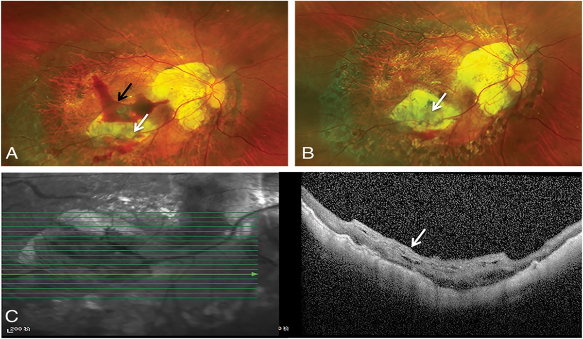
 Fig. 4.:
 The multimodal imaging of patient 14 diagnosed with cone–rod dystrophy (CORD). A. SLO revealed retinal pigmentary changes and a tessellated fundus of the left eye. B. Autofluorescence was found to be abnormal. C. OCT revealed a disrupted interdigitation zone and discontinuous ellipsoid zone. D. An ERG showed a slightly decreased rod response and a severely reduced cone response. SLO, scanning laser ophthalmoscopy.
Table 2. - Clinical Data of the Probands ID Gender First Symptom BCVA-OD BCVA-OS Fundus OD Fundus OS ERG-Rod Response ERG-Cone Response b/a Wave Ratio OD b/a Wave Ratio OS OCT-OD OCT-OS Diagnosis 1 M Poor vision 20/50 20/63 DWP DWP Reduced b wave Severely reduced 0.873 0.795 IIZ IIZ CSNB 2 M Photophobia 20/63 20/50 TF TF Reduced b wave Severely reduced 0.696 0.68 IIZ IIZ CSNB 3 M Strabismus 20/63 20/100 TF TF Normal Severely reduced 5.21 0.985 IIZ IIZ CORD 4 M Nystagmus NA NA — — Extinguished Extinguished NA NA IIZ IIZ LCA 5 M Nystagmus 20/100 20/100 TF TF Reduced b wave Severely reduced 0.515 0.345 IIZ IIZ CSNB 6 M Poor vision 20/25 20/25 TF TF Normal Moderately reduced 1.173 2.229 NA NA CORD 7 M Poor vision 20/50 20/50 TF TF Reduced b wave Mildly reduced 0.591 0.804 IIZ IIZ CSNB 8 M Poor vision NA NA TF TF NA NA NA NA IIZ IIZ CSNB 9 M Poor vision 20/50 20/40 TF TF Reduced b wave Normal 0.412 0.432 IIZ IIZ CSNB 10 M Abnormal color vision 20/25 20/25 Normal Normal Reduced b wave NA NA NA NA NA Achromatopsia 11 F Poor vision 20/63 20/63 TF TF Reduced b wave NA NA NA ORA ORA RP 12 F Poor vision, strabismus 20/32 20/50 TF TF Reduced b wave Severely reduced 1.947 1.951 Normal Normal CORD 13 M Poor vision 20/40 20/40 RPC RPC Reduced b wave Extinguished 1.324 3.647 ORA ORA RP 14 M Poor vision NA NA RPC, TF RPC, TF Reduced b wave Severely reduced 3.078 3.496 DEZ, DIZ DEZ, DIZ CORD 15 M Night blindness 20/32 20/40 RPC RPC Reduced b wave Extinguished NA NA ORA ORA RP 16 M Poor vision 20/50 20/50 NA NA Reduced b wave Normal 2.289 3.068 IIZ IIZ High myopia 17 M Night blindness 20/63 20/63 RPC RPC Reduced b wave Extinguished NA NA ORA ORA RPDEZ, discontinuous ellipsoid zone; DIZ, disrupted interdigitation zone; DWP, Dark without pressure; F, female; IIZ, indistinct interdigitation zone; LCA, leber congenital amaurosis; M, Male; NA, not available; OD, right eye; ORA, Outer retinal atrophy; OS, left eye; RP, Retinitis pigmentosa; RPC, retinal pigmentary changes; TF, Tessellated fundus.
Among the 17 patients, 88.2% (15/17) had varying degrees of myopia, and 64.7% (11/17) had high myopia in at least one eye. Gene analysis showed that high myopia was present in 80% (4/5) of patients with a CACNA1F mutations, 100% (3/3) of patients with an NYX mutation, and 57.1% (4/7) of patients with RPGR mutations (Figure 2C), whereas the patient with the OPN1MW mutation had mild-to-moderate myopia. According to the ATN classification, which considers the atrophic (A), tractional (T), and neovascular (N) components,10 64.7% (11/17) of patients were A1T0N0, and 35.3% (6/17) were A0T0N0 (Table 3). Staphyloma was presented in eight (47.1%) patients. By analyzing the refractive error follow-up data, it was determined that the myopia progressed over time. The spherical equivalent change was −0.388 ± 0.515 diopters within 6 months, −1.104 ± 0.578 diopters within 1 year, and −1.219 ± 0.598 diopters within 2 years (Figure 2D). Even in patients with CSNB, the refractive error still progressed.
Table 3. - Refractive Errors and Myopic Maculopathies of Patients ID Gene Spherical Equivalent-OD, Diopter Spherical Equivalent-OS, Diopter Axial Length-OD, mm Axial Length-OS, mm ATN Stage -OD ATN Stage -OS 1 CACNA1F −4.25 −4.50 25.27 — A0T0N0 A0T0N0 2 CACNA1F −8.75 −8.50 — — A1T0N0 A1T0N0 3 CACNA1F −7.63 −5.88 26.12 26.22 A1T0N0 A1T0N0 4* CACNA1F — — — — A0T0N0 A0T0N0 5 CACNA1F −5.00 −6.25 — — A1T0N0 A1T0N0 6 CACNA1F −8.50 −7.88 — — A1T0N0 A1T0N0 7 NYX −6.50 −6.50 24.83 24.76 A1T0N0 A1T0N0 8 NYX −6.25 −6.13 24.89 24.94 A1T0N0 A1T0N0 9 NYX −6.50 −6.25 — — A0T0N0 A1T0N0 10 OPN1MW −2.75 −3.88 — — A0T0N0 A0T0N0 11 RPGR −6.88 −8.00 — — A1T0N0 A1T0N0 12 RPGR −6.00 −7.88 — — A1T0N0 A1T0N0 13 RPGR −1.38 0.13 24.74 24.25 A0T0N0 A0T0N0 14 RPGR −8.13 −8.13 24.62 24.55 A1T0N0 A1T0N0 15 RPGR 1.50 0.88 — — A0T0N0 A0T0N0 16 RPGR −4.88 −4.75 — — A0T0N0 A0T0N0 17 RPGR −9.75 −10.75 — — A1T0N0 A1T0N0*Patient four was too young to cooperate with the refractive error and axial length examination at the age of one.
Two female patients of preschool age were found to have de novo heterozygous RPGR mutations; that is, mutations were absent in their family members. Patient 11 had a c.739C > T (p.Gln247*) mutation in exon 7, and her BCVA was 20/63. Fundus examination revealed a tessellated fundus and OCT revealed bilateral outer retinal atrophy, which supported the diagnosis of RP. Meanwhile, Patient 12 had a c.2237_2241del (p.Glu746Glyfs*22) mutation in exon 15, and her BCVA was 20/32 in the right eye and 20/50 in the left. The scanning laser ophthalmoscope revealed a tessellated fundus, OCT showed an indistinct interdigitation zone, and ERG showed a moderately reduced rod response and a severely reduced cone response, which support the diagnosis of CORD (Tables 1–3). Patient 11 and Patient 12 had high myopia.
Discussion&#
Comments (0)