
記住我


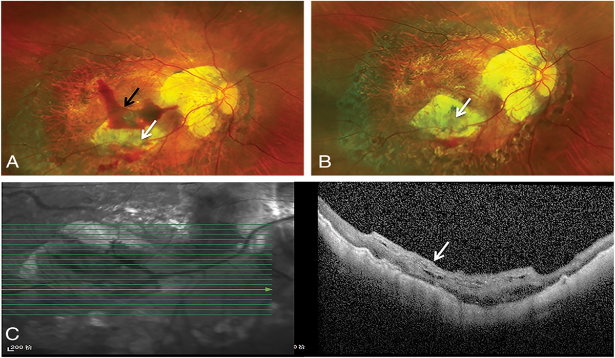
Anti–vascular endothelial growth factor (anti-VEGF) therapies have become the standard of care for neovascular age-related macular degeneration (nAMD) and are highly efficacious in reducing intraretinal fluid (IRF) and subretinal fluid (SRF).1–4 Pigment epithelial detachment (PED) is the most common lesion type in nAMD and can develop as the result of exudation originating from type 1 or type 3 macular neovascularization.5,6 Anatomically, PED represents a separation of the retinal pigment epithelium (RPE) and its basal lamina from the underlying Bruch membrane.7–9 Although the pathophysiology of PED development in nAMD is still a topic of investigation, it commonly arises as a result of the in-growth of type 1 macular neovascularization through the Bruch membrane into the sub-RPE space with subsequent fluid extravasation.8,10–12 Pigment epithelium detachments associated with type 1 macular neovascularization often develop a multilayered morphology with a sub-RPE fibrovascular component and a deeper fibrocellular layer.11 Thus, the PED in eyes with nAMD can be composed of varying proportions of neovascular and stromal tissue, and sub-RPE fluid, hemorrhage, and exudation. Leakage of fluid and blood into the sub-RPE or subneurosensory compartments typically lead to significant vision loss.
Subretinal fluid and IRF are tried and true biomarkers of exudation and are the targets of anti-VEGF therapy in eyes with nAMD. However, analysis of the quantitative effect of anti-VEGF therapy on PED outcomes in most pivotal anti-VEGF clinical trials is limited. Furthermore, inconsistent relationships between PED reduction and visual outcomes have been reported by several studies.13–19 Large PED thickness or height (>400–500 µm), however, is associated with an elevated risk of RPE tear in anti-VEGF-treated eyes.20–22 Although a reduction of various PED dimensions, including PED thickness, is a known outcome of certain anti-VEGF therapies, a commensurate improvement in visual acuity has not been documented.8,14,23,24
The Phase 3 HAWK and HARRIER trials demonstrated noninferior best-corrected visual acuity (BCVA) gains and superior anatomical outcomes with brolucizumab 6 mg dosed every 12 or 8 weeks (q12w/q8w) versus aflibercept 2 mg q8w, with more than 50% of brolucizumab-treated patients maintained on a q12w dosing interval up to the primary endpoint at week 48.3,4 The aim of this treatment-agnostic, post hoc, pooled analysis was to investigate the impact of baseline and week 12 PED thickness and PED thickness variability on BCVA outcomes through week 96 in anti-VEGF-treated eyes from HAWK and HARRIER.
Methods Study Design and Post Hoc Analysis PopulationHAWK (NCT02307682) and HARRIER (NCT02434328) were pivotal 96-week, double-masked, multicenter Phase 3 clinical trials that randomized patients with nAMD 1:1:1 to brolucizumab 3 mg, brolucizumab 6 mg, or aflibercept 2 mg (HAWK) or 1:1 to brolucizumab 6 mg or aflibercept 2 mg (HARRIER). After three monthly loading doses at weeks 0, 4, and 8, brolucizumab study eyes were given an intravitreal injection every 12 weeks (q12w) and the dose was adjusted to every 8 weeks (q8w) for the remainder of the study period if disease activity was detected at any of the predefined assessment visits; aflibercept was dosed q8w after three monthly loading doses, as per label at study initiation.3,4
This post hoc analysis pooled all patients from the brolucizumab 6 mg arms and the aflibercept 2 mg arms from HAWK and HARRIER to create one treatment-agnostic patient cohort. The final data set includes all randomized and treated patients from HAWK and HARRIER with at least two evaluable postbaseline optical coherence tomography (OCT) images.
The studies adhered to the tenets of the Declaration of Helsinki, International Conference on Harmonization E6 Good Clinical Practice Consolidated Guidelines, and other regulations as applicable and was compliant with the Health Insurance Portability and Accountability Act of 1996. Institutional review board/ethics committee approval was obtained at each study site and all study participants provided written informed consent.
Clinical AssessmentsMasked investigators in HAWK and HARRIER conducted anatomical and visual assessments at baseline and every 4 weeks. Best-corrected visual acuity was measured using Early Treatment Diabetic Retinopathy Study charts. For this post hoc analysis, OCT images from baseline, and at weeks 4, 8, 12, 16, 20, 24, 32, 48, 68, and 96 (where available) were transferred to the Doheny Image Reading and Research Lab (Los Angeles, CA) for assessment. PEDs were measured from the RPE inner border to the Bruch membrane. Graders reviewed all B scans in the volumetric cube to determine and measure the maximum PED thickness (or height) across the entire macula (OCT scan area 6 mm × 6 mm) for each patient at each time point.
Study Outcome MeasuresIn this pooled, treatment-agnostic analysis, the 2-year BCVA outcomes were compared in patients with different PED thickness cut-off thresholds at baseline (200, 300, and 400 µm) and at week 12 (100, 200, 300, and 400 µm), when the full impact of the loading phase (three monthly loading doses) is observed. The ocular adverse events, including RPE tears, were also investigated in the subgroup of eyes with week 12 PED ≥400 µm.
The association between fluctuations in PED thickness in the maintenance phase and visual outcomes was also investigated by calculating the mean and SD of a patient's maximum PED thickness using all measurements available from week 12 to week 96. The SD [PED] was then taken as a metric of individual PED variability. Patients were grouped into quartiles of increasing SD [PED], designated as Q1 (lowest variability) to Q4 (highest variability) and the BCVA outcomes in each of these quartiles compared. Finally, the association between PED thickness and the presence of IRF and SRF was evaluated by creating four PED thickness quartiles at week 48 and comparing the prevalence of IRF or SRF in each quartile.
Statistical AnalysesThe post hoc analysis was descriptive in nature, and the groups analyzed were defined postbaseline. Best-corrected visual acuity change from baseline was analyzed using the analysis of variance model with baseline BCVA categories (≤55, 56–70, ≥71 letters), age categories (<75, ≥75), and fluid-free status as fixed effect factors. Missing values were imputed using the last observation carried forward method.
Results Patient PopulationThe anti-VEGF treatment-agnostic cohort comprised 1,459 patients pooled from HAWK and HARRIER and treated with brolucizumab 6 mg or aflibercept 2 mg. Baseline PED status data were available and analyzed in a total of 95% (1,383/1,459) of all patients and these were subgrouped and classified according to the maximum PED thickness at baseline as follows: <200 µm (n = 870), ≥200 µm (n = 513); <300 µm (n = 1,115), ≥300 µm (n = 268); and <400 µm (n = 1,235), ≥400 µm (n = 148). The demographic and clinical characteristics at baseline for each of these subgroups are presented in Table 1. These subgroups were all well-balanced with no significant differences in baseline characteristics.
Table 1. - Demographic and Clinical Characteristics at Baseline of Pooled HAWK and HARRIER CohortCNV, choroidal neovascularization; CST, central subfield thickness; ETDRS, Early Treatment Diabetic Retinopathy Study.
Best-corrected visual acuity gains through week 96 were lower in eyes with greater PED thickness at baseline. At week 96, eyes with PED ≥200 µm (n = 513) gained a least square mean (LS mean) standard error (SE) of +4.6 (0.6) letters compared with +7.0 (0.5) letters in eyes with PED <200 µm (n = 870) (Figure 1A). Similarly, eyes with PED ≥300 µm (n = 268) gained an LS mean (SE) of +4.0 (0.9) letters at week 96 compared with +6.6 (0.4) letters in eyes with PED <300 µm (n = 1,115) (Figure 1B) and eyes with PED ≥400 µm (n = 148) gained an LS mean (SE) of +3.0 (1.2) letters at week 96 compared with +6.5 (0.4) letters in eyes with PED <400 µm (n = 1,235) (Figure 1C).
 Fig. 1.:
 Change from baseline in BCVA in patient subgroups with baseline PED thickness cut-offs of A. 200 µm; B. 300 µm; C. 400 µm. Eyes with baseline PED thickness >400 µm show the lowest vision gains through 48 and 96 weeks. BL, baseline; EDTRS, early treatment diabetic retinopathy study.
Impact of Week 12 Pigment Epithelial Detachment Thickness on Visual Outcomes to Week 96At weeks 48 and 96, eyes with week 12 PED thickness ≥100 µm (n = 763) exhibited lower LS mean (SE) BCVA change from baseline than eyes with PED thickness <100 µm (n = 594) (week 48: 6.8 [0.5] vs. 7.6 [0.5]; week 96: 5.6 [0.5] vs. 6.6 [0.6]; Figure 2A). Stratifying eyes with week 12 PED thickness ≥100 µm into 100 µm PED thickness interval groups (≥100 µm to <200 µm; ≥200 µm to <300 µm; ≥300 µm to <400 µm; and ≥400 µm) revealed that eyes with week 12 PED thickness ≥400 µm exhibited a net loss of vision over time in contrast to eyes with PED thickness <400 µm, which showed a net gain of vision over time (Figure 2B).
 Fig. 2.:
 The influence of week 12 PED thickness on BCVA outcomes over time. A. Week 12 PED thickness cut-off at 100 µm; B. Week 12 PED thickness cut-offs at 100 µm, 200 µm, 300 µm, and 400 µm; C. PED thickness cut-off at ≥400 µm with presence or absence of RPE tear. Eyes with PED thickness >400 µm at 12 weeks exhibit the lowest vision gains through weeks 48 and 96. When eyes with RPE tears are excluded, visual outcomes improve, but are still remarkably lower than the group of eyes with thickness <400 µm at 12 weeks. BL, baseline; EDTRS, Early Treatment Diabetic Retinopathy Study.
Because RPE tears are common in patients with large PEDs and can contribute to overall vision loss, visual outcomes were also assessed in patients with week 12 PED ≥400 µm with and without the presence of an RPE tear. One-third (33% [12/36]) of eyes with PED thickness ≥400 µm at 12 weeks developed an RPE tear (although this rate was 10% [15/148] in the group with PED thickness >400 µm at baseline), and BCVA outcomes over time were lower in this group compared with eyes without an RPE tear (Figure 2C). At week 96, LS mean (SE) BCVA change from baseline in the subgroup of eyes with PED ≥400 µm and RPE tear present was −9.7 (5.5) letters compared with −0.5 (4.1) letters in the subgroup of eyes with PED ≥400 µm and no RPE tear. All ocular adverse events that were reported in eyes with week 12 PED thickness ≥400 µm and which may have contributed to the net vision loss observed in this subgroup are listed in Supplemental Digital Content 1 (see Table, https://links.lww.com/IAE/C81). Retinal hemorrhage was reported in 5 (13.9%) eyes and 1 (2.8%) eye experienced a retinal artery thrombosis, but there were no cases of endophthalmitis, retinal detachment, or retinal tear.
Impact of Pigment Epithelium Detachment Thickness Variability on Best-Corrected Visual Acuity Outcomes over TimeGrouping eyes into quartiles based on their PED thickness variability between week 12 and week 96 showed that higher fluctuations in PED thickness resulted in lower BCVA gains over time (Figure 3). In particular, after an initial LS mean (SE) BCVA gain of +5.2 (0.5) letters postloading at week 12, mean BCVA change from baseline in the quartile with highest variability (Q4, >33 µm) decreased to +3.3 (0.8) letters at week 96. By contrast, the mean gain in BCVA in eyes with <33 µm variability was between +6.2 (0.8) and +7.9 (0.8) letters at week 96. No clear association was found between PED thickness variability and the presence of RPE tears.
 Fig. 3.:
 The influence of PED thickness variability on BCVA outcomes over time. BCVA over time for the four week 12–week 96 PED thickness variability quartiles, N = 1,383. Eyes with the greatest PED thickness variability show the lowest vision gains through 96 weeks. BL, baseline; EDTRS, early treatment diabetic retinopathy study.
Association of Pigment Epithelium Detachment Thickness with Intraretinal Fluid and Subretinal FluidWith the evaluation of one time point (in this case week 48, the primary endpoint of the studies), increasing PED thickness was associated with increased presence of IRF and SRF. Compared with the reference quartile of week 48 PED thickness of <71 µm, the odds ratios [95% confidence interval] were 3.9 [2.4, 6.3] and 5.6 [3.6, 8.6] for the presence of IRF (Figure 4A) and SRF (Figure 4B), respectively, in the quartile of eyes with week 48 PED thickness >165 µm.
 Fig. 4.:
 Association of PED thickness at week 48 with presence of A. IRF and B. SRF. Greater PED thickness at 48 weeks correlates with higher rates of intraretinal and subretinal fluid detection. OR, odds ratio.
DiscussionIn this treatment-agnostic, post hoc analysis of HAWK and HARRIER, vision gains through week 96 in patients with nAMD were lower in those with greater PED thickness, with the poorest BCVA outcomes observed in patients with a PED thickness ≥400 µm at baseline or postloading at week 12. Grouping of the anti-VEGF-treated nAMD patients into four quartiles based on PED thickness variability during the maintenance phase (week 12–week 96) revealed that poorer BCVA outcomes also occurred in the quartile with the greatest PED thickness fluctuations. Furthermore, increased PED thickness at week 48 was associated with higher prevalence of IRF and SRF.
Previous studies investigating an association between PEDs and visual outcomes in patients with nAMD have yielded varying results with several studies concluding that the presence of PED does not impact vision, whereas others show a negative correlation.18 For example, in a study of eyes with nAMD or polypoidal choroidal vasculopathy, Cheong et al13 found that PED-associated OCT parameters at baseline did not influence month 12 BCVA, whereas Lai et al19 showed that in a real-world setting, the presence of PED at baseline and at month 12 were significantly correlated with less BCVA improvement at month 12. Larger studies such as HARBOR14 and VIEW17 did not identify a significant impact of PEDs, but assessed only the presence or absence of PED at baseline in contrast to the data presented herein from HAWK and HARRIER, which quantitate PED thickness and variability and correlate greater values with worse visual outcomes. Even with a PED thickness ≥400 µm at baseline, patients still attained a mean increase in vision with anti-VEGF therapy (albeit lower than in those with PED <400 µm), but in the subgroup in which the PED did not initially flatten and was still ≥400 µm at 12 weeks, there was a mean vision loss at week 96. Thus, the initial response of PEDs to anti-VEGF therapy could be useful as an early predictor of longer-term functional outcomes.
Retinal pigment epithelium tears were identified in 33.3% of eyes with week 12 PED thickness ≥400 µm and this complication was a major contributor to BCVA deterioration over time in this subgroup of patients. This RPE tear rate contrasts with a rate of 10.1% (15/148) in eyes with PED thickness ≥400 µm at baseline and 2.0% (29/1,459) in the overall HAWK and HARRIER population (brolucizumab 6 mg and aflibercept 2 mg arms only).4 The association of PED thickness with RPE tears is well known and several studies have shown that a thickness or height >400 µm to 500 µm is especially high risk.20–22 The pathogenesis of RPE tear in these eyes may relate to a skew in the balance of antiangiogenic versus profibrotic mediators and therefore administration of agents to reduce fibrosis and contraction of type 1 macular neovascularization after anti-VEGF injection may serve an important role.25 Nevertheless, eyes with week 12 PED thickness ≥400 µm did not gain vision through week 96, even when RPE tears were excluded. These may represent eyes with more aggressive exudative or neovascular disease or those at greater risk of RPE ischemia because of the greater separation of the RPE from the underlying choriocapillaris.26
The provision of more robust prognostic information for the clinician and patient can enhance understanding of potential disease outcomes. Greater PED thickness is emerging as a biomarker of nAMD disease activity. In fact, in this study, PED thickness correlated positively with the rate of presence of IRF and SRF and with reduced visual outcomes. In addition, greater fluctuations in PED thickness during the maintenance phase in HAWK and HARRIER were also associated with poorer visual outcomes through week 96. This finding aligns with previous analyses demonstrating that fluctuations in other nAMD parameters, such as retinal fluid volume and central subfield thickness,27–29 also contribute to worse visual outcomes in patients. Pigment epithelium detachment thickness throughout treatment may therefore represent an easily measured and practical biomarker to predict nAMD disease outcomes.
The strengths of this study include the large dataset from two double-blinded clinical trials and the assessment of the OCT images by masked certified readers. A limitation is that the PED measurements were based on thickness rather than volumetric assessments, although PED thickness measurements correlate reasonably well with PED volume. Pigment epithelium detachment thickness measurements are also an easier and more practical parameter to quantify in the clinic to assess prognosis and risk.30 Another limitation is that missing values were imputed using the last observation carried forward method, which may be a source of bias in the analysis. Finally, although we did observe a correlation between greater PED thickness and the presence of IRF and SRF, no multivariate analysis was conducted to investigate this further.
In conclusion, this post hoc analysis of a large, treatment-agnostic patient cohort from the HAWK and HARRIER trials demonstrates that in patients with nAMD, increased PED thickness at baseline, or at week 12, and greater PED thickness variability, may be associated with reduced vision gains. This indicates that more aggressive anti-VEGF therapy to reduce absolute PED thickness and PED thickness fluctuations may result in better long-term visual outcomes in patients with nAMD, although this approach should consider the potential downsides such as RPE tear and geographic atrophy.
AcknowledgmentsMedical writing support, under the guidance of the authors, was provided by Stefan Amisten, PhD and Susan Simpson, PhD (Novartis Ireland Ltd), in accordance with Good Publication Practice (GPP3) guidelines (http://www.ismpp.org/gpp3). Medical writing support was funded by Novartis Pharma AG. Dr. David Sarraf received financial support from a Research To Prevent Blindness (RPB, New York, NY) grant.
References 1. Ricci F, Bandello F, Navarra P, et al. Neovascular age-related macular degeneration: therapeutic management and new-upcoming approaches. Int J Mol Sci 2020;21:8242. 2. Heier JS, Brown DM, Chong V, et al. Intravitreal aflibercept (VEGF trap-eye) in wet age-related macular degeneration. Ophthalmology 2012;119:2537–2548. 3. Dugel PU, Koh A, Ogura Y, et al. HAWK and HARRIER: phase 3, multicenter, randomized, double-masked trials of brolucizumab for neovascular age-related macular degeneration. Ophthalmology 2020;127:72–84. 4. Dugel PU, Singh RP, Koh A, et al. HAWK and HARRIER: ninety-six-week outcomes from the phase 3 trials of brolucizumab for neovascular age-related macular degeneration. Ophthalmology 2021;128:89–99. 5. Nagiel A, Sarraf D, Sadda SR, et al. Type 3 neovascularization: evolution, association with pigment epithelial detachment, and treatment response as revealed by spectral domain optical coherence tomography. Retina 2015;35:638–647. 6. Chen X, Al-Sheikh M, Chan CK, et al. Type 1 versus type 3 neovascularization in pigment epithelial detachments associated with age-related macular degeneration after anti-vascular endothelial growth factor therapy: a prospective study. Retina 2016;36:S50–S64. 7. Ashraf M, Souka A, Adelman RA. Age-related macular degeneration: using morphological predictors to modify current treatment protocols. Acta Ophthalmol 2018;96:120–133. 8. Khanani AM, Eichenbaum D, Schlottmann PG, et al. Optimal management of pigment epithelial detachments in eyes with neovascular age-related macular degeneration. Retina 2018;38:2103–2117. 9. Tan ACS, Simhaee D, Balaratnasingam C, et al. A perspective on the nature and frequency of pigment epithelial detachments. Am J Ophthalmol 2016;172:13–27. 10. Au A, Hou K, Davila JP, et al. Volumetric analysis of vascularized serous pigment epithelial detachment progression in neovascular age-related macular degeneration using optical coherence tomography angiography. Invest Ophthalmol Vis Sci 2019;60:3310–3319. 11. Rahimy E, Freund KB, Larsen M, et al. Multilayered pigment epithelial detachment in neovascular age-related macular degeneration. Retina 2014;34:1289–1295. 12. Spaide RF, Jaffe GJ, Sarraf D, et al. Consensus nomenclature for reporting neovascular age-related macular degeneration data: consensus on neovascular age-related macular degeneration nomenclature study group. Ophthalmology 2020;127:616–636. 13. Cheong KX, Grewal DS, Teo KYC, et al. The relationship between pigment epithelial detachment and visual outcome in neovascular age-related macular degeneration and polypoidal choroidal vasculopathy. Eye (Lond) 2020;34:2257–2263. 14. Sarraf D, London NJS, Khurana RN, et al. Ranibizumab treatment for pigment epithelial detachment secondary to neovascular age-related macular degeneration: post hoc analysis of the HARBOR study. Ophthalmology 2016;123:2213–2224. 15. Schmidt-Erfurth U, Waldstein SM, Deak G-G, et al. Pigment epithelial detachment followed by retinal cystoid degeneration leads to vision loss in treatment of neovascular age-related macular degeneration. Ophthalmology 2015;122:822–832. 16. Shu Y, Ye F, Liu H, et al. Predictive value of pigment epithelial detachment markers for visual acuity outcomes in neovascular age-related macular degeneration. BMC Ophthalmol 2023;23:83. 17. Waldstein SM, Simader C, Staurenghi G, et al. Morphology and visual acuity in aflibercept and ranibizumab therapy for neovascular age-related macular degeneration in the VIEW trials. Ophthalmology 2016;123:1521–1529. 18. Cheong KX, Teo KYC, Cheung CMG. Influence of pigment epithelial detachment on visual acuity in neovascular age-related macular degeneration. Surv Ophthalmol 2021;66:68–97. 19. Lai TT, Hsieh YT, Yang CM, et al. Biomarkers of optical coherence tomography in evaluating the treatment outcomes of neovascular age-related macular degeneration: a real-world study. Sci Rep 2019;9:529. 20. Sarraf D, Chan C, Rahimy E, Abraham P. Prospective evaluation of the incidence and risk factors for the development of RPE tears after high- and low-dose ranibizumab therapy. Retina 2013;33:1551–1557. 21. Chiang A, Chang LK, Yu F, Sarraf D. Predictors of anti-VEGF-associated retinal pigment epithelial tear using FA and OCT analysis. Retina 2008;28:1265–1269. 22. Sarraf D, Joseph A, Rahimy E. Retinal pigment epithelial tears in the era of intravitreal pharmacotherapy: risk factors, pathogenesis, prognosis and treatment (an American Ophthalmological Society thesis). Trans Am Ophthalmol Soc 2014;112:142–159. 23. Sarici K, Lunasco L, Le TK, et al. Assessment of retinal pigment epithelial detachment treatment response and impact of size on outcomes in neovascular age-related macular degeneration from the OSPREY study. Invest Ophthalmol Vis Sci 2021;62:435. 24. Broadhead GK, Hong T, Chang AA. Treating the untreatable patient: current options for the management of treatment-resistant neovascular age-related macular degeneration. Acta Ophthalmol 2014;92:713–723. 25. Van Geest RJ, Lesnik-Oberstein SY, Tan HS, et al. A shift in the balance of vascular endothelial growth factor and connective tissue growth factor by bevacizumab causes the angiofibrotic switch in proliferative diabetic retinopathy. Br J Ophthalmol 2012;96:587–590. 26. Hilely A, Au A, Freund KB, et al. Non-neovascular age-related macular degeneration with subretinal fluid. Br J Ophthalmol 2021;105:1415–1420. 27. Chakravarthy U, Havilio M, Syntosi A, et al. Impact of macular fluid volume fluctuations on visual acuity during anti-VEGF therapy in eyes with nAMD. Eye (Lond) 2021;35:2983–2990. 28. Evans RN, Reeves BC, Maguire MG, et al. Associations of variation in retinal thickness with visual acuity and anatomic outcomes in eyes with neovascular age-related macular degeneration lesions treated with anti-vascular endothelial growth factor agents. JAMA Ophthalmol 2020;138:1043–1051. 29. Dugel PU, Jhaveri CD, Chakravarthy U, et al. Effect of retinal thickness variability on visual outcomes and fluid persistence in neovascular age-related macular degeneration: a post hoc analysis of the HAWK and HARRIER studies. Retina 2022;42:511–518. 30. Heussen FM, Ouyang Y, Sadda SR, Walsh AC. Simple estimation of clinically relevant lesion volumes using spectral domain-optical coherence tomography in neovascular age-related macular degeneration. Invest Ophthalmol Vis Sci 2011;52:7792–7798.
留言 (0)