Survey of interpretation practices of unbalanced MYC break-apart results
Fifty-four responses were obtained to the survey querying laboratory practices regarding FISH strategies and interpretation of unbalanced MYC BAP results. The survey participants were derived from ≥ 31 different institutions located in ≥4 different countries (23 responders did not provide information related to their work institution). Twenty-three of 54 laboratories (43%) only performed the MYC BAP probe and 30/54 (56%) laboratories performed the MYC BAP and IGH/MYC D-FISH probes upon initial investigation. One of 54 laboratories (2%) performed the MYC BAP, IGH/MYC, IGK/MYC and IGL/MYC D-FISH probe sets upon initial investigation. BCL2 and BCL6 rearrangements were sought upfront by 36/54 (67%) responders and 14/54 (26%) only queried these rearrangements in the event of a MYC-R. Thirty-six percent and 42%, respectively, reported interpreting RF- and GF-type patterns as a MYC-R, 58 and 56% reported RF- and GF-type patterns as equivocal, respectively, and 6 and 2% reported RF and GF-patterns as negative for a MYC-R, respectively (Supplementary Fig. 1). These data demonstrate significant variability in the interpretation of unbalanced MYC BAP results with most laboratories reporting an equivocal result.
Cohort description and associated FISH analysis results
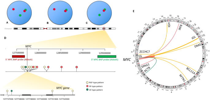
Our cohort included 14 cases of DLBCL/HGBCL evaluated in our clinical FISH laboratory between 2019 and 2021 and selected sequentially in inverse chronological order of sampling (with preference given to internal cases for clinical correlation). Seven cases had an unbalanced MYC BAP result, including 5 cases with a RF-type pattern and 2 cases with a GF-type pattern on MYC BAP analysis in the absence of an IGH partner by D-FISH. Seven specimens with a typical RGF-type pattern with a known MYC-IGH (n = 1) or unknown (n = 6) partner were also included. These served as controls to verify the ability of the WGS methodology from FFPE sections used in this study to identify rearrangements and dissect the genomic architecture at the MYC locus.
Whole genome sequencing results
A MYC-R was confirmed in all control cases with a balanced pattern on BAP FISH (Table 1, Fig. 1). These involved previously reported rearrangement partners/loci (IGH (n = 1), IGL (n = 2), ZCCHC7 (n = 1), RFTN1 (n = 1), DMD (n = 1) and BCL11A (n = 1)). A structural variant (SV) involving the MYC region was also detected in all 5 cases with a RF-type pattern. In line with the higher number of R signal(s) observed on BAP FISH, a relative gain of genomic material 5′ of the MYC gene or relative loss of material 3′ of the MYC gene was detected with WGS in all these cases. Putative fusion events juxtaposing MYC to (1) an intergenic region upstream of TG, (2) TG, (3) IRF8, and (4) SPAG1 was detected in 4/5 cases, while case 5 involved a copy number (CN) gain of MYC. WGS also allowed to resolve the unbalanced FISH results for cases with a GF-type pattern and revealed SVs leading to a higher copy number (CN) of the 3′MYC BAP FISH probe-binding sequence in comparison with the 5′ region in both cases with a GF-type pattern, thereby also reconciling BAP FISH results. The first case with a GF-type pattern juxtaposed MYC with ACTB and the second involved a CN loss including MYC and the 5’ FISH probe-binding sequence. Of all cases with unbalanced FISH results, this case represented the only one in which WGS revealed a deletion involving MYC in our study cohort. MYC overexpression by immunohistochemistry ( ≥ 40%) was detected in 4/5 and 2/2 cases with RF- and GF-type patterns, respectively, suggesting that in 6/7 cases, the unbalanced MYC rearrangement was associated with increased MYC expression. In 7 cases with a typical pattern, overexpression of MYC was documented by IHC.
Table 1 Correlation of fluorescence in situ hybridization and whole-genome sequencing results at the MYC locus in 14 patients with DLBCL/HGBL.Clinical correlation
Clinical information was available for 3 cases with a RF-type pattern (Supplementary Table 2). While one case exhibited a favorable response to MR-CHOP chemotherapy, the other two cases remained refractory to therapy (R-CHOP and R-CHOP followed by R-ICE, polatuzumab+bendamustine respectively) and expired from disease. In the first case with a GF-type pattern, a favorable response to R-CHOP chemotherapy was exhibited; nonetheless, the patient expired from sepsis and multi-organ failure. In the second case, no systemic therapy was provided to the patient.


留言 (0)